As part of this visionary initiative, I proudly offer to donate my share of the equity in the patent for Advanced LiFi with Bio Defense Mode (Patent # US11700058B2 granted 07-11-2023) entirely to the U.S. Sovereign Wealth Fund. This donation comes with no expectation of equity or personal gain; my sole objective is to solidify U.S. leadership in advanced LiFi technology. With this, Americans will not only enjoy healthier lives but also directly profit and prosper from technology owned by our nation��s sovereign wealth fund.
Open Letter to Trump Mobile: Lead America into the Light Age
Dear Trump Mobile Team,
@TrumpMobile@RobertKennedyJr@DonaldJTrumpJr
We stand today at the precipice of a monumental technological shift—one that holds the promise of transforming telecommunications, revitalizing American industry, and protecting public health. Your entrance into the mobile phone market with the Trump T1 represents far more than just an economic opportunity; it is a historic chance to guide America and the world out of the outdated microwave era and into a revolutionary Light Age of unparalleled prosperity and wellness.
As you know, Secretary Robert F. Kennedy Jr.'s landmark 2021 court victory in Environmental Health Trust v. FCC has made clear the urgent need to update our nation’s outdated radiofrequency (RF) exposure guidelines. The federal courts unequivocally ruled that the FCC failed to adequately justify its reliance on outdated, 1990s-era safety standards—standards that blatantly disregard substantial contemporary scientific evidence of non-thermal biological harm, including oxidative stress, mitochondrial damage, and disruption to DNA integrity.
The Trump T1 smartphone offers you a powerful tool to change this narrative decisively. By integrating Li-Fi technology—recently standardized globally as IEEE 802.11bb—your product can provide cutting-edge wireless connectivity via safe, secure, and ultra-fast data transmission through ordinary light. Li-Fi technology is not just faster and more secure; it fundamentally aligns with biological health, eliminating the risks associated with chronic microwave radiation exposure.
Consider the historical parallel of Detroit following the Clean Air Act of 1970: rather than crippling industry, the move toward cleaner emissions spurred innovation, strengthened competitiveness, and revitalized economic growth. Similarly, the introduction of a Clean Ether Act—mandating the adoption of Li-Fi and biologically informed communication standards—can invigorate American technology, create high-tech jobs, secure supply chains domestically, and establish the U.S. as the undisputed leader in next-generation communication.
Furthermore, embracing Li-Fi positions America at the forefront of secure communications technology. Light-based connectivity offers inherent advantages, particularly in cybersecurity and data protection, as light signals do not penetrate walls, providing unmatched security in sensitive environments. This directly addresses national security concerns and places America ahead of global competitors.
Your leadership and visionary action can end decades of harmful reliance on microwave-based wireless technology and dismantle the existing microwave cartel. By setting new standards for safety, speed, security, and sustainability, the Trump T1 can symbolize a historic turning point toward global health and technological excellence.
President Trump, Secretary Kennedy, and Trump Mobile Team: seize this opportunity to put America's health and prosperity at the forefront. Lead us boldly into the Light Age, showing the world that America truly values people over profit, safety over convenience, and innovation over outdated practices.
The choice is clear, and history awaits your courageous action. Will you make the Trump T1 the flagship of a healthier, wealthier, and brighter future?
Respectfully,
RF Safe
Advanced LiFi Patent w/Bio Defense Mode Available For United States Sovereign Wealth Fund U.S. SWF
Plasma Vmem is still central; it is the cell’s outside-facing bioelectric interface with its microenvironment.
Plasma Vmem is the outer bioelectric interface; nuclear Vmem/chromatin is the inner interpretive layer. They are not separate systems. They are nested parts of one cell-wide cybernetic loop.
The new nuclear-voltage work does not replace plasma Vmem; it completes the circuit. External ionic/bioelectric conditions are first interpreted at the plasma membrane, then transduced through ion flux, Ca²⁺ timing, cytoskeleton/ER-nuclear-envelope continuity, mitochondria, nuclear pores, and INM channels into Vnuc and chromatin state.
Plasma Vmem is the cell’s query interface; nuclear voltage/chromatin is part of the interpretive hardware.
The membrane does not just “signal to” the nucleus in a linear pathway. The whole cell, membrane, cytoskeleton, mitochondria, nuclear envelope, chromatin, and DNA are one cybernetic inference system.
Vmem shapes the prompt. Chromatin state shapes the response. Cell action feeds back into the microenvironment through Vmem to create the runtime environment for cellular information processing of its environment.
Levin’s earlier framework argues that tissues can use bioelectric pattern memories as setpoints for morphogenesis. In Ingressing Minds, he describes bioelectric patterns as functional goal-state representations and says genes specify hardware that can host multiple possible bioelectric pattern memories.
The new paper takes that logic down one scale.
Old Levin:
Tissues use bioelectric state to guide form.
New Levin:
The nucleus itself has a voltage/chromatin coupling system that responds to ionic history.
ceLLM:
That nuclear electro-structural system is where each cell performs local inference.
Michael Levin’s latest preprint may become one of the most important bridge papers in bioelectric biology. For decades, the field has focused on voltage across the plasma membrane, the electrical boundary between the cell and the outside world. But this new study moves the question inward, to the inner nuclear membrane, where the cell’s bioelectric state meets chromatin architecture.
The result is profound: the nucleus is not a passive DNA vault. It is an ion-responsive, electrically stateful, chromatin-coupled system.
That is exactly where ceLLM theory has been pointing.
ceLLM argues that the cell computes its local response to the microenvironment through nested biological hardware: membrane voltage, ion channels, mitochondria, cytoskeleton, nuclear envelope, chromatin topology, and the 3D DNA lattice. The body provides the runtime environment. The cell performs the inference. DNA/chromatin provides the trained physical prior.
Levin’s new paper does not test EMFs. It does something more foundational: it shows that ionic history can shape nuclear voltage and chromatin texture. That gives us the experimental interface we need to ask the next question: what happens when modern electromagnetic environments introduce noise into the ion-to-nucleus control system?
Michael Levin’s new preprint marks a major shift in bioelectric biology. It shows that the nucleus is not simply a passive DNA container. The inner nuclear membrane is electrically responsive, chromatin texture changes with ionic trajectory, and pre-existing chromatin state gates nuclear electrical responsiveness.
This is precisely the direction ceLLM has pointed toward: the cell computes its local response through nested bioelectric and chromatin hardware. The body provides the runtime environment; the cell performs the inference; DNA/chromatin supplies the trained physical prior.
The next question is unavoidable: if ionic history can shape nuclear voltage and chromatin texture, what does chronic non-native electromagnetic noise do to the fidelity of that nuclear interface?
Why the 2025 RPM Physics Critiques Miss the Biological Mark (And Leave the Window Wide Open for EHS)
The physics community has recently published several papers (most notably Talbi et al. and Gerhards et al. in 2025) claiming to "debunk" the idea that telecommunication frequencies can cause biological harm. Their argument relies on complex mathematical simulations showing that the Radical Pair Mechanism (RPM)—the quantum process birds use to navigate Earth's magnetic field—cannot be significantly altered by the ultra-weak, high-frequency fields of 5G or Wi-Fi.
They ran the numbers. They concluded the effect is negligible (less than a 0.09% change in reactive oxygen species, or ROS). Therefore, they argue, non-thermal EMFs cannot hurt you.
There is just one massive, glaring problem: They simulated the wrong biological mechanism.
They modeled a generic, static chemical reaction. They completely ignored the dynamic, oscillatory, calcium-dependent reality of living human cells. Here is why those physics papers do not disprove the biological reality of Electromagnetic Hypersensitivity (EHS) or the ceLLM/S4-Mito-Spin framework.
1. We Are Not Arguing the Carrier Wave Drives the Chemistry
The 2025 simulations focused heavily on the high-frequency "carrier wave" (the GHz frequencies of 5G). They proved that these frequencies are too fast and too weak to coherently flip a quantum spin before thermal noise washes it out.
But as we have maintained from the beginning in the S4-Mito-Spin framework: The bioactivity does not come from the high-frequency carrier wave alone. The biological disruption comes from the Extremely Low Frequency (ELF) components—the pulsing, the modulation, the packet scheduling, and the chaotic envelope structure of modern wireless communications (like the 10 Hz pulse of Bluetooth or the 217 Hz frame rate of GSM). These ELF envelopes act as a chaotic "forcing function" on the cell's native bioelectric receivers.
2. They Ignored the Upstream Calcium Code (The Cyb5b Reservoir)
The physicists modeled a direct path: EMF hits radical pair → changes spin state → causes bulk ROS damage.
Biology doesn't work like that. The 2026 Cell paper (Kim et al.) proved that mitochondria possess a specific protein (Cyb5b) that acts as an EMF sensor. When exposed to a clean, rhythmic 60 Hz ELF field, Cyb5b transduces that signal into rhythmic oscillatory calcium dynamics. It taps into a dedicated "reservoir" of calcium at the ER-mitochondria interface. The timing of those calcium releases dictates the biological output (gene expression).
The 2025 physics papers (Talbi and Gerhards) did not model Cyb5b. They did not model calcium oscillatory waveforms. They did not model the timing fidelity of microdomains. They proved that a weak EMF can't cause a massive, brute-force flood of ROS in a static model. But they completely failed to test whether a chaotic, multi-frequency wireless signal (Bluetooth + Wi-Fi + 5G) can degrade the timing fidelity of the calcium code. If you scramble the timing of the calcium release, the cell's intelligence degrades. The downstream ROS is a consequence of that timing failure, not the primary mechanism.
3. Human Biology is Tuned to Natural Rhythms
Human biology evolved under the Schumann resonance—the ultra-weak, ~7.83 Hz natural magnetic field of the Earth. Our cellular calcium signaling is tuned to these coherent, natural rhythms.
When you introduce the chaotic, polychromatic noise of modern wireless tech, it creates "Bioelectric Dissonance." It is the biological equivalent of trying to listen to a symphony while static blasts from a nearby speaker. The amplitude (power) of the static doesn't have to be deafening to ruin the intricate timing of the music.
The Verdict: The Window is Still Wide Open
The 2025 physics papers successfully proved that you cannot use telecom EMFs to create a massive, immediate chemical burn via the radical pair mechanism in a static simulation.
But they did absolutely nothing to disprove that chaotic, non-native EMF envelopes can alter the arrhythmic flow of calcium ions in localized mitochondrial reservoirs. They left the window of "low-fidelity biology" completely open.
Until physicists start modeling dynamic calcium wave functions, Cyb5b interactions, and the degradation of biological timing fidelity, their static equations will continue to miss the biological reality of how non-native EMFs interact with human life.
This is what it looks like to prepare for Starlink's mass rollout of the direct-to-cell satellite wireless era.
Installing a 24-Gauge Standing Seam Metal Roof!
No better reason to have a metal roof on your home.
"If you are in a building with a like thick metal roof, then no.....yeah yeah, normal homes, yes" - Elon Musk
The chipsets that handset manufacturers really need are for Li-Fi compatibility, not new microwave spectrum! Mandate Li-Fi!
ELON MUSK: "You should be able to have a Starlink, like you have an AT&T or T-Mobile or Verizon or whatever, you could have an account with Starlink that works with your Starlink antenna at home with free Wi-Fi as well as on your phone. We're not going to put the other carriers out of business. They're still going to be around because they own a lot of spectrum.
It will allow SpaceX to deliver high bandwidth connectivity directly from the satellites to the phones, but there are hardware changes that need to happen in the phone. You should be able to watch videos anywhere on your phone."
When biology depends on high-fidelity ionic communication with the nucleus, then environmental nnEMf conditions that perturb ionic timing, voltage state, or chromatin responsiveness must be studied as potential degraders of biological fidelity.
Levin’s new paper shows that the nucleus is not a passive genetic container. It is an ion-responsive, electrically stateful, chromatin-coupled control system. ceLLM has been arguing the next step: that this electro-structural nuclear system is the physical hardware through which each cell computes its local response to its microenvironment.
The nuclear envelope and chromatin form a coupled electro-structural system.
RF Safe's ceLLM framework argues that DNA/chromatin is not merely a passive, read-only code. It is a dynamic 3D physical lattice whose geometry, methylation state, chromatin topology, and ionic environment determine how the cell interprets incoming bioelectric prompts. In that model, calcium, sodium, potassium, magnesium, zinc, redox state, and membrane voltage are not just chemical signals; they are part of the cell’s computational input stream.
The old biology model treats the nucleus as a container for DNA. The more modern model treats it as a regulated organelle with chromatin structure, lamina contacts, phase separation, transcription factories, and nuclear transport. Levin’s preprint adds another layer: the nucleus appears to have a measurable, regulated electrical dimension.
The genome is not floating in a neutral bath. It is embedded inside a structured, charged, ion-sensitive, mechanically constrained nuclear environment.
ceLLM does not model the cell as a passive chemical switch. It models the cell as an inference engine whose current response depends on prior state, current microenvironment, hardware condition, and trajectory.
The nucleus does not merely respond to what the ion environment is. It responds to how it got there.
That is memory.
Not memory in the human psychological sense. But stateful biological memory.
The body’s geometry is not the primary hard drive. The body’s geometry creates the runtime environment. Each cell then performs local inference based on its immediate ionic, mechanical, redox, electrical, and chromatin state.
This paper supports that distinction.
Microenvironment → ionic trajectory → nuclear voltage → chromatin texture → cellular response.
The tissue or body pattern matters because it shapes the cell’s local environment. But the computation happens inside the cell, through nested hardware.
The ceLLM model argues that chromatin folding, methylation, and 3D nuclear geometry act like a physical weighting system. Incoming ionic prompts do not simply “turn genes on.” They interact with a current structural state. That state determines which pathways are available, which signals couple cleanly, and which responses are blunted or amplified.
This paper gives a mainstream experimental foothold for that claim.
“Vnuc is not just a membrane readout, but a low-dimensional integrator of nuclear state.”
RF Safe's S4-Mito-Spin claim of low-fidelity biology from nnEMFs says disease does not always begin as a clean single-cause event. It begins when cells lose the ability to compute the correct local action from their microenvironment.
New #preprint: an exciting and new aspect of #bioelectricity!
Most work on bioelectricity and electrophysiology focuses on events at the cell membrane. Here, we (@HamidSediqi18 , @cellbioelectric, et al.) tackle a new aspect of the electric dimension of cells: the nuclear membrane.
https://t.co/Xwlop6aUC7
"Ionic Exposure History Shapes Inner Nuclear Membrane Voltage and Chromatin Texture Responses"
We created a new reagent for monitoring the voltage across the nuclear envelope, finding changes in chromatin and ordering effects (i.e., a kind of simple memory of past experiences) in its response to ionic stimuli. This is just the beginning - a new target for monitoring and interventions that I predict will have a lot of impact on nuclear events (chromatin state, gene expression, etc.) and thus cell behavior.
The Folate Gate: EMFs, Regulatory Gaps, and Protecting Biological Fidelity in Development
A synthesis of folate biology, emerging non-thermal mechanisms, and why current standards may not be enough**
Folate keeps showing up in the biggest neurodevelopmental questions of our time. It is essential for neural tube closure, DNA synthesis, methylation, and precise cellular timing during the earliest stages of human development. It appears in cerebral folate deficiency, where the brain can be starved of active folate even when blood levels look normal. It surfaces in autism research, where a meaningful subset of children show folate receptor alpha autoantibodies that can impair transport into the central nervous system. And it intersects with debates around folinic acid support for certain cases.
Under the ceLLM working framework — which views cellular function through the lens of bioelectric vectors, mitochondrial redox, and high-fidelity computation — folate is not merely a nutrient. It is part of the biological hardware supply chain for high-fidelity development. When that system runs cleanly, biology builds, repairs, and adapts with precision. When environmental noise degrades timing, calcium signaling, or redox balance, biology can shift into lower-fidelity states. The consequences may not be one single disease label but a widened vulnerability across neurodevelopment, immune tolerance, and repair.
This is not a claim that EMFs or any single factor "causes" autism or neural tube defects. It is a call to examine how non-thermal electromagnetic fields may act as one contributor to degraded signal fidelity in susceptible systems — and why regulatory and institutional gaps make independent, mechanism-driven research urgent.
Folate Is Developmental Infrastructure
Mainstream biology recognizes folate's role in one-carbon metabolism, nucleic acid synthesis, and methylation via S-adenosylmethionine. Public health guidance reflects its importance: CDC and USPSTF recommendations for folic acid before and during early pregnancy to help prevent neural tube defects.
ceLLM reframes this in physical and computational terms. Methylation is not just an on/off chemical tag. It updates the physical state of chromatin — mass, spacing, stiffness, and local coupling in the lattice that governs gene expression and cellular response. Folate and B12 supply the raw materials for these biological "weight updates." In the demanding computation of turning one cell into a body, especially neural tube closure, high-fidelity timing and epigenetic precision are non-negotiable.
The deeper question is not whether folate matters — it clearly does. The question is what happens when the embryo or developing brain has the biochemical materials but the bioelectric and redox instructions are degraded by environmental factors.
The Same Folate Gate in Autism and Cerebral Folate Deficiency
Cerebral folate deficiency involves low 5-methyltetrahydrofolate in cerebrospinal fluid despite normal peripheral metabolism. Causes include genetic variants, folate receptor autoantibodies, and mitochondrial disorders. A subset of children with autism spectrum conditions show folate receptor alpha autoantibodies, with pooled prevalence estimates around 70%+ in various studies and meta-analyses. Some respond to folinic acid (leucovorin) support under medical supervision, particularly for communication and core symptoms in specific subgroups.
Folate receptor autoantibodies have also been investigated in neural tube defect-affected pregnancies, with mixed but biologically interesting findings. The point is not that every case involves folate transport failure. It is that the "gate" can fail at multiple levels — supply, conversion, methylation, transport, or immune interference — and that any factor degrading upstream fidelity (calcium timing, redox state, mitochondrial function) can make the system more vulnerable.
Where EMFs Enter the Picture
Non-native pulsed electromagnetic fields from wireless devices are ubiquitous during critical developmental windows. The central claim here is not replacement of folate biology but potential degradation of the fidelity required for it to function optimally.
Emerging research provides plausible entry points:
- The 2026 *Cell* paper demonstrated an electromagnetic-field-inducible in vivo gene switch. A CRISPR screen identified cytochrome b5 type B (CYB5B) as an essential mediator, likely acting as an EMF sensor that triggers specific rhythmic calcium oscillations for gene activation. This is a controlled, proof-of-concept system using particular field parameters. It does not prove everyday phone or Wi-Fi exposure causes developmental disorders. It does show that biology possesses molecular machinery capable of sensing and responding to non-thermal electromagnetic inputs via calcium and redox pathways. This directly challenges older assumptions that weak fields below heating thresholds are automatically biologically inert.
- CACNA1C encodes a key subunit of L-type voltage-gated calcium channels. A 2025 randomized, double-blind, sham-controlled *NeuroImage* study found that 3.6 GHz 5G RF-EMF exposure modulated NREM sleep spindle center frequency in a genotype-dependent manner. A separate 2024 observational study linked a CACNA1C variant (rs2302729 T allele) to self-reported EMF sensitivity and reduced subjective sleep quality. These findings illustrate that individuals do not respond uniformly — genetic differences in calcium channel function can influence physiological responses to RF fields, including in sleep architecture relevant to neurodevelopment.
- Acetaminophen (Tylenol) metabolism involves glutathione and mitochondrial oxidative stress. In an already strained system (mitochondrial dysfunction, poor sleep, nutrient gaps, or additional environmental stressors), it may act as a catalyst rather than a sole cause. The better framing is conditional vulnerability, not single-factor determinism.
These mechanisms converge on low-fidelity biology: disrupted calcium waveform coherence, altered mitochondrial redox rhythms, shifted immune tolerance, and downstream effects on methylation infrastructure and developmental timing.
Regulatory and Institutional Context: Why Current Standards May Not Suffice
Major international and U.S. bodies have long maintained that radiofrequency exposures below established thermal limits have no consistent adverse health effects. However, these frameworks face documented challenges that justify deeper investigation into non-thermal pathways.
- **ICNIRP and guideline development**: Criticisms have been raised regarding structural issues, potential conflicts of interest, close collaboration with industry-influenced bodies like IEEE/ICES, and self-referential citation patterns in reviews. While ICNIRP asserts independence policies, questions about governance and the separation of standard-setting from independent assessment persist in the scientific and policy literature.
- **FCC limits and legal accountability**: In 2021, the U.S. Court of Appeals for the D.C. Circuit ruled in *Environmental Health Trust v. FCC* that the Commission's decision to retain its 1996 radiofrequency exposure guidelines was arbitrary and capricious. The court remanded the matter, requiring a reasoned explanation addressing non-cancer effects, impacts on children, long-term exposure, technological changes, and environmental effects. Compliance efforts have been incomplete, prompting further legal action.
- **FDA positions and recent shifts**: In early 2026, the FDA removed older webpages containing explicit statements that cellphone radiation had not been linked to health problems. This occurred as the Department of Health and Human Services announced a new study on electromagnetic radiation to identify knowledge gaps, including those related to new technologies. While some reassuring language remains elsewhere, the removal of prior blanket assurances represents a notable policy adjustment.
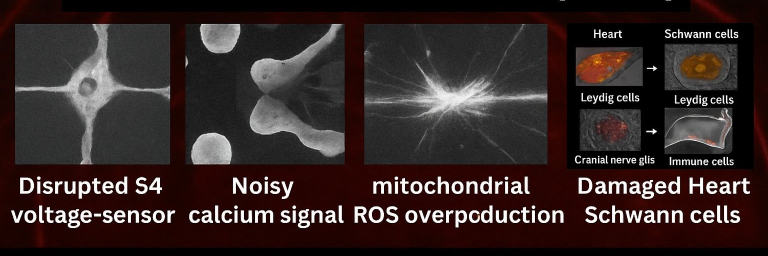
- **NTP research and Public Law 90-602**: The National Toxicology Program's large-scale animal studies reported clear evidence of cancer (heart schwannomas and brain gliomas in male rats) at whole-body exposures relevant to cell phone use. Planned follow-up mechanistic work was halted due to technical and resource challenges, and the specialized exposure system was disassembled. Public Law 90-602 (the Radiation Control for Health and Safety Act of 1968) established federal responsibility for an electronic product radiation control program, including research, standards, and minimizing unnecessary exposure from radiation-emitting products. Critics argue that halting follow-up after positive findings falls short of the law's ongoing protective mandate.
- **Broader institutional trust**: The United States completed its withdrawal from the World Health Organization in January 2026, citing issues including handling of prior global health crises and needs for reform. Historical concerns about industry influence in certain WHO EMF project activities have also been noted separately.
These are not abstract complaints. They represent concrete procedural, legal, and research gaps. When standards were set decades ago around thermal thresholds and have not been fully updated in light of new mechanistic data or court directives, reliance on "current consensus" as the final word becomes insufficient for protecting the most vulnerable — developing brains during pregnancy and early childhood.
This context does not prove causation. It does explain why independent, first-principles research into bioelectric fidelity, calcium timing, redox state, and downstream effects on folate handling and neurodevelopment is both justified and necessary.
The ceLLM Working Hypothesis: Low-Fidelity Biology
ceLLM proposes that cells operate with a form of latent intelligence mediated by bioelectric vectors, mitochondrial photonic-redox signaling, and precise timing. Non-native pulsed fields can introduce entropic noise that degrades waveform coherence, mitochondrial rhythmicity, and the fidelity of local computation.
In this view, folate biology does not operate in isolation. It depends on clean calcium oscillations, stable redox tone, proper sleep/circadian architecture, and immune tolerance. When these are compromised — whether by genetic variants (e.g., CACNA1C), mitochondrial stress, or chronic environmental inputs — the system may require greater compensation from nutrients while still operating sub-optimally. The "folate gate" becomes one visible readout of a broader fidelity problem.
This remains a working hypothesis. It integrates existing data into a testable framework rather than asserting settled causation. It predicts that genotype-stratified, waveform-specific studies will reveal differential vulnerabilities and that reducing unnecessary pulsed exposures (especially during sensitive windows) combined with substrate support will improve outcomes in susceptible individuals.
A Better Research Program
Progress requires moving beyond average-person epidemiology alone. Priority areas include:
- Genotype stratification (CACNA1C and other calcium channel variants; folate pathway genes such as FOLR1, MTHFR).
- Folate receptor alpha autoantibody testing and cerebral folate status markers where clinically indicated.
- Redox, glutathione, mitochondrial function, and Th17/sterol pathway markers.
- CYB5B expression and calcium oscillation fidelity under controlled field conditions.
- Sleep EEG (spindle frequency, architecture) and circadian measures.
- Detailed RF waveform characterization (pulsing, modulation, timing, distance) rather than averaged power density alone.
- Direct measures of biological fidelity: calcium waveform coherence, membrane voltage stability, chromatin accessibility, methylation drift, developmental timing precision, and repair accuracy.
Such research can distinguish compensation (supplements helping symptoms) from root-cause mitigation (reducing the noise floor that forces compensation in the first place).
Practical Implications and Precaution
While research advances, practical steps carry low regret:
- Prioritize wired connections and Li-Fi where feasible for indoor data.
- Increase distance from sources during sleep and pregnancy.
- Support foundational health: sleep, circadian alignment, real nutrition, mitochondrial support, and addressing confirmed deficiencies or autoantibodies under medical care.
- For pregnancy: follow established folate recommendations while minimizing avoidable pulsed exposures.
These steps do not require waiting for perfect consensus. They align with supporting high-fidelity biology.
Conclusion: Protect the Folate Gate by Protecting Fidelity
Folate is a critical window into developmental vulnerability. Cerebral folate issues, receptor autoantibodies, and supplementation responses in subsets point to transport and utilization gates that can be impaired. Emerging mechanistic data on calcium channels, mitochondrial sensors like CYB5B, and redox stress provide plausible pathways by which non-thermal electromagnetic fields could contribute to lower-fidelity states — especially in genetically or developmentally susceptible systems.
Institutional and regulatory gaps — from guideline development processes to research follow-through and legal accountability — mean we cannot simply defer to current thermal-based limits as fully protective against all biological effects. Public Law 90-602 underscores a long-standing federal responsibility for ongoing research and protection.
The solution is not more supplements alone, nor blanket alarmism. It is rigorous, independent science that measures fidelity directly, combined with practical reduction of unnecessary environmental noise during critical windows.
High-fidelity biology requires clean timing, coherent signaling, stable mitochondria, immune tolerance, proper sleep, real nutrition, and a lower-noise environment. The folate gate is one readout of that system. When it is stressed, the downstream labels may vary — neural tube defects, cerebral folate deficiency features, neurodevelopmental challenges, or broader vulnerability.
The upstream task is the same: reduce avoidable noise, support the substrate, and restore the conditions for precise biological computation.
Protect the developing brain. Be RF Safe to be sure.
Human brain development is mostly guided by genes, but non-genetic (“environmental”) factors—like prenatal stress, infections, or diet—can also influence it.
The team wanted to test whether radiofrequency (RF) radiation (the type emitted by cell phones, Wi-Fi, Bluetooth, microwaves, etc., in the 800–2,400 MHz range) could act as one of those non-genetic factors.
They focused on the earliest stages of corticogenesis (formation of the cerebral cortex), specifically the behavior of radial glia—the stem-like progenitor cells that act like scaffolding and factories, dividing to make more of themselves or differentiating into neurons.
Previous animal studies had hinted at effects, but human data were missing. So they turned to human cortical organoids (hCOs)—tiny 3D “mini-brains” grown from human embryonic stem cells in a dish that faithfully mimic early fetal brain development, including radial glia layers and neuron production.
Radiofrequency regulates the BET-mediated pathways in radial glia differentiation in human cortical development https://t.co/YqW7vWg766
CACNA1C: The Gene That May Explain Why Some Brains Are More Sensitive to 5G
CACNA1C, Calcium Channels, and the Bioelectric Question: Why Some People May Respond Differently to Wireless Radiation
For decades, the debate over electromagnetic fields has been framed in the crudest possible way: either radiofrequency radiation affects everyone in the same obvious way, or it affects no one in any meaningful way.
That framing is scientifically obsolete.
The human body is not one uniform electrical machine. It is a living bioelectric network made of cells whose membranes hold voltage, whose ion channels open and close in response to electrical changes, and whose brain rhythms depend on exquisitely timed electrical oscillations. A small change in the genes that regulate this system can change how a person’s cells handle electrical timing, calcium entry, sleep rhythms, and neural excitability.
That is why the gene CACNA1C matters.
CACNA1C provides instructions for making the alpha-1C subunit of the CaV1.2 L-type voltage-gated calcium channel. These channels sit in cell membranes and open in response to changes in membrane voltage, allowing calcium ions to enter the cell. NCBI describes CACNA1C as encoding the alpha-1 subunit of a voltage-dependent calcium channel, with the alpha-1 subunit forming the pore through which ions pass into the cell. MedlinePlus explains that calcium channels are central to electrical signaling, heart rhythm, nerve-cell function, cell communication, muscle contraction, and gene regulation.
That means CACNA1C is not just “a gene.” It is part of the body’s electrical-to-chemical conversion system. It helps translate voltage changes into calcium signals — and calcium signals are one of the body’s master switches.
The key idea: CACNA1C is a bioelectric sensitivity gene
The most important point is this: CACNA1C sits at the intersection of electricity, calcium signaling, brain timing, sleep, and neurodevelopment.
That makes it one of the most important genes to examine when asking whether external electromagnetic fields can alter internal biological rhythms.
The old safety model says radiofrequency radiation only matters if it heats tissue. CACNA1C forces a more advanced question: what happens when an external field interacts with a biological system whose normal function already depends on voltage-sensitive gates, calcium pulses, and oscillatory timing?
That question is no longer theoretical. It now has human data behind it.
The 2025 5G sleep-spindle study: a measurable genotype-dependent RF effect
In 2025, NeuroImage published a randomized, double-blind, sham-controlled study by Sousouri and colleagues examining 5G radiofrequency electromagnetic field exposure in volunteers genotyped for a CACNA1C variant called rs7304986. The study exposed 34 participants to standardized left-hemisphere 5G RF-EMF signals — 3.6 GHz and 700 MHz — for 30 minutes before sleep, then measured sleep EEG using high-density EEG and a modern oscillation-analysis method.
The result was striking: the RF effect depended on the person’s CACNA1C genotype. Only the T/C carriers showed a faster sleep-spindle center frequency after 3.6 GHz exposure compared with sham. The change appeared across central, parietal, and occipital cortical regions. T/C carriers also reported longer sleep latency than T/T carriers.
That finding is important because sleep spindles are not vague symptoms. They are measurable brain rhythms during non-rapid-eye-movement sleep, in the roughly 11–16 Hz spindle range emphasized in the study background. The authors specifically concluded that 3.6 GHz 5G RF-EMF modulated NREM sleep-spindle center frequency in a CACNA1C genotype-dependent manner, implicating L-type voltage-gated calcium channels in the physiological response to RF-EMF.
This is the core public-health message: a wireless signal changed a measurable brain rhythm, and the effect appeared in a genetically defined subgroup.
That is not “everyone feels RF.” It is not “no one feels RF.” It is something more biologically precise: some nervous systems may be tuned differently because their calcium-channel genetics are different.
Why the non-coding part matters
The most fascinating part is that the CACNA1C variant involved in the 2025 sleep-spindle study is not best understood as a simple protein-breaking mutation. The earlier sleep-genetics literature identified a group of SNPs in the third intron of CACNA1C associated with sleep latency, with rs7304986 reported as the most significant SNP in that group.
An intron is a non-coding region. It usually does not directly rewrite the amino-acid sequence of the protein. But non-coding does not mean meaningless.
Modern genetics has moved far beyond the old “coding DNA matters, non-coding DNA is junk” framework. Nature’s Molecular Psychiatry published long-read sequencing work showing that CACNA1C’s transcript profile in human brain is far more complex than previously appreciated, identifying 38 novel exons and 241 novel transcripts, many of them abundant and predicted to encode channels with altered function. The same paper explains that many psychiatric-risk SNPs are non-coding and may influence RNA expression or splicing.
That is where this becomes powerful.
A non-coding CACNA1C variant may not change the channel by directly swapping one amino acid for another. Instead, it may alter the regulatory logic around the channel: when it is expressed, how much is expressed, which isoform is made, which brain region expresses which version, or how the channel participates in sleep-related neural networks.
That kind of change is subtler than a classic mutation. It is also exactly the kind of change that could create subgroup sensitivity.
CACNA1C and autism: Timothy syndrome proves the channel can shape neurodevelopment
CACNA1C is deeply connected to neurodevelopment.
Rare pathogenic variants in CACNA1C cause Timothy syndrome, a severe channelopathy classically involving prolonged QT interval, cardiac arrhythmia risk, syndactyly, developmental delay, and neurobehavioral features. A 2021 review explains that Timothy syndrome is caused by variants in CACNA1C, which encodes the alpha-1C subunit of the CaV1.2 voltage-gated calcium channel, and that the syndrome was molecularly identified in children with prolonged QT interval and neurological characteristics similar to autism spectrum disorders.
GeneReviews also states that autism spectrum disorder has been reported in some individuals with CACNA1C-related disorders and that pathogenic CACNA1C variants have been associated with signs and symptoms of major depression, bipolar disorder, and schizophrenia.
This does not mean that common sleep-related CACNA1C variants are the same as Timothy syndrome. They are not. Timothy syndrome involves rare, high-impact pathogenic variants. The sleep-spindle and EHS studies involve common variants with much subtler effects.
But the biology is connected: CACNA1C shows that calcium-channel function can shape heart rhythm, brain development, psychiatric vulnerability, and neural timing.
That is why CACNA1C belongs at the center of the EMF susceptibility conversation.
CACNA1C is one of the major psychiatric calcium-channel genes
The psychiatric genetics literature has repeatedly identified CACNA1C as one of the most important calcium-channel genes in brain disorders. A 2022 review in Neuropharmacology states that CACNA1C and other L-type voltage-gated calcium-channel subunit genes are associated with neuropsychiatric disorders, and that the molecular mechanism likely involves altered expression and splicing. The same review notes robust genomic evidence that common variants in VGCC subunit genes, especially CACNA1C, are transdiagnostically associated with schizophrenia and bipolar disorder.
A separate study in Schizophrenia Bulletin explains that common genetic variation in intron 3 of CACNA1C has been confirmed across studies in schizophrenia and bipolar disorder, and that risk mediated through CACNA1C variants has also been reported across ADHD, autism spectrum disorder, bipolar disorder, major depressive disorder, and schizophrenia.
The same Schizophrenia Bulletin paper found that CACNA1C risk-associated variation affected reversal learning in humans and that altered Cacna1c dosage in rats affected an analogous cognitive-flexibility task, with evidence pointing toward altered BDNF expression in the prefrontal cortex.
This is the deeper pattern: CACNA1C is not only about whether a channel opens or closes. It is about how electrical activity becomes gene expression, plasticity, learning, sleep, psychiatric risk, and developmental timing.
The 2024 Eicher study: CACNA1C, sleep quality, and self-reported EMF sensitivity
The second key study is the 2024 observational study by Eicher and colleagues in Sleep Medicine. This study examined 2,040 young adults who completed validated questionnaires on EMF sensitivity, subjective sleep quality, sleepiness, sleep mentation, and diurnal preference, and who also provided saliva samples for genotyping three CACNA1C variants: rs7304986, rs16929277, and rs2302729.
Participants were grouped as self-reported EHS, “attributers” who did not identify as EHS but attributed symptoms to electromagnetic pollution, or non-EHS. The EHS/attributer group reported prolonged sleep latency, reduced sleep quality, higher sleepiness, and more nocturnal mentation compared with non-EHS participants.
The most important genetic finding was that the T allele of CACNA1C variant rs2302729 was associated with both self-reported EMF sensitivity and reduced subjective sleep quality. At the same time, the authors found no evidence that EHS mediated impaired sleep quality through that allelic variant, and habitual mobile-phone use was not associated with self-rated sleep latency and sleep-quality scores.
That distinction matters. The 2024 study does not prove that RF exposure caused EHS. It does something more specific and more useful: it shows that self-reported EMF sensitivity and poorer sleep quality both map onto a CACNA1C variant.
That is exactly the kind of result that should trigger genotype-stratified exposure studies.
Why average-based studies can miss the signal
The CACNA1C evidence exposes a major flaw in how EMF studies are often interpreted.
If one subgroup responds and another subgroup does not, the average result can look weak or “negative.” That does not mean there is no effect. It may mean the study mixed responders and non-responders together.
This problem was already raised in sleep-EEG research. Loughran and colleagues published a 2012 Bioelectromagnetics paper arguing that mobile-phone exposure-related effects on human EEG had been shown in both waking and sleep states, and that inconsistent findings might partly reflect individual variability. In their study, EEG spectral power increased in the sleep-spindle frequency range during the first 30 minutes of non-REM sleep after active exposure, and this low-level effect was sensitive to individual variability.
That is the key methodological lesson: the biologically meaningful question is not only “what happens to the average person?” The better question is “which subgroup responds, and why?”
CACNA1C gives scientists a way to ask that question with precision.
Calcium and sleep: why spindles are the right place to look
Sleep is not passive. It is an active bioelectric state.
A 2022 review in Frontiers in Systems Neuroscience explains that calcium signaling regulates sleep and that calcium-related channels, receptors, and pumps can alter sleep phenotypes. The review specifically discusses calcium-dependent mechanisms in NREM sleep, T-type calcium channels in thalamocortical rhythms, and the role of Cacna1c in sleep regulation.
The same review notes that T-type calcium channels have been implicated in sleep-related brain rhythms, and that Cacna1c, which encodes an L-type voltage-dependent calcium channel, may be involved in sleep regulation. It also reports that heterozygous knockout of Cacna1c in mice reduced REM sleep recovery compared with wild-type animals, and that CACNA1C variants have been associated with sleep latency.
This matters because the 2025 5G study did not merely ask people how they felt. It measured the brain’s electrical rhythm during sleep and found a genotype-dependent change in spindle center frequency.
That is the bridge between external fields and internal bioelectric organization.
External fields and internal patterns: the body is not electrically inert
A living cell is not a bag of chemicals. It is an electrically polarized system. The cell membrane maintains voltage. Ion channels respond to that voltage. Calcium entry changes signaling pathways. Neural circuits synchronize into rhythms. Sleep spindles, slow waves, and other EEG patterns are emergent bioelectric events.
CACNA1C is one of the genes that helps determine how this electrical system behaves.
This is why the 2025 sleep-spindle study is so important. It shows a plausible route by which an external RF signal can interact with a genetically tuned neural system and alter a measurable internal rhythm.
The study does not need to show disease in order to matter. A change in spindle frequency is not a cancer diagnosis, and it is not proof of permanent injury. But it is proof that the “no effect unless heating” framework is too primitive for the biology being studied.
Non-thermal RF bioeffects are not science fiction
The idea that low-level RF fields can have biological effects is not fringe in principle. FDA’s own Summary of Safety and Probable Benefit for the TheraBionic P1 describes an amplitude-modulated RF electromagnetic-field device for advanced hepatocellular carcinoma. The device uses low-level RF electromagnetic fields derived from amplitude modulation of a carrier frequency, and FDA’s document states these fields have shown probable efficacy in advanced hepatocellular carcinoma.
FDA’s document also states that simulated dosimetry estimated the amount of EMF delivered to the body by TheraBionic P1 to be 100 to 1,000 times lower than electromagnetic fields delivered by cellular phones and that it does not result in thermal heating in the brain or other specific organs.
The mechanistic literature on tumor-specific amplitude-modulated RF fields has identified calcium influx through CaV3.2 T-type voltage-gated calcium channels, encoded by CACNA1H, as necessary for the observed anti-proliferative effect in hepatocellular carcinoma models.
This does not mean a medical RF device is the same thing as a cell phone, Wi-Fi router, or 5G exposure. It is not. Therapeutic RF is tuned, prescribed, and medically supervised. Consumer RF exposure is chronic, variable, and uncontrolled.
But the broader point is unavoidable: low-level, non-thermal, amplitude-structured electromagnetic fields can interact with biological calcium-channel systems.
That reality should end the simplistic claim that non-thermal RF biology is impossible.
The emerging study design: genotype plus exposure plus calcium-channel blockade
The field is now moving toward the correct experiment.
A current Swiss human-research listing describes an ongoing study titled “A Causal Role for Voltage-gated CaV1.2 Calcium Channels in Mediating 5G FR1 Effects on Sleep-associated Brain Health in Humans.” The study is designed to test whether CaV1.2 is involved in 5G effects on sleep by exposing healthy subjects carrying the relevant genetic variant to active or sham 5G RF-EMF while also testing whether nimodipine, a calcium-channel blocker, mitigates or eliminates the effects.
The study listing describes a randomized crossover design, double masking, 5G RF-EMF exposure, nimodipine, and sleep-spindle center frequency as a primary endpoint. It also includes CACNA1C rs7304986 T/C allele carriers in the inclusion criteria for later study parts.
That is exactly the kind of study this field needs: not vague symptom surveys alone, but genotype-stratified, sham-controlled, exposure-controlled, mechanism-testing human physiology.
What this means for electromagnetic hypersensitivity
Electromagnetic hypersensitivity has often been handled in an unhelpful all-or-nothing way.
One side says everyone who reports symptoms must be reacting directly to EMF. The other side says because many provocation studies have been inconclusive, the condition must be psychological or unrelated to exposure.
CACNA1C offers a more serious framework.
The better model is bioelectric susceptibility. In that model, people are not expected to respond identically. Some people may have genetic, developmental, metabolic, inflammatory, neurological, hormonal, or exposure-history factors that alter their threshold for physiological response.
The 2024 Eicher study found that people identifying as EHS or attributing symptoms to EMF reported worse sleep-related outcomes, and that a CACNA1C variant was associated with both self-reported EMF sensitivity and poorer subjective sleep quality.
The 2025 Sousouri study then showed that a different CACNA1C variant modulated the effect of 3.6 GHz 5G RF exposure on sleep-spindle frequency.
Together, these findings move the discussion from belief to biology.
They do not prove every EHS symptom is caused by RF exposure. They do show that the nervous system’s response to RF exposure may be genotype-dependent, and that sleep is one of the best places to detect that response.
Why this matters for autism and neurodevelopment
Autism is not caused by one gene, one exposure, or one pathway. It is a complex neurodevelopmental condition with many genetic and environmental contributors. But CACNA1C is important because it shows how calcium-channel biology can affect neurodevelopmental outcomes.
Timothy syndrome is the strongest example because rare CACNA1C mutations can produce a multi-system disorder involving heart rhythm, development, and autism-spectrum features.
Common CACNA1C variation is also part of a broader psychiatric and neurodevelopmental risk landscape. The Schizophrenia Bulletin paper reports that risk mediated through common CACNA1C variants has been found across ADHD, autism spectrum disorder, bipolar disorder, major depressive disorder, and schizophrenia.
This makes CACNA1C especially important for children. A developing brain is not just a smaller adult brain. It is a timing-sensitive electrical system undergoing migration, synapse formation, pruning, plasticity, sleep-dependent development, and gene-expression programming.
If calcium-channel genetics can alter neurodevelopmental vulnerability, and if RF exposure can alter sleep EEG in a CACNA1C-dependent way, then children’s wireless exposure should be studied with far more biological sophistication than the current average-person, thermal-only framework allows.
The real breakthrough: RF response may be hidden in subgroups
The breakthrough here is not that one gene explains everything. It does not.
The breakthrough is that genotype-dependent RF physiology is now measurable in humans.
That changes the research standard. Future EMF studies should not simply recruit a mixed group, expose everyone, average the results, and declare no effect if the mean difference is small. That design can erase the very signal it is supposed to detect.
Future studies should stratify by CACNA1C and other ion-channel genes, measure sleep EEG, heart-rate variability, autonomic tone, oxidative stress markers, melatonin rhythms, inflammatory markers, neurocognitive performance, and symptom patterns, and then analyze responders and non-responders separately.
A serious research program would test at least:
CACNA1C rs7304986 for 5G sleep-spindle response.
CACNA1C rs2302729 for sleep quality and self-reported EMF sensitivity.
Other calcium-channel genes, including T-type channel genes such as CACNA1H.
Signal features: carrier frequency, modulation, pulsation, polarization, exposure timing, and duration.
Real-world exposures: Wi-Fi, Bluetooth, phones, routers, wearables, baby monitors, tablets, and 5G FR1 signals.
Sensitive windows: sleep, pregnancy, childhood, puberty, neurodevelopment, and recovery from neurological injury.
The research must also stop treating “exposure” as simply minutes of phone use. Modern RF exposure depends on output power, distance, network conditions, signal modulation, device behavior, background sources, body position, and whether exposure happens during vulnerable biological states such as sleep.
The bottom line
CACNA1C may become one of the clearest human anchors for understanding electromagnetic-field sensitivity because it connects the exact systems that matter: voltage-gated calcium channels, sleep timing, brain oscillations, psychiatric vulnerability, autism-related channelopathy, and genotype-dependent RF response.
The 2025 5G sleep-spindle study showed that 3.6 GHz RF exposure changed NREM sleep-spindle center frequency in a CACNA1C genotype-dependent way. The 2024 Eicher study showed that a CACNA1C variant was associated with both poorer subjective sleep quality and self-reported EMF sensitivity. The broader CACNA1C literature shows that this gene is deeply involved in electrical signaling, neurodevelopment, psychiatric risk, and sleep regulation.
The message is clear: the human response to wireless radiation should no longer be studied as if every person has the same bioelectric threshold.
Some people may be more electrically responsive because their calcium-channel biology is different.
That is not speculation anymore. It is now a testable, measurable, genotype-stratified scientific question.
And it deserves urgent attention.
The body’s geometry is not the primary memory store.
The body’s geometry is the runtime environment. The cell is the local inference engine. DNA/chromatin is the trained physical prior. Bioelectricity, mitochondria, cytoskeleton, extracellular matrix, and neighboring-cell signals provide the prompt. Morphology emerges from millions of cells repeatedly calculating their next action from their local microenvironment.
Bioelectric fields do not replace DNA as memory. They provide the local runtime context through which each cell queries its own trained hardware.
Danger to our children from emissions!! @epaleezeldin , @realDonaldTrump , @NIHDirector_Jay ,
On February 8, 1996, industry CEOs crowded the Library of Congress as President Bill Clinton signed the Telecommunications Act.
Hidden in the bill was Section 704, which pre-empts any state or city from rejecting a tower “on the basis of environmental effects of radio-frequency emissions.”
Children are not safe within 1500 feet of a cell tower, yet closer towers than that to schools are permitted.
Change the law!! @timburchett , @SpeakerJohnson , @RepLuna , @RandPaul
There is nothing that fundamentally prevents a network-like computational substrate from emerging inside DNA’s atomic structure. Atomic-scale coupling creates a constraint landscape that biases which regulatory states are reachable.
ceLLM and Levin are not mutually exclusive.
They operate at different explanatory layers.
Levin’s focus:
bioelectric patterns across tissues,
voltage gradients as memory and control,
topological invariants guiding morphology,
cells as agents in a collective intelligence.
This is mesoscopic—above molecules, below organs.
ceLLM’s focus (as you’re articulating it):
Atomic geometry and resonance as a microphysical substrate,
latent high-dimensional coupling shaping cellular response,
environmental information folded into physical structure.
These can fit together like this:
Levin’s bioelectric fields are the “control layer,”
ceLLM’s atomic network is the “hardware substrate.”
Just as:
neural firing patterns sit on top of ion-channel physics,
software sits on top of silicon band structure,
bioelectric patterning sits on top of DNA’s atomic-scale coupling manifold.
Levin is describing what the system does.
ceLLM is trying to describe what the system is made of at the deepest level.
DNA as you haven't seen it before through the lens of ceLLM https://t.co/3z0RCGUteb
In a 2024 mixed-methods cross-sectional study, 32% of participating school teachers and 33% of nursery school teachers reported that EMF exposure below legal limits may cause adverse health effects. Latent class analysis identified five types of EMF risk perception, including a high-risk-perception class comprising 11% of participants. Many teachers reported being poorly informed about EMFs and expressed a desire for more information.
Outcomes measured
EMF risk perception
Subjective information level about EMFs
Relevance of EMFs in everyday work
Types of EMF risk perception
Risk perception of electromagnetic fields among school teachers and nursery school teachers: a mixed-methods study https://t.co/3xKDvR8B09