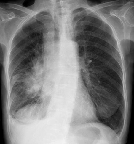
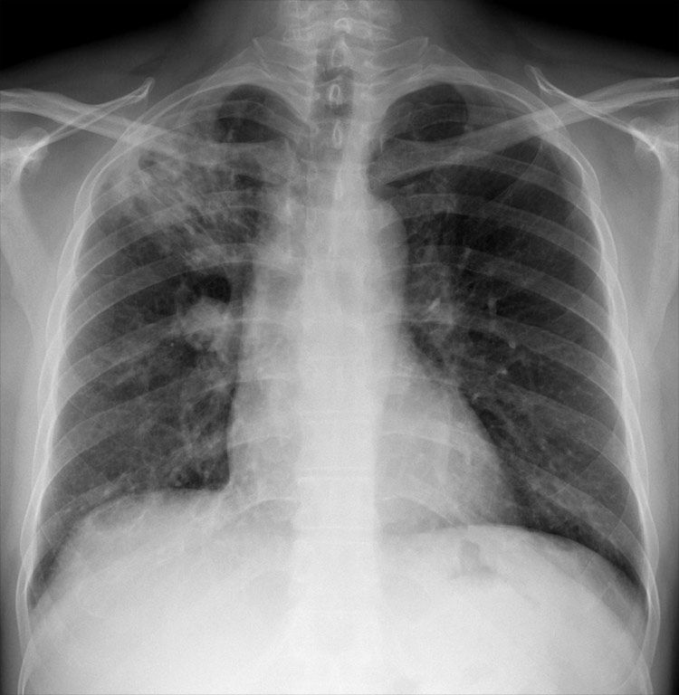
CHEST X-RAY 33: Shortness of breath and haemoptysis. Describe as many abnormalities as you can. What is the likely diagnosis? Answer https://t.co/qTT88AgczM
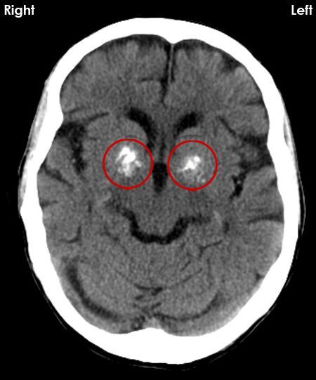
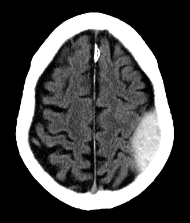
CT BRAIN 3: What are the white (calcified) structures ringed and what other structures seen on CT brain images can be considered normal if calcified? Answer https://t.co/g5Ypjyk1Bs
CHEST X-RAY 32: Why are the right heart border (white arrows) and the left heart border (black arrows) displaced? What would you do next? Answer https://t.co/bi6XuZrbuA
@TDekkersPhysio Any half decent radiologist would avoid the term ‘Modic’ for the very reason that patients don’t understand it and neither do doctors, not even bone doctors (neither orthopaedic surgeons nor rheumatologists).
Eponyms are rarely helpful in radiology.
Plain language is best.
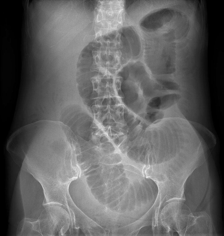
X-RAY QUIZ: 24-year-old patient with suspected appendicitis. What does the image show?
A: Caecal volvulus
B: Normal appearances
C: Pneumoperitoneum
D: Small bowel obstruction
E: Toxic megacolon
Start Quiz https://t.co/YfAknCatxy
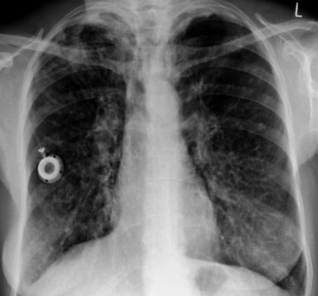
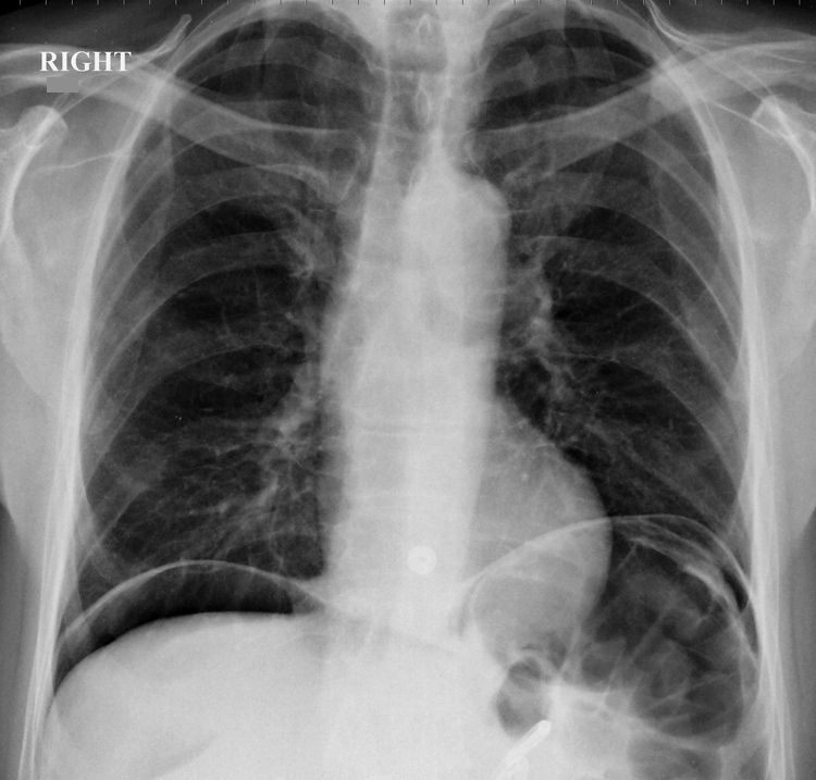
CHEST X-RAY 31: Patient with repeated chest infections. What is the medical device and what is the underlying diagnosis? Answer https://t.co/jBdJAdC8lV
X-RAY QUIZ: Acute abdominal pain. What would you do next?
A: Place an abdominal drain
B: Request abdominal ultrasound
C: Request abdominal MRI
D: Resuscitate the patient and inform the surgeons
E: Take a break
Start Quiz https://t.co/mgvgKXJC0i
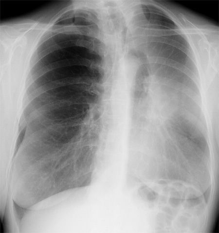
CHEST X-RAY 30: Cough, shortness of breath, weight loss, smoker. What is the radiological abnormality and what is the likely underlying cause? Answer https://t.co/R8e7NQsRiS
X-RAY QUIZ: Cough. What is the most likely cause of the chest X-ray appearances?
A: Pulmonary oedema
B: Lung metastases
C: Tuberculosis infection
D: Previous spontaneous pneumothorax
E: Sarcoidosis
Start Quiz https://t.co/v6wtWrkJot