Juega y aprende con los casos @Kahoot de #radiología musculoesquelética de #SERME
👨💻👩🏼💻👩🏾💻
#MSKRad@MskSerme
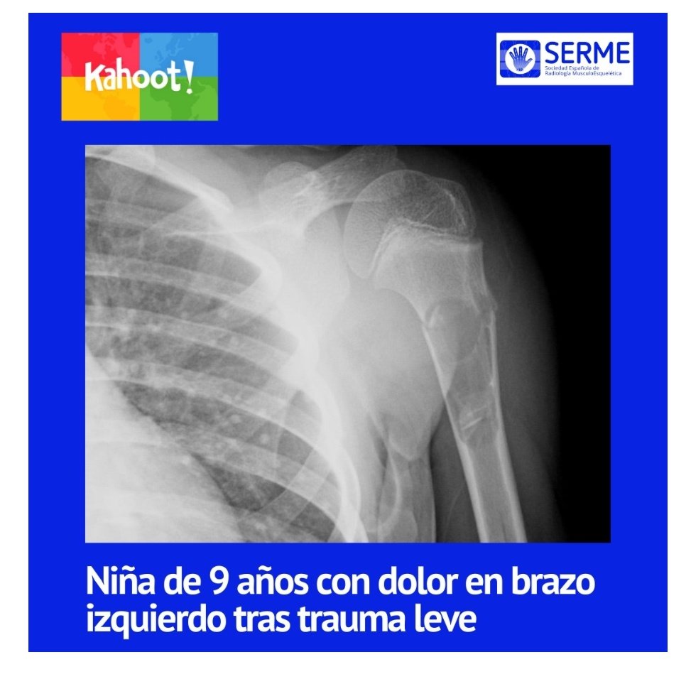
Niña de 9 años
Dolor en brazo tras trauma leve
👇🏼
https://t.co/BhOqe47irf
Update on Adult Acquired Flat Foot Deformity Using MR Imaging https://t.co/7qSq4DBpz3
Fresh out of the oven: New paper in MRI Clinics with Montefiore (NY) and HUNSC Tenerife on adult acquired flatfoot and the value of MRI in assessing PTT and spring ligament failure.
Challenge time.
Here’s an MRI (3 planes FSPD and sagital PD) of the knee from a patient with medial compartment pain. One image, one diagnosis.
What do you see? Drop your answers below! 👇 #Radiology#MedTwitter#FoaMed#orthotwitter
Young adult male sustains trauma to the dorsum of the hand while trying to break a fall. History of old distal radius and ulnar styloid fracture
Hand AP & oblique views.
Diagnosis? What would be your best management option?
#FoaMed#Trauma
Posterior interosseous nerve syndrome:
There is a compression of the posterior interosseous branch of the radial nerve at the arcade of Frohse, with focal thickening of the nerve.
#mskrad#radres#mskus@SermeMsk
👇🏻
Right ischial apophysitis in a 14-year-old male showing significant bone marrow edema in the ischial tuberosity without avulsion, periosteal reaction, soft tissue edema, and hamstring tendinopathy with peritendinitis. Likely due to repetitive traction forces during football ⚽ 🏃
💭If bone edema is present in the lateral compartment, assess for MCL injuries (valgus mechanism)
💭If it is in the medial compartment, assess for LCL injuries (varus mechanism)
#mskrad#radres@SermeMsk
1 year has passed between these two radiographs. Initially, suspected sprained ankle.
Name the initial injury. Any complications? Can you explain the pathophysiology responsible for the evolution of the injury?
Radiofrecuencia pulsada e infiltracion corticonanestesica de nervio pudendo(bupi 5 ml+trigon)en paciente con neurofibroma perineal guiada con TC m. ¿Dudas?
Injury and trauma mechanism on the same x-ray. CT 3D rendering for surgical planning. Diagnosis? Lesion classification according to combined Weber and Lauge-Hansen systems?
Share your insights!
🦴 Historia de la Lesión de Hill-Sachs 🦴
La lesión de Hill-Sachs es un defecto óseo, que afecta a la cabeza del húmero, está estrechamente asociado con las luxaciones del hombro. Fue descrita en 1940 por los radiólogos de San Francisco (USA) Harold Hill y Maurice Sachs.
Recent ankle sprain, pain in the dorsal region of the midfoot. Classic sign on X-ray (interpreted as normal in the ER), complementary ultrasound for persistent pain. Diagnosis?
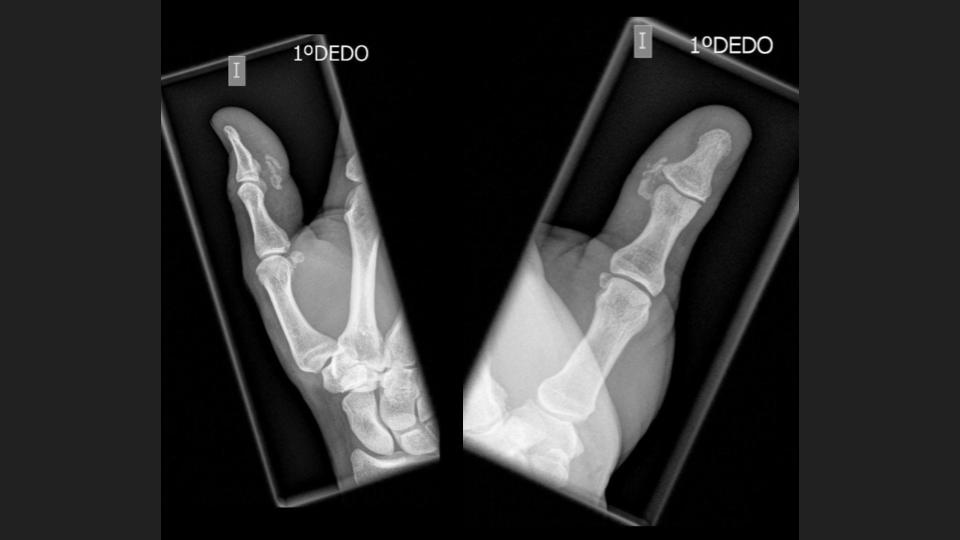
🖐 Varón de 51 años, dolor en 1er dedo.
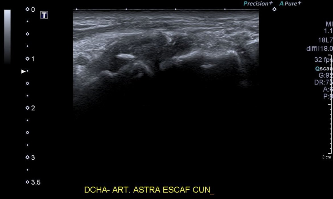
🔍 Dx: Periartritis calcificante en fase de reabsorción espontánea, con inflamación en el borde palmorradial de la articulación interfalángica.
💡 La periartritis calcificante causa dolor por depósitos de calcio (hidroaxiapatita calcica).