Every massive shift in scientific history started exactly like this: a single, dissenting voice challenging an established paradigm #glaucoma#ophthalmology#optometry
Glaucomatous field defects, RNFL thinning, splinter hemorrhages at the disc margin, and the end-stage histology of the glaucomatous disc devoid of nerve fibers suggest that the nerve fibers along with the vasculature are being severed in glaucoma #glaucoma#ophthalmology #optometry
Left-eye fundus of 48 y/o male with advanced glaucoma. Note the nasal shifting of blood vessels due to the unequal severance of nerve fibers #glaucoma#ophthalmology#optometry
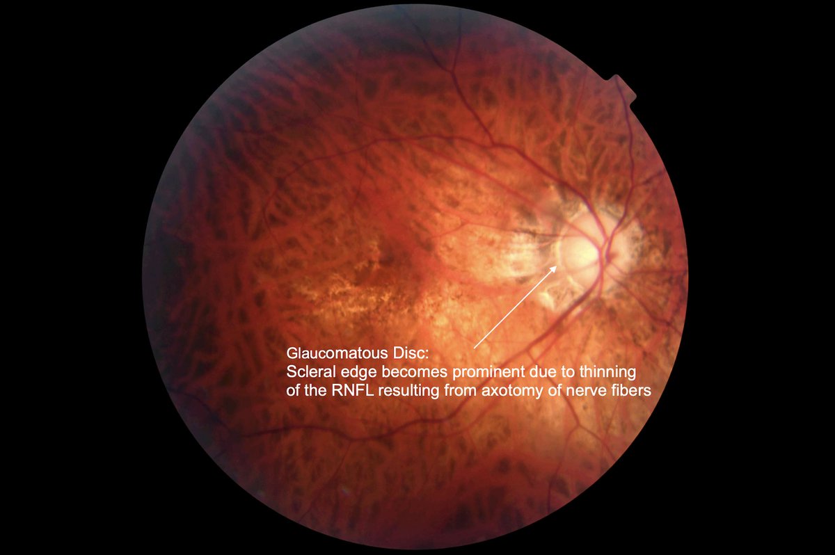
Left-eye fundus of 48 y/o male with advanced glaucoma. Prominent scleral edge due to RNFL thinning as a result of the severance (axotomy) of nerve fibers at the scleral edge #glaucoma#ophthalmology#optometry
Right eye of 37 year-old male with advanced glaucoma.
Notable observations:
- Prominent scleral edge (Hasnain sign) due to thinning of the retinal nerve fiber layer (RFNL), resulting from the AXOTOMY of nerve fibers.
- Sinking of the entire lamina cribrosa resulting in stretching and the axotomy of nerve fibers at the scleral edge.
- Absence of smaller vasculature (baldness) around the optic disc resulting from their severance (Hasnain sign).
Bottom Line: Chronic glaucoma may not be an optic neuropathy, but an optic disc axotomy #glaucoma #ophthalmology #optometry
Schematic (cross-sectional) view of the retinal nerve fibers after being severed at the scleral edge due to sinking of the lamina cribrosa occurring in chronic glaucoma #glaucoma#ophthalmology#optometry