Remember the #MiracleBerry that would make sour foods sweet? Wang et al (2018) used optogenetics (light stimulation) to trigger the same effects with mice and tracked their taste bud recognition in the amygdala, as well as how mice identify tastants. #neuroscience#optogenetics
6) The amygdala does not control tastant recognition, and silencing the amygdala does not silence gustatory (taste) neurons. The gustatory neurons want their 15 minutes of fame 🤭
fun fact: the amygdala probably links taste valence to other brain areas like learning and memory! In the future we can use this study to help is understand how these different brain areas work together to create innate and learned responses #neuroscience
https://t.co/XnuzYRbNeK
@GdeLavilleon So to conclude, the researchers induced explicit memory traces, leading to a goal-directed behavior toward the PF. Their findings demonstrate that place cells’ activity during sleep still conveys relevant spatial information that is functionally significant for navigation.
Ever wondered how neurons firing during #sleep can influence #memories? In 2015, @GdeLavilleon et al. created reward-based memories in mice when #placecells fired during sleep. So, place cell activity during sleep can still convey relevant spatial information! doi:10.1038/nn.3970
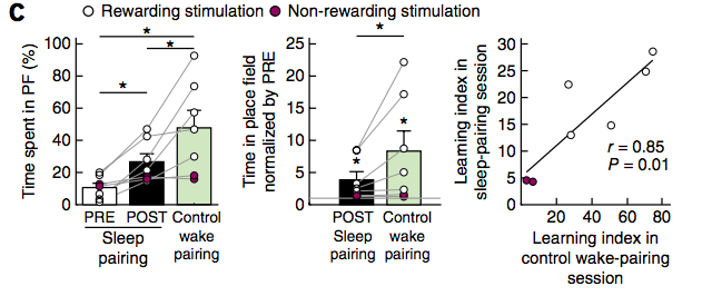
@GdeLavilleon When mice did not get rewarding stimulation via the MFB (purple dots), they did not show place preference for the PF. There was also a significant correlation between sleep-learning 😴🧑🏫(measured as an increase in time spent in PF from PRE to POST trials) and stimulation of MFB.
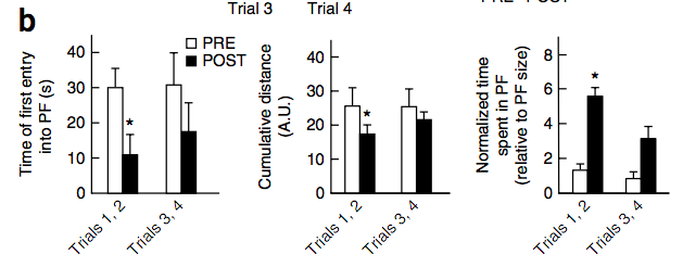
@GdeLavilleon Mice spent significantly more time in the PF during earlier trials (1 + 2) from PRE to POST, but not during later trials (3 + 4). Lavilleon et al. say that this is consistent with results “classically observed during unrewarded probe tests” (pg. 494).
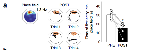
@GdeLavilleon Compared to the PRE sessions, mice took significantly less time to run to the PF in POST trials (when pairing trials). However as trials went on, the mice became less interested in the PF and ran around more. 🏃🐁
There was low/non-significant correlation between PRE/POST occupancy maps (grey) and between PRE occupancy and firing maps (white). However, both POST sleep-pairing (black) and wake-pairing (yellow) placement cells fired significantly in correlation to increased occupancy time.
@GdeLavilleon The wakeful🌞 (B) and sleep 😴(C) animals both showed strong preference for the associated PFs post-spike placement. The PF as a percentage (blue) was equivalent to pre-placement time spent in PF (white). Post-spike placement, time spent in PF almost quadrupled (black). 🐀🧠⚡️
@GdeLavilleon Their hypothesis? “The transient decorrelation of place cell activity from current location during sleep could provide a way to directly test the function of place cells in navigation.” (Lavilléon et al., pg. 493)