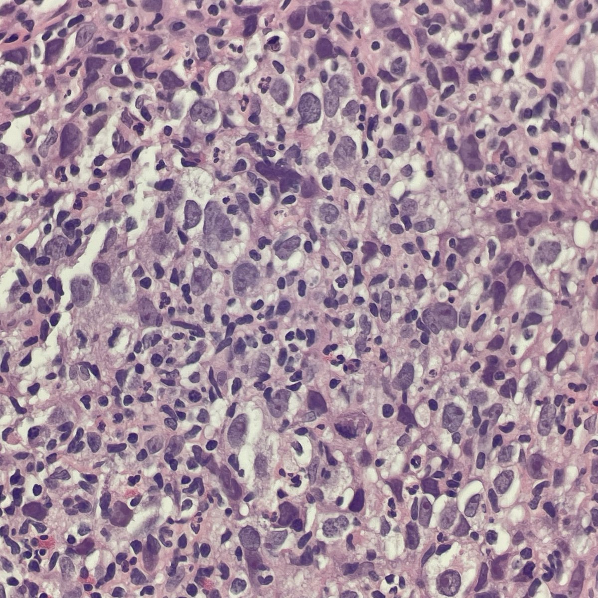
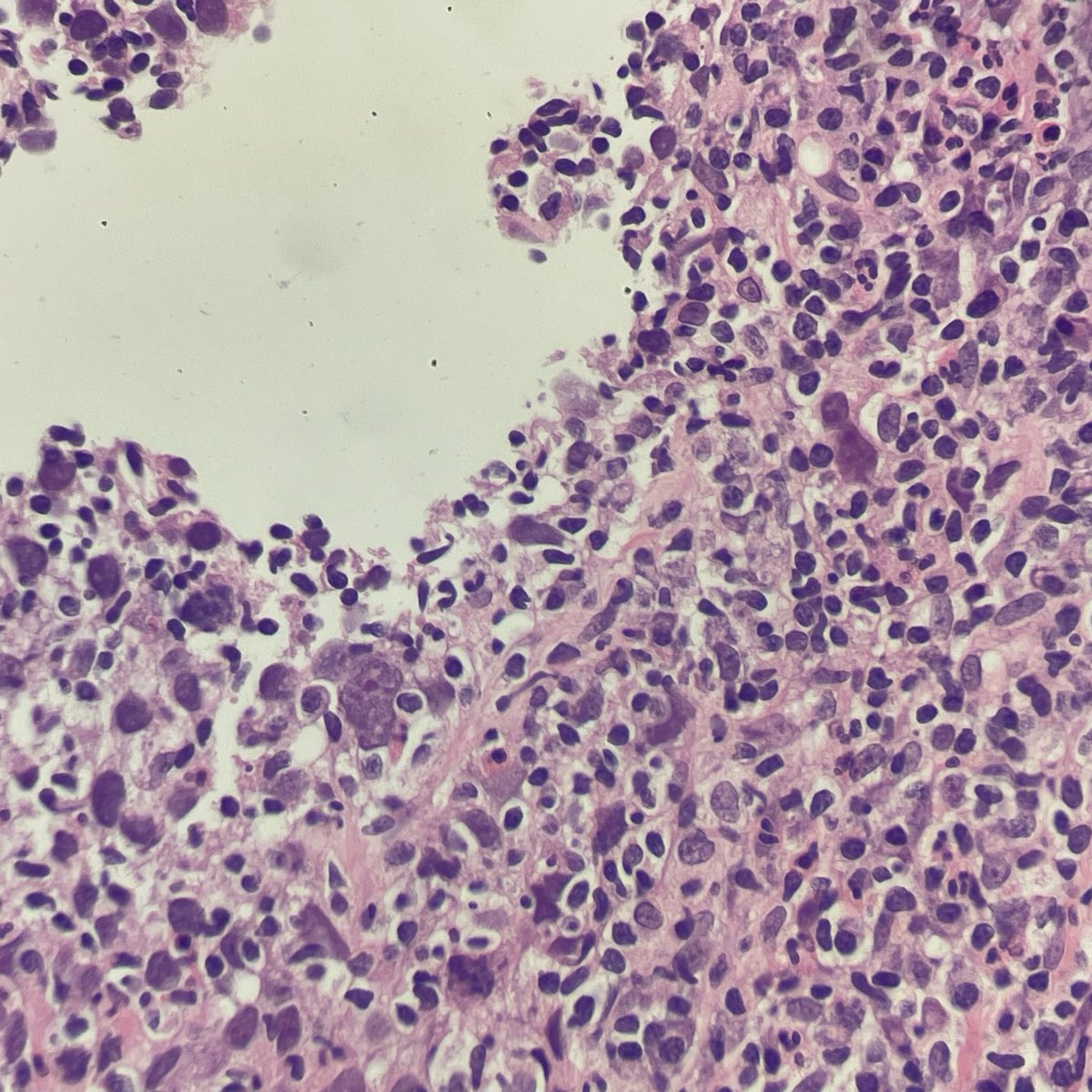
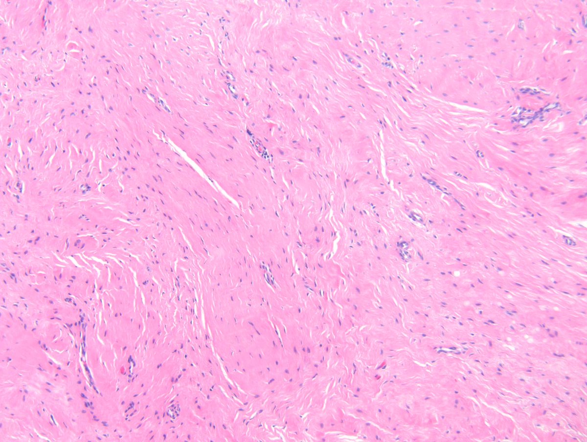
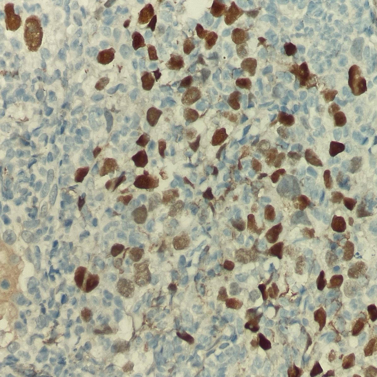
Both RANBP2-ALK inflammatory myofibroblastic tumor (IMT, a rare sarcoma) & anaplastic large cell lymphoma (ALCL) are (+) ALK*, CD30, and even SMA*!
*ALCL does not show nuclear membrane (+) ALK allowing for the distinction of IMT. SMA is focally (+) (PMID: 26893756)
Animal models demonstrate that combining an ALK inhibitor with anti-CD30 upfront might help prevent ALK-inhibitor-resistant IMT (PMID: 32684628)
For 🔬 example of HE, CD30, ALK1 for ALCL https://t.co/BMteHuoWNW
#HemePath #BSTPath #PathTwitter #PathResidents
🖼️🙏@UMichPath
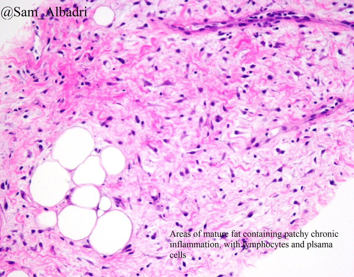
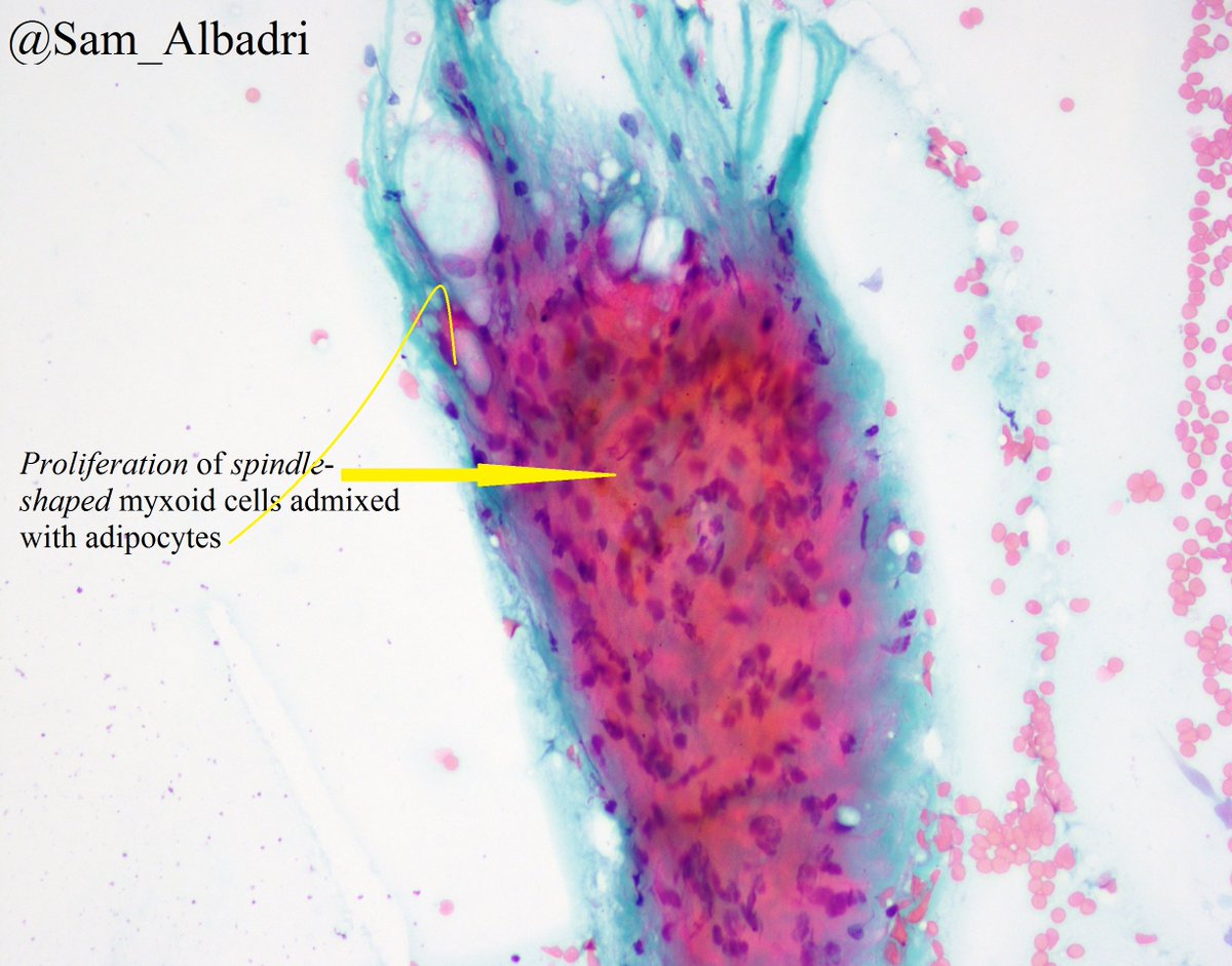
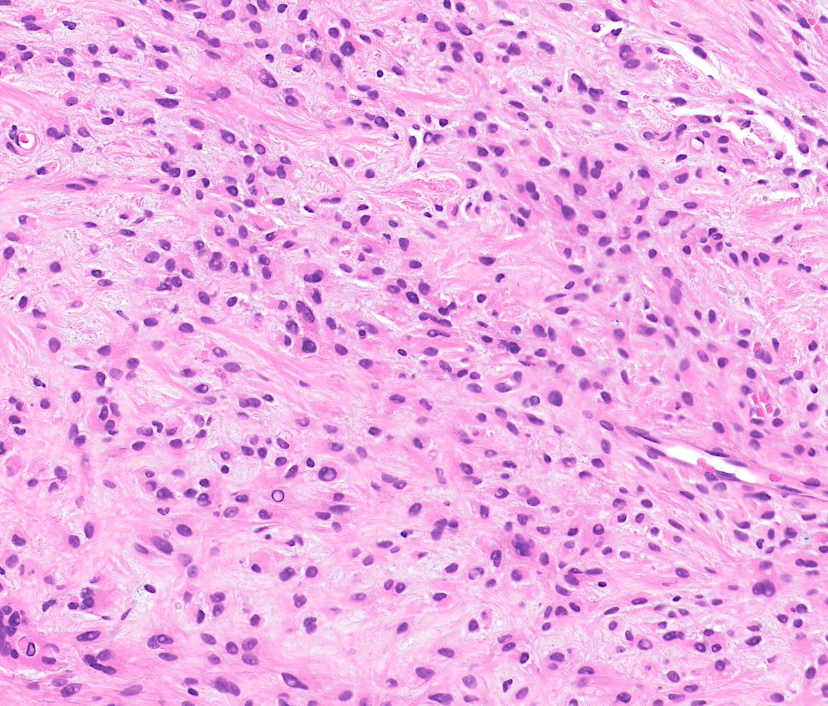
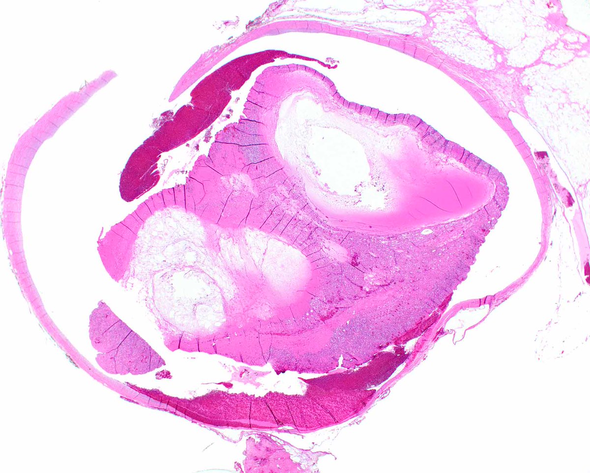
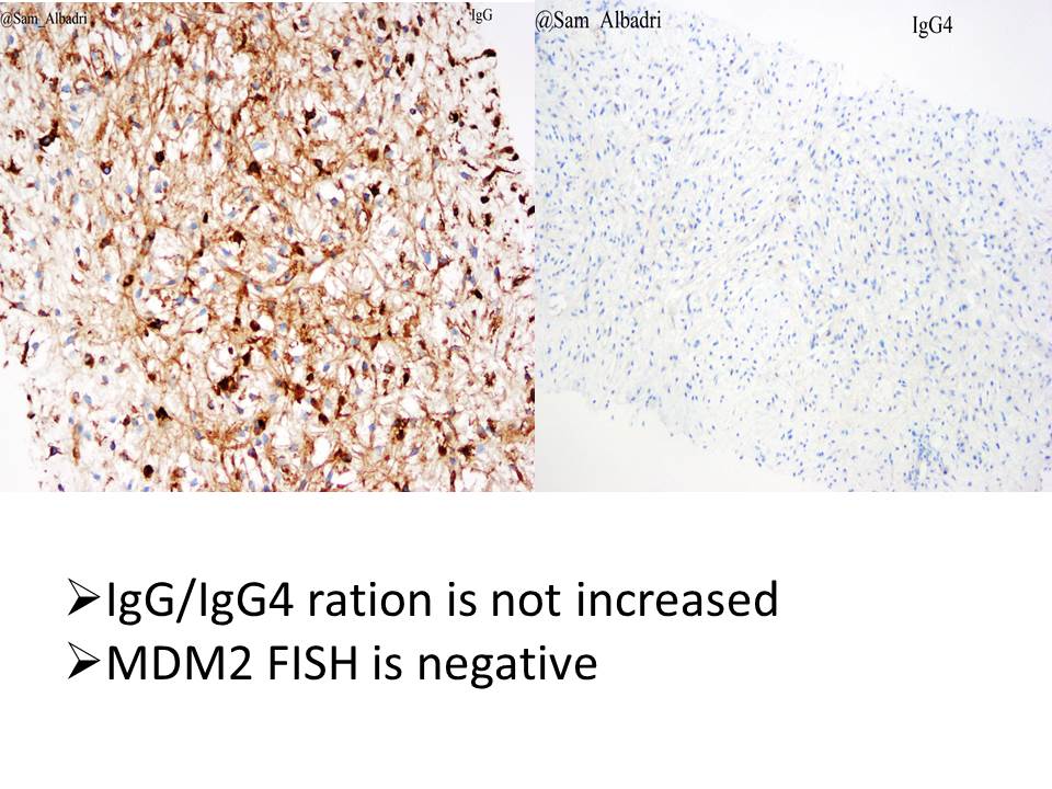
✅Sarcoma
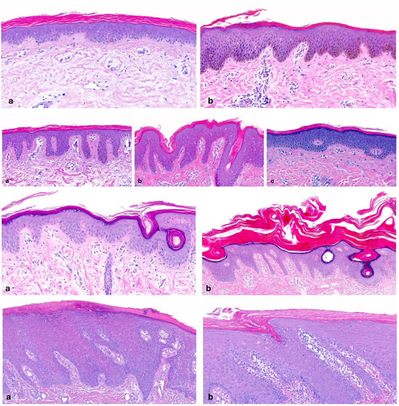
2024 Wk31 RAC9148 F60s. 4 years h/o intermittent / transient subcutaneous small sometimes tender / firm nodules that can self resolve over days / weeks.
No skin rash. Affects hands (dorsal & palmar areas). Help please #Dermpath A)Right thumb
#WhatsNewInDermpath
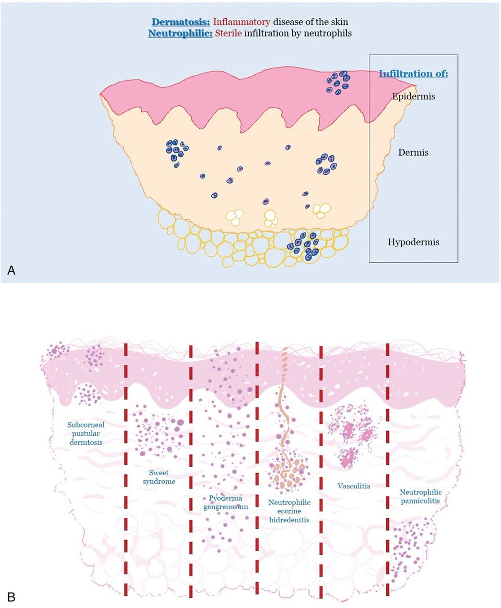
"Conceptual contextualization of neutrophilic dermatoses" by @ngelFernndezFl1
https://t.co/5epFC3Mmml
This article discusses neutrophilic dermatoses & describes the connection b/w neutrophilic dermatoses, autoinflammatory diseases, & autoimmune diseases.
Squamous epithelium with a sharply-demarcated area of necrosis is a typical finding in external/mechanical injury. This biopsy was from the oral cavity of a man with a history of chewing tobacco use.
WSI:
https://t.co/oerA6U6bwS
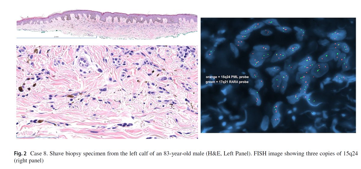
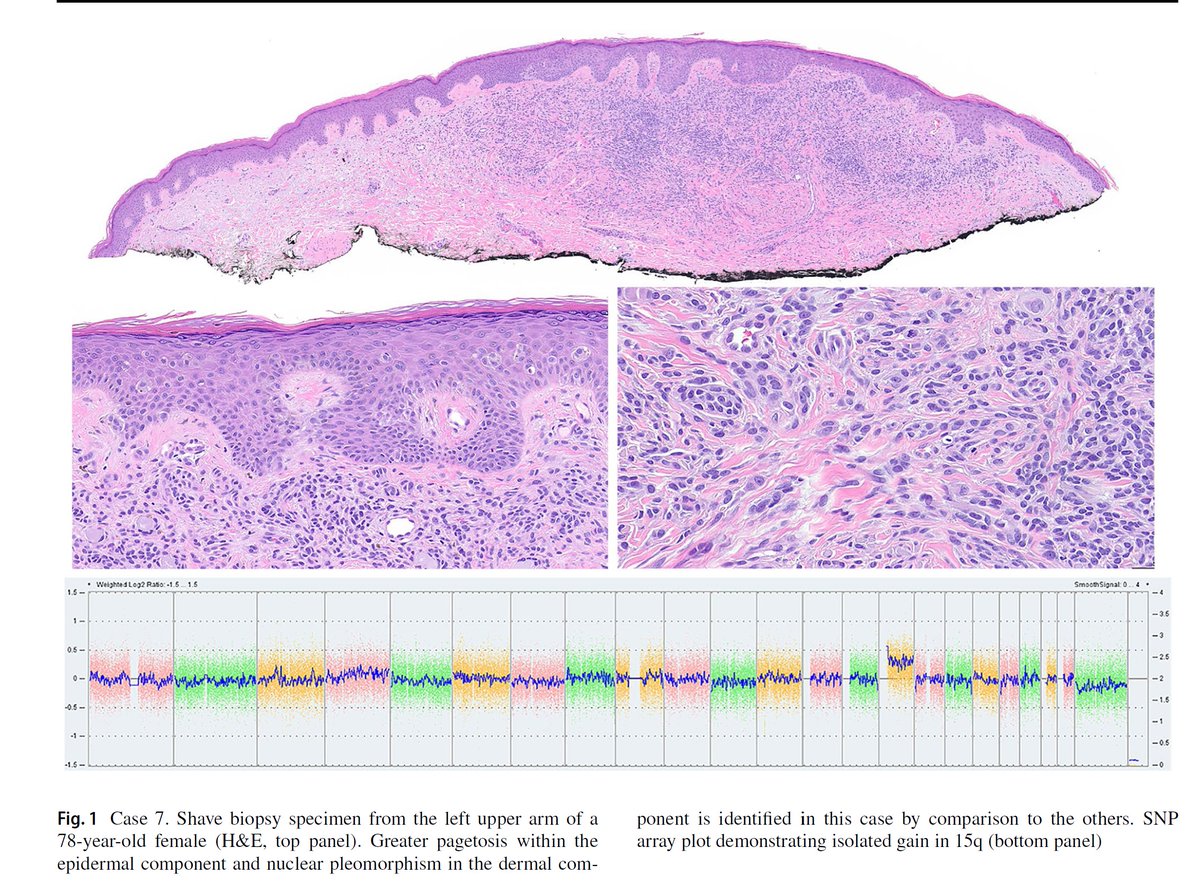
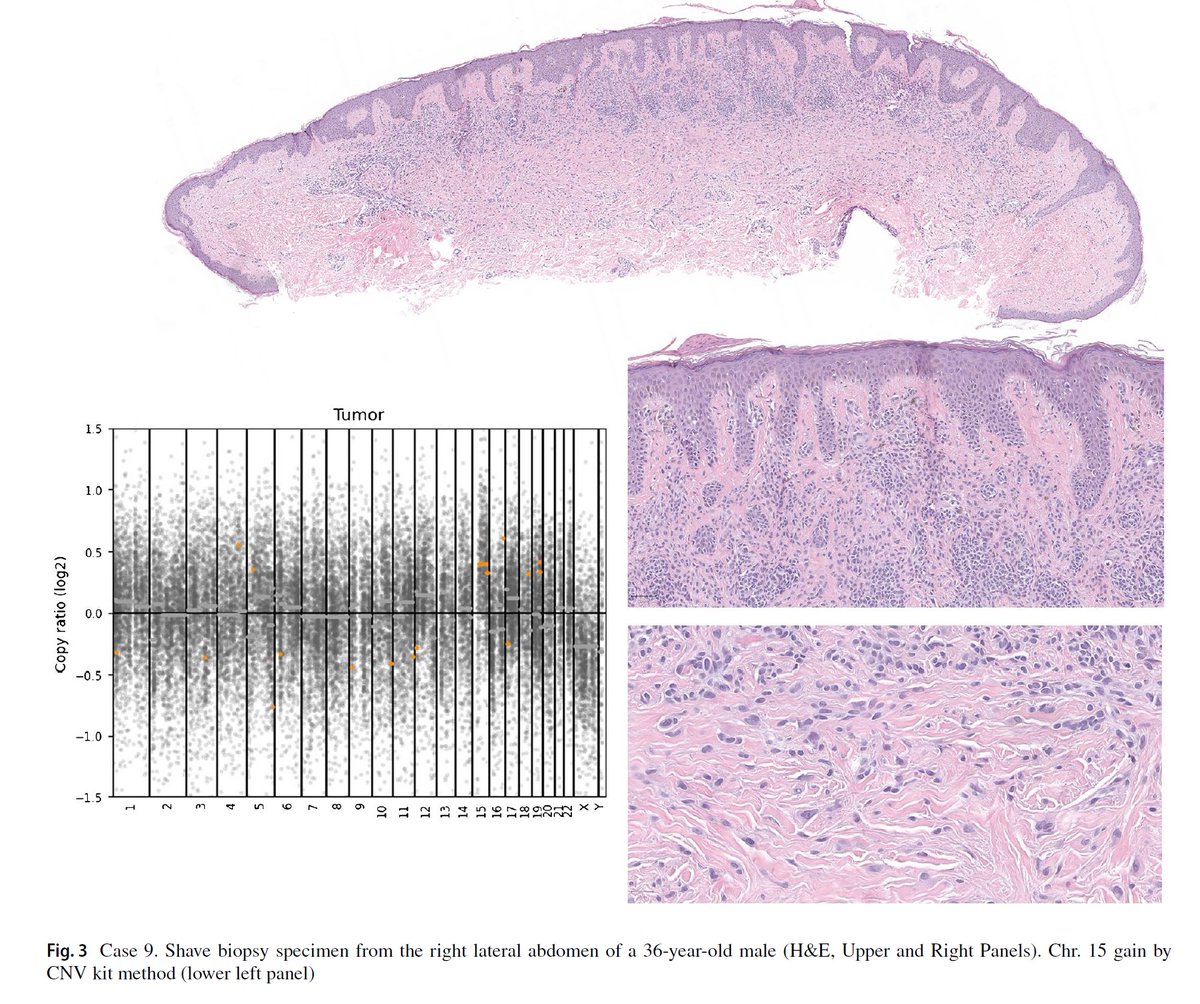
Our new work has just been published 📄
"Sclerosing melanocytic tumors with MAP2K1 in-frame deletions and 15q gains: A distinctive pathway of nevogenesis with reproducible morphology."
#dermpath#pathology
Free access to the article here: https://t.co/7QXCZCcgVB
Evolving Landscape of Cutaneous Mesenchymal Tumors — our latest publication at journal Dermatologic Clinics (Elsevier) a. Free access to the article until April 22 at the link below.
https://t.co/ijK1pokTjV
#pathology#dermpath#dermatology#neoplasia
AML-defining cytogenetic and molecular abnormalities (regardless of blast count)between the WHO 5th Edition (2022) and the International Consensus Classification (ICC 2022). #leusm#MedTwitter#hemonctrainees#when_on_service
Intravascular anastomosing hemangioma.
John I, Folpe AL. Anastomosing Hemangiomas Arising in Unusual Locations: A Clinicopathologic Study of 17 Soft Tissue Cases Showing a Predilection for the Paraspinal Region. Am J Surg Pathol. 2016 Aug;40(8):1084-9.
@Neur0tic Amióta kamera előtt lesúgták neki a kötelező mondanivalót a HírTV-ben, azóta még mindig tényező ez az ember? Egy megélhetési pribék, akarcsak a Hont, semmi több.
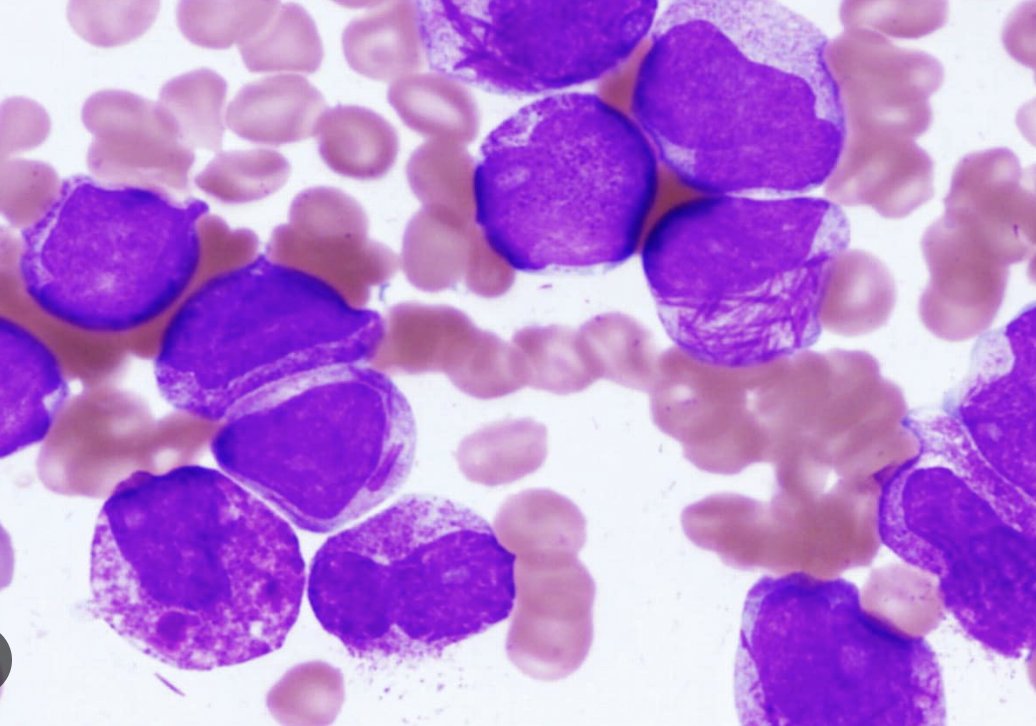
Bone marrow aspirate. Has anyone seen cells like this before? Who knows what these cells are? What do you think is going on? I'll post the answer in a day or two... :) #hemepath#pathx#leusm#pathology#pathtwitter
Classic anastomosing hemangioma. I most commonly see these lesions in the retroperitoneum and paraspinal regions. Anastomosing thin walled vascular channels are lined by hobnail endothelial cells. Helpful features: thrombi and extramedullary hematopoiesis.