📖 Radiology textbooks: “CT and MRI have taken over.”
👨🏫 Examiners: “Anyway… identify this barium meal follow-through.”
Some things age, but exam questions don’t.
🎯 High-yield. Old-school. Guaranteed to show up when you least expect it.
#MedStudent#MedEd
🚨 1 in a Million Case | Arterio-Colic Fistula 🚨
CT reveals an extraordinary finding; a thin channel communicating directly from the LEFT external iliac artery to sigmoid colon producing a hyperdense intraluminal appearance.
#RadTwitter#MedicalImaging#FOAMed#MedEd
⚠️ Imaging pearl:
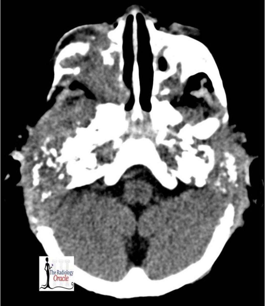
Marked osseous destruction out of proportion to the apparent infectious process.
What would be your top differential diagnosis before biopsy?👇
📌 Please Consider Sharing to Spread the Word !
https://t.co/TSF2PBuR8i
18-month-old boy with bilateral post-auricular swelling 👶🦴
Clinical presentation:
🔹 Otorrhea with mastoiditis
🔹 Poor response to sounds
🔹 Gradual hearing loss reported by parents
#FOAMRad#MedEd@_the_SRT@ESHNRSociety@spinacademics
💡 Diagnosis:
Skull Base Langerhans Cell Histiocytosis (LCH)
Temporal bone LCH in children can closely mimic aggressive otomastoiditis or neoplastic pathology. Bilateral destructive temporal bone lesions with hearing symptoms should always prompt consideration of LCH.
🧠 Rare Pelvic Case | Radiology Spotlight
A well-encapsulated lesion with innumerable internal cysts is seen in the RIGHT adnexal region displacing the urinary bladder without perifocal infiltration.
👉 Classic imaging appearance raises strong suspicion for Hydatid Disease
📌 Diagnosis
🔎 Pelvic Hydatid Cyst
🩺 Post-op: Primary Giant RIGHT Ovarian Hydatid Cyst
💡 Why is this interesting?
Hydatid cysts (caused by Echinococcus granulosus) most commonly involve the liver and lungs.
👉 Ovarian involvement is extremely rare !
I dedicate this honor to the cherished memory of my father. His values, sacrifices, and unwavering belief in me have shaped my journey and made me who I am today; this achievement stands as a reflection of his enduring influence on my life.
With immense pride and sincere gratitude, I wish to share that I have been conferred the Dr. Kishor Taori Gold Medal at the IRIA Annual National Conference, Hyderabad 2026.
This prestigious award was presented during the IRIA Oration Session, along with a Medal and Citation.
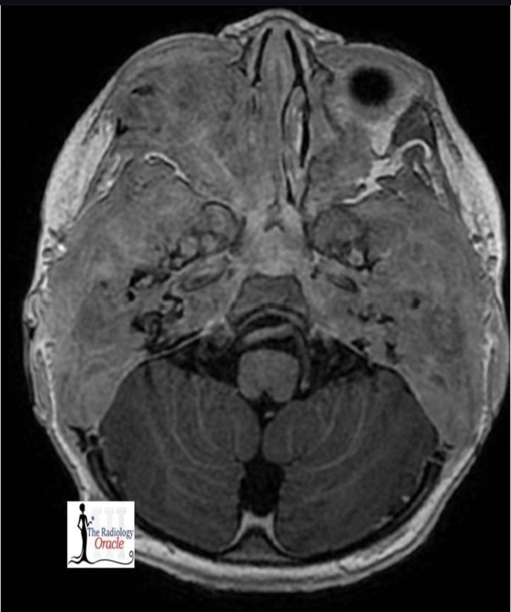
On T2, FLAIR, T1 C+ and DWI Images:
There are 2 distinct thick walled rim enhancing areas with heterogeneous morphology within both frontal lobes. Significant restriction is seen on diffusion acquisition in keeping with liquefied contents.
"Light bulb Sign of a Different Kind"
Fortunately; Foreign Body was Removed Successfully, No Internal Injuries
But Bulb Did Not Illuminate; Post Surgery 😜
Share if You Care 😉
#radres#foamrad#FOAMed#radiology#radlife#MedTwitter
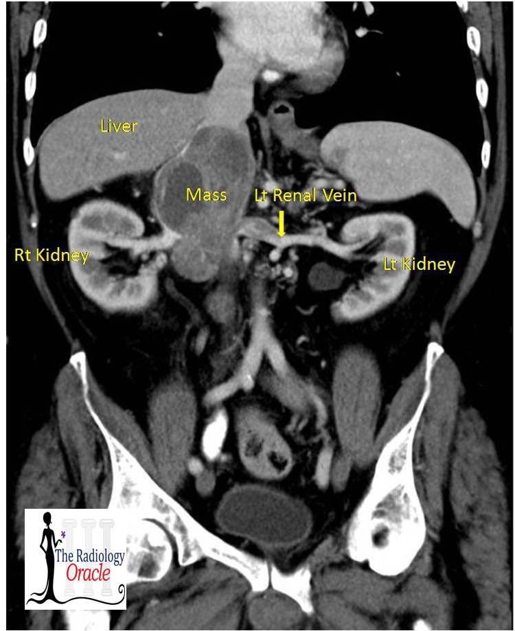
Findings:
Large lobulated heterogeneously enhancing intraluminal mass in the suprarenal inferior vena cava distending and obliterating the lumen with necrotic changes. Left renal vein is distended with hypodense non enhancing thrombus
45 YO Male, with complains of Severe Epigastric Pain.
•Pneumobilia.
•Lower segment of the neck of gallbladder is seen communicating with the 2nd part of duodenum.
•Small Bowel Obstruction
•Ectopic Calcified Gallstone
Diagnosis:
Gall stone Ileus with Riglers Triad