CD30⁺ lymphomas: from biology → diagnosis → therapy 🎯
CD30 is not a diagnosis.
It’s a shared activation marker across distinct lymphomas.
🧬 EBV can be + in BOTH:
• Hodgkin lymphoma
• DLBCL
So CD30⁺ + EBV⁺ ≠ one disease.
👉 Diagnosis requires:
• Morphology
• Immunophenotype (CD20 / CD15)
• Clinical context (mediastinum?)
⚠️ The risk:
Misclassification → wrong therapy
💊 Why this matters:
CD30 is targetable
→ Brentuximab vedotin (including FDA-approved R² combination in R/R DLBCL)
💡 Take-home:
Same marker ≠ same disease ≠ same treatment
CD30 is a bridge—not a diagnosis.
#lymphoma #Hemetwitter
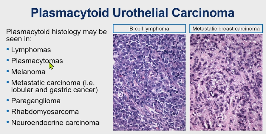
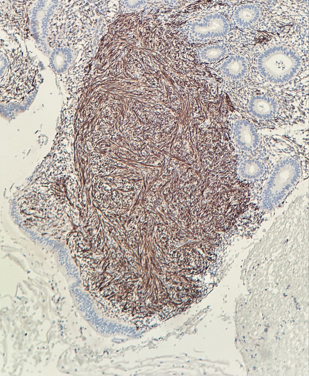
Bladder Plasmacytoid Urothelial Carcinoma
Pitfall ⚠️: It can stain CD138 positive (CD138 can stain epithelium).
Run a panel of IHC (pic 2).
Plasmacytoid histology can be seen in different neoplasms (see pic 3).
Dr. Magi-Galluzzi – 2025 Diagnostic Pathologic Update #USCAP #pathology #PathX
Stunning contrast between flat urothelial carcinoma in situ (right side of each image) and reactive urothelium (left side)
CIS➡️enlarged, irregular, & hyperchromatic nuclei
Reactive➡️small, uniform nuclei w/vesicular chromatin + intraepithelial acute & chronic inflammation
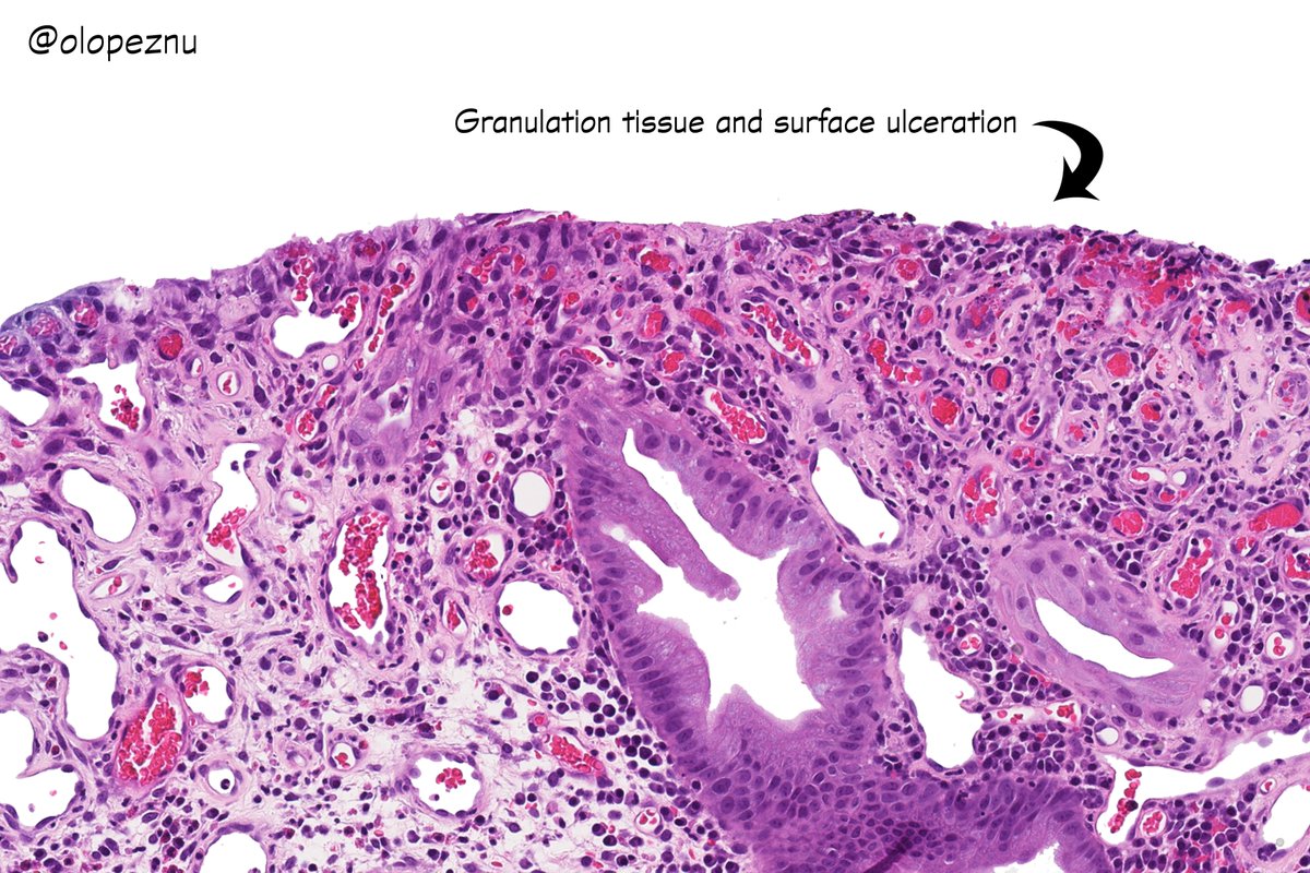
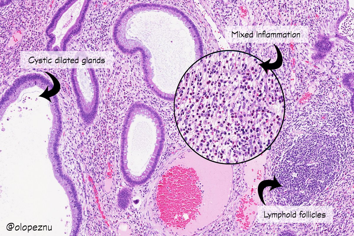
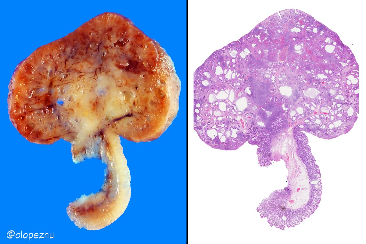
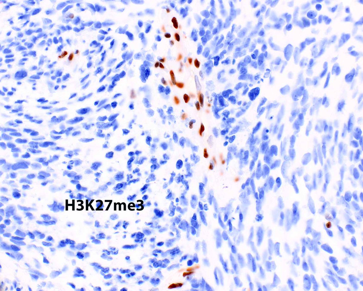
Juvenile Polyp
✔️Most common polyp in children
✔️More than 1/2 in the rectosigmoid colon
✔️Can be sporadic or syndromic (JPS)
✔️SMAD4/BMPR1A mutations ~ 50% of JPS families
✔️Histologically identical polyps can be seen in other hamartomatous polyposis syndromes
#PediPath#GIpath
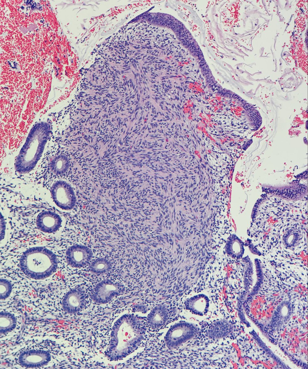
Breast-When evaluating possible ADH or DCIS involving a papilloma:
- Look for areas of pure epithelial proliferation with few fibrovascular cores.
- Examine the duct wall or neighboring glands outside the papilloma; if involved by DCIS or ADH, compare their morphology to the proliferation that inside the papilloma to make the diagnosis.
- Papilloma involved by ADH: <3 mm.
- Papilloma involved by DCIS: ≥3 mm.
- Pitfall: Myoepithelial markers are not reliable for distinguishing papilloma with UDH from those with ADH or DCIS (papillomas with ADH and DCIS can retain myoepithelial cells).
Dr. Lerwill - 46th Annual Current Concepts in Surgical Pathology #pathology #PathX #Pathtwitter
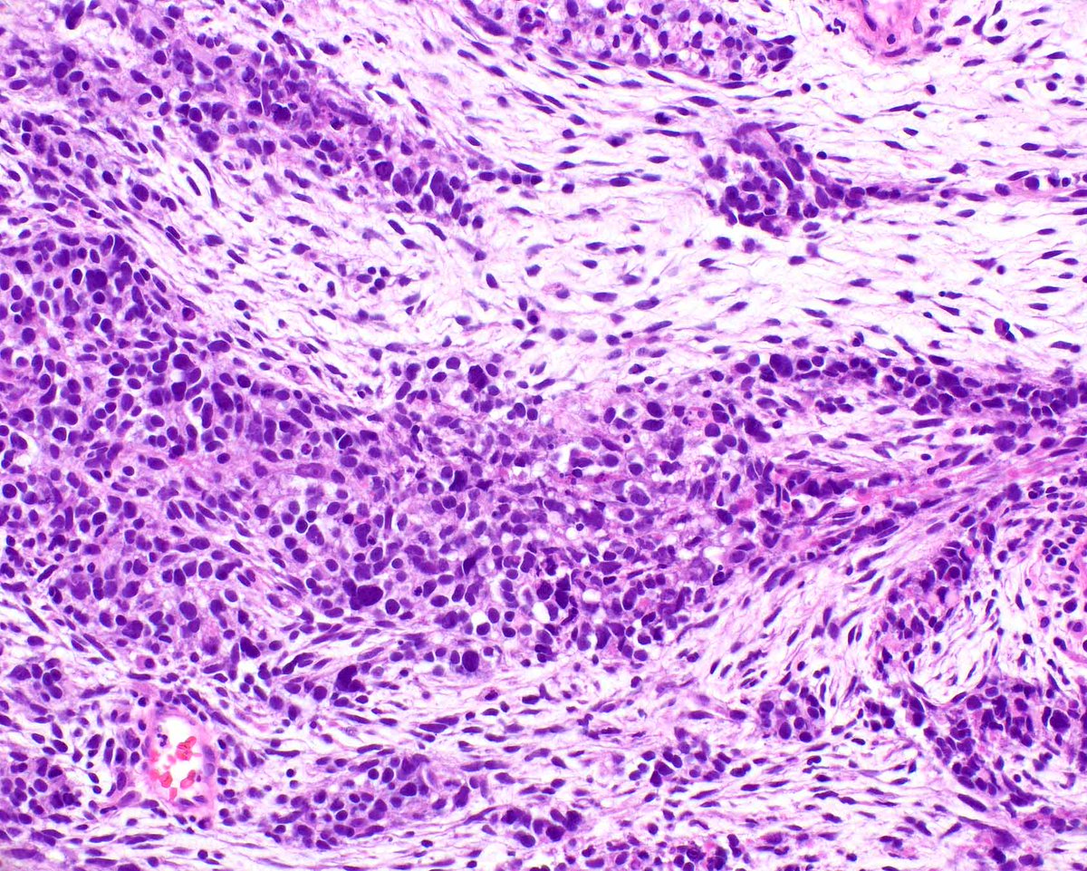
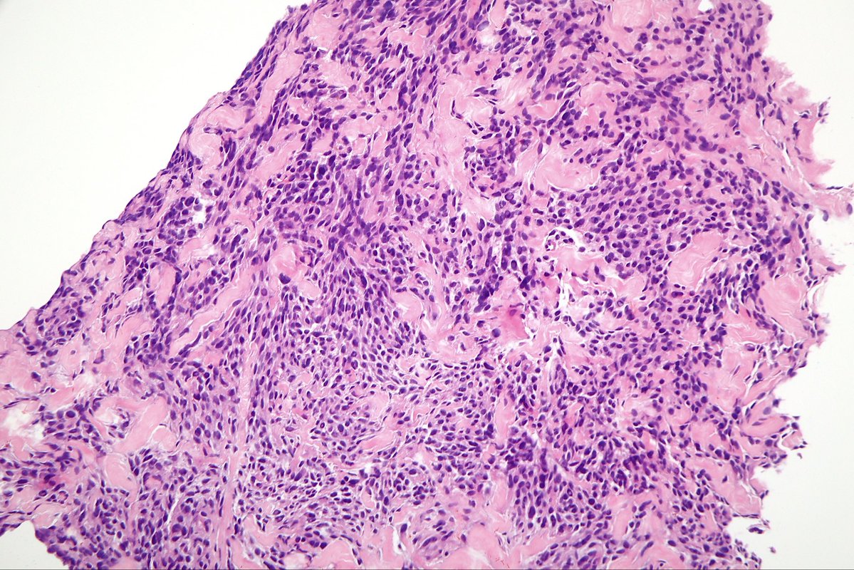
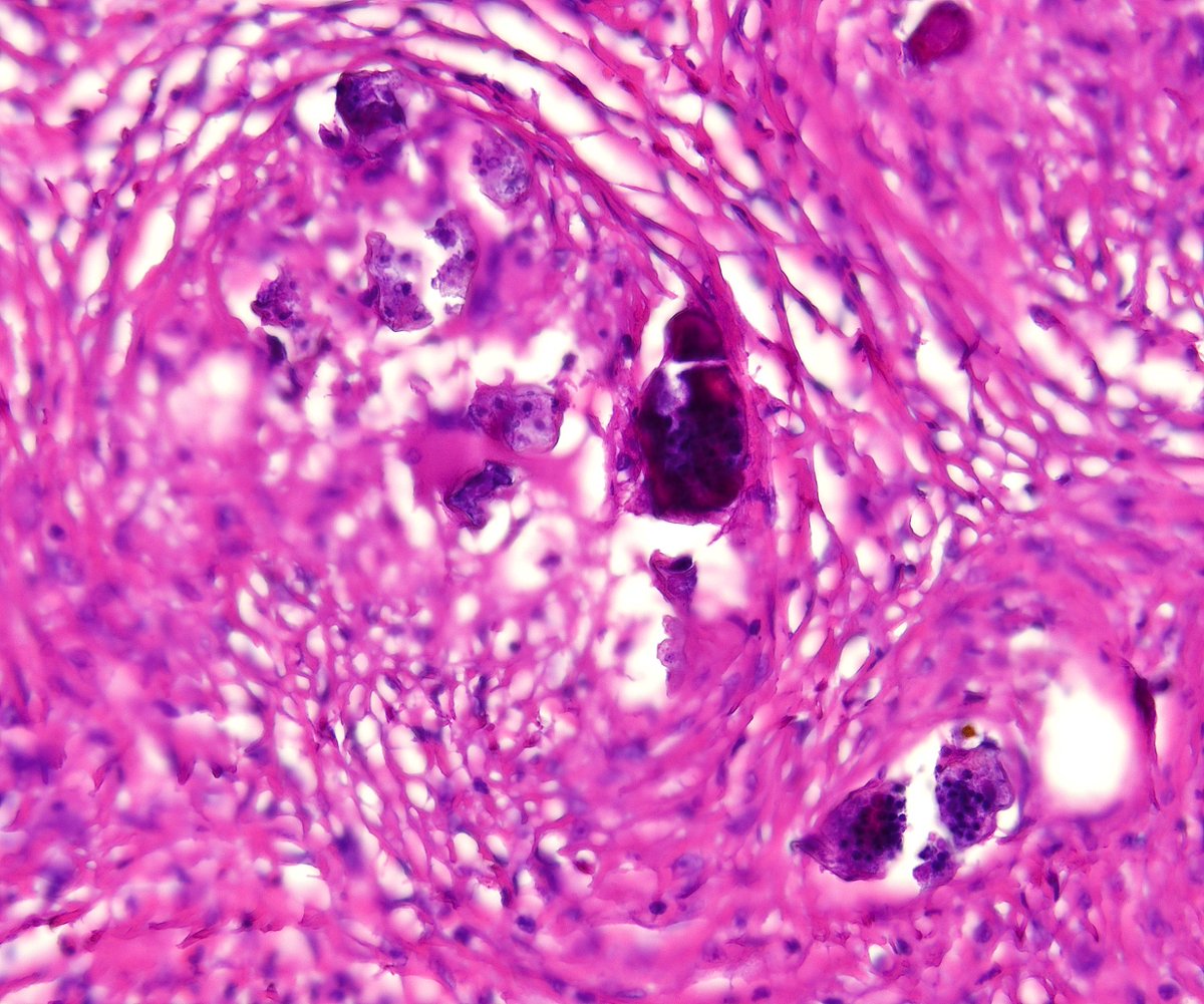
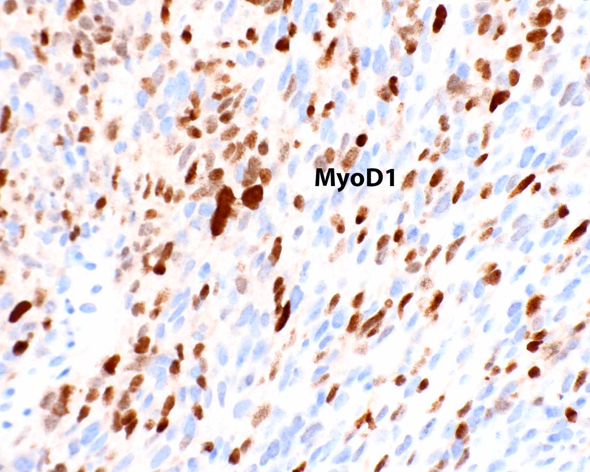
Dr. Sarah Dry has published a wonderful review of fatty tumors and nerve sheath tumors. Check out this malignant triton tumor (malignant peripheral nerve sheath tumor with rhabdomyosarcomatous differentiation) - the muscle differentiation is obvious on H&E.
Dry S. Updates in Peripheral Nerve Sheath and Adipocytic Tumors. Mod Pathol. 2025 Nov 1;39(1):100929. PMID: 41183597.
This adrenal cortical carcinoma presented as a liver mass. The CK8/18 highlights a tiny rim of compressed liver at the periphery. On a prior needle biopsy it was a mimicker of hepatocellular carcinoma but we figured it out thanks to modern immunohistochemistry.