60s patient presented with tearing chest pain that woke them from sleep.
✔️#echofirst#POCUS is performed right away
✔️Wide aortic root is noted (>4cm)
✔️Sonographer switches to suprasternal notch view and sees:
(1/3)
Quiz time: What is this #POCUS finding?
Probe location: Right neck
Context: #heartfailure patient, #Nephrology fellow @khaledsidiya wants to do #VExUS#IMPOCUS
Patient denies shortness of breath or chest pain; but does complain of back pain.
#MedEd
Will post answer tomorrow.
Cuando la ventana paraesternal es de mala calidad …la visión subcostal nos puede entregar información cualitativa importante … #echofirst#POCUS@UltrasonidoEm
Thanks to @OSUultrasound for allowing me to speak at Ultrafest 2020! I had a whole lot of fun trying a "Choose your own adventure" style virtual lecture. I am here to report on how it went. Overall a success! 1/6
⚡#POCUSCase ⚡
A 25yo female patient was brought to the ED after a traffic accident. A quick #eFAST#POCUS exam was performed in the ED. The image is a transabdominal view of the pelvic region in the mid-sagittal plane.
What do you see?
#POCUSEducation#ECHOFIRST#Ultrasound
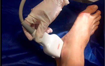
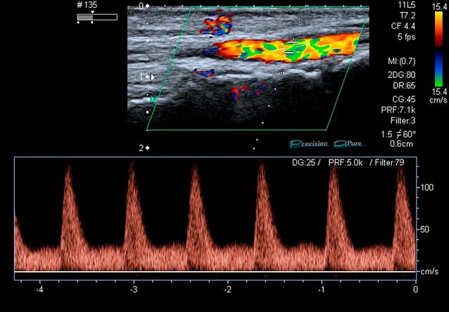
The FAST D protocol: a simple method to rule out traumatic vascular injuries of the lower extremities. Sens and spec 100% #POCUS#FOAMed @ResaELewiss @PittIMPOCUS @NephroP@Wilkinsonjonny@POCUSAcademy https://t.co/lUFnBKlI6l