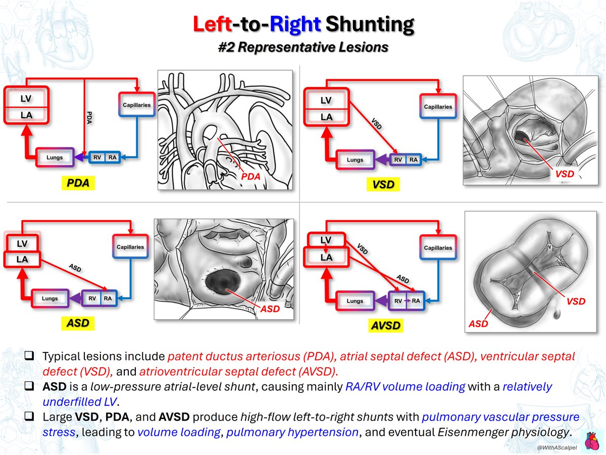
Left-to-Right vs Right-to-Left Shunting
👉Left-to-right shunts increase pulmonary blood flow and produce pulmonary overcirculation with volume loading of the involved chambers.
👉Right-to-left shunts bypass effective pulmonary oxygenation, causing systemic desaturation and reduced systemic oxygen delivery.
👉Shunt direction and magnitude depend on anatomy, pressure gradients, ventricular compliance, PVR/SVR balance, and pulmonary blood flow.
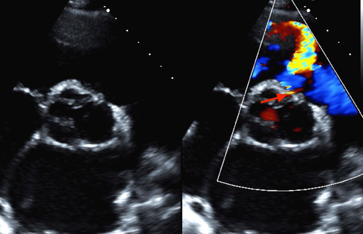
Outlet VSD Spectrum💡
👉Outlet VSDs include perimembranous, muscular, and doubly committed juxta-arterial defects, distinguished by their anatomical relationships to the arterial valves and ventricular septum.
🫀 CardioNugget™: congenital VSD location on echo
Perimembranous vs outlet VSD can look very similar on PLAX → you often can’t reliably differentiate them there.
🔄 Go to PSAX at the aortic valve level and use the clock face:
🕙 ~10 o’clock → Perimembranous VSD
🕑 ~2 o’clock → Outlet (supracristal/subpulmonary) VSD
💡 Why it matters: Outlet VSDs are particularly associated with aortic cusp prolapse → aortic regurgitation.
#CardioNugget #EchoBoards #Echocardiography #Cardiology
Fontan Circulation #5: Fontan Fenestration
👉Fontan fenestration creates a right-to-left shunt from the Fontan pathway to the atrium, acting as a pressure-relief mechanism.
👉It reduces systemic venous pressure while preserving ventricular preload and cardiac output, particularly when PVR is elevated.
👉Fenestration may improve early postoperative hemodynamics and reduce venous congestion, at the cost of lower oxygen saturation.
Ross Procedure—Pulmonary Autograft Replacement🎯
👉The pulmonary autograft replaces the diseased aortic valve, with coronary reimplantation and RVOT reconstruction using a pulmonary homograft.
What are outcomes of the Ross procedure in patients older than 60 years? Read the two-center retrospective study from the Canadian Ross Registry in #JTCVS: https://t.co/UEqdJfDfQO
See more at #Aortic2026: https://t.co/RrhOMz8zrr
Fontan Circulation #4: Pressure Gradient and Systemic Venous Hypertension
👉Fontan flow relies on elevated systemic venous pressure (CVP often 12–18 mmHg) to drive blood through the pulmonary circuit.
👉The transpulmonary gradient (CVP − atrial pressure) is typically small (~3–6 mmHg), requiring low PVR for adequate flow.
👉Elevated PVR, atrial pressure, or ventricular EDP worsens venous congestion and compromises ventricular preload and cardiac output.
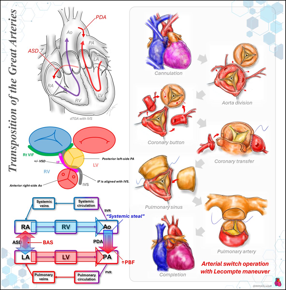
Arterial Switch Operation for d-TGA🔀
👉The arterial switch operation restores ventriculoarterial concordance through great artery transection, coronary artery transfer, and reconstruction of the neoaorta and neopulmonary artery.
@eddyjoemd@fabio_taccone@PhilGuerci Very comprehensive overview! I agree that capillary leak can be difficult to assess objectively and is often judged clinically🤔 Thank you for sharing!🙏
Fontan Circulation #3: Transition from Glenn to Fontan Circulation
👉Fontan completion incorporates IVC return into the Glenn circulation, establishing total cavopulmonary circulation.
👉Low PVR, low atrial pressure, and an adequate systemic venous-to-atrial pressure gradient are essential for maintaining cardiac output.
👉Fontan completion improves systemic oxygen saturation but increases systemic venous pressure, predisposing to venous congestion.
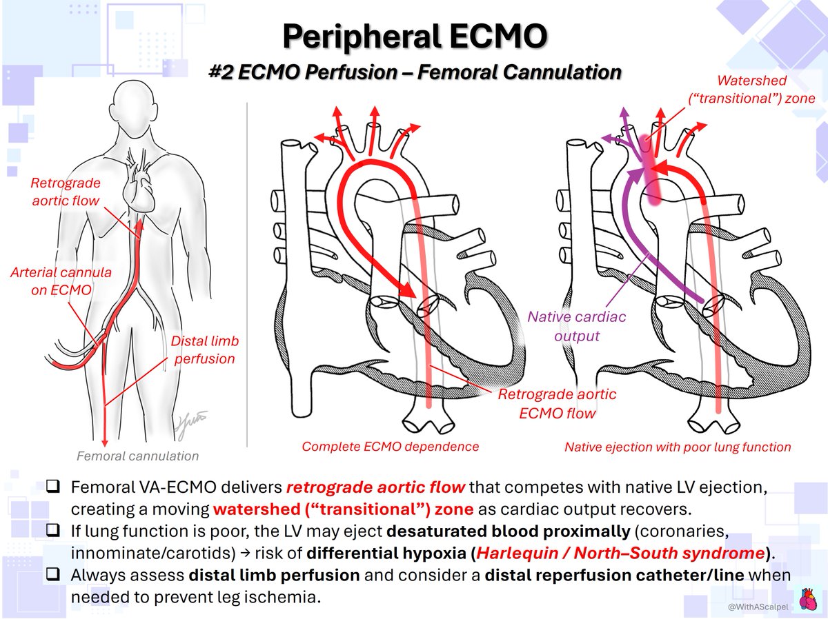
Harlequin syndrome isn't a flow-rate problem. The brain + coronaries can desat while the legs look fine. Check a right-hand sat, then change the circuit config. 🎩 tip to the authors including @DrMohEldiasty, @ElgudinYakov, @ProfDrPopov & @adham_sherbini https://t.co/sSr8PFAqru
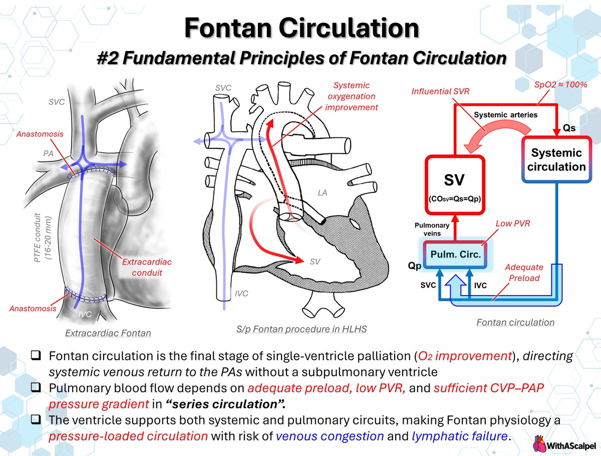
Fontan Circulation #2: Fundamental Principles
👉Fontan completion directs SVC and IVC flow to the pulmonary arteries without a subpulmonary ventricle.
👉Pulmonary blood flow depends on the CVP–atrial pressure gradient, low PVR, and unobstructed pulmonary pathways.
👉Low PVR supports ventricular preload and output, while elevated CVP predisposes to venous congestion and lymphatic failure.