The IVC has long been used to estimate "volume status." Multiple studies have demonstrated that it performs poorly.
The Venous Excess Ultrasound (VExUS) score was developed as a more accurate alternative — and it predicts outcomes that the IVC alone cannot.
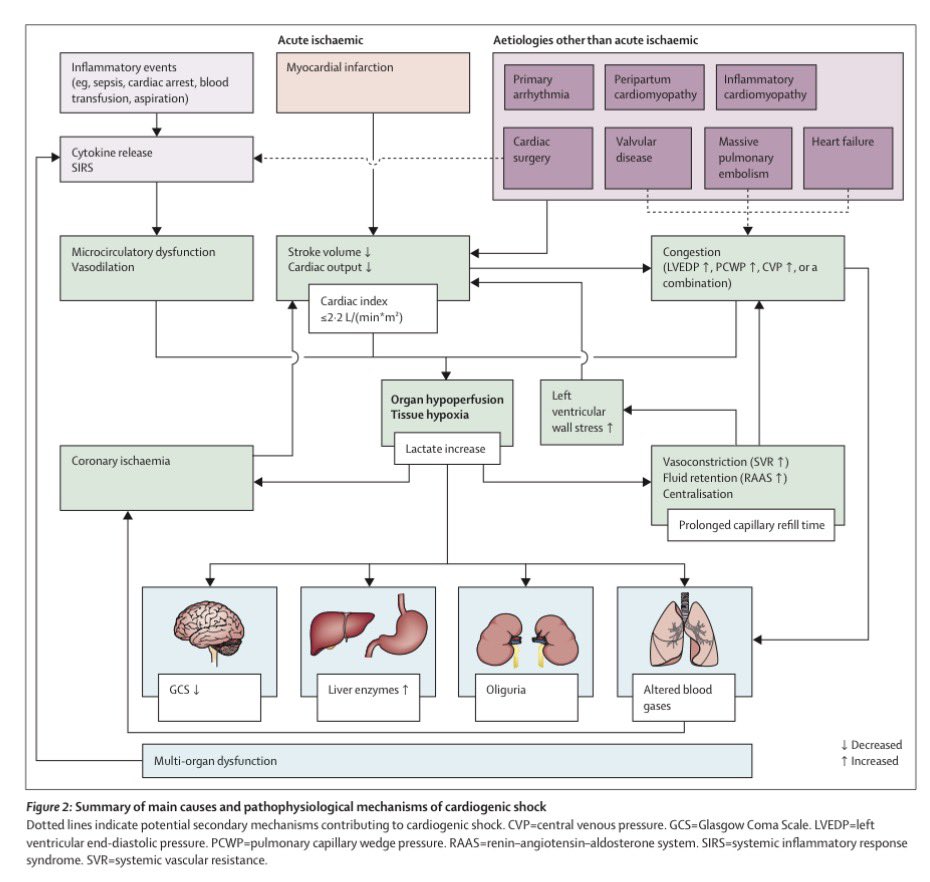
#POCUS#echofirst#Nephpearls
RVOT pulse-wave Doppler can provide useful clues about pulmonary vascular resistance.
In normal individuals (A), the waveform has a smooth, dome-shaped appearance, with peak velocity occurring in mid-systole, reflecting a compliant, low-resistance pulmonary circulation.
As RV afterload increases, the waveform gradually becomes more triangular. The RVOT acceleration time shortens, and the peak velocity shifts earlier into systole (B).
With further increases in pulmonary vascular impedance and reduced arterial compliance, a characteristic mid-systolic notch may appear (C), creating the classic "W sign."
In advanced pulmonary hypertension with RV failure, the Doppler envelope becomes smaller and more abbreviated, with a very short and steep AccT (D). This reflects rapid equilibration of RVOT and proximal pulmonary artery pressures due to severe afterload elevation.
Like most POCUS findings, RVOT Doppler should be interpreted in the context of the overall echocardiographic picture rather than in isolation.
The Ischemic Cascade: Why Angina Is a Late Sign
Myocardial ischemia doesn't begin with chest pain.
The sequence starts with a reduction in myocardial perfusion, leading to a mismatch between oxygen supply and demand. As ischemia progresses, the heart undergoes a predictable cascade:
1. Perfusion defect
2. Metabolic changes
3. LV diastolic dysfunction
4. LV systolic dysfunction
5. ECG changes
6. Angina
Key clinical pearl:
Because perfusion abnormalities occur before wall-motion abnormalities, ECG changes, or symptoms, stress imaging techniques that assess myocardial perfusion are generally more sensitive for detecting ischemia than tests relying solely on ECG changes or regional wall-motion abnormalities.
Angina is often the last manifestation of the ischemic cascade, not the first.
Follow @CardiovascularCorner for daily cardiovascular insights.
Reference: Braunwald's Heart Disease: A Textbook of Cardiovascular
Today's Paper of the Day is:
Heart-Lungs interactions: the basics and clinical implications
https://t.co/JKgcYjlUQ5
Join us to read 1 paper per day and stay up-to-date as we cover the spectrum of critical care across 2026
Today's Paper of the Day is:
Acute Respiratory Distress Syndrome and Fluid Management: Finding the Perfect Balance
https://t.co/JKgcYjlUQ5
Join us to read 1 paper per day and stay up-to-date as we cover the spectrum of critical care across 2026
⚡🫀 CAUSES OF ST ELEVATION — Know Them All
Use mnemonic MI-PERICARD-BEN-EARTH-VAMPI to master every cause — from STEMI & pericarditis to Takotsubo, hyperkalemia & pulmonary embolism.
ST elevation isn't always a heart attack.
#STElevation#STEMI#ECG
Transthoracic Echocardiographic (TTE) Views: A Complete Guide
Understanding TTE views is essential for accurate cardiac imaging. Let’s break down the 5 core views:
1️⃣ Parasternal Long Axis
2️⃣ Parasternal Short Axis
3️⃣ Apical
4️⃣ Subxiphoid
5️⃣ Suprasternal
A thread 🧵
Reminder: the original #VExUS grading does not use RVSI. It relies on qualitative waveform assessment (biphasic, monophasic), which is adequate for most real-world clinical use. RVSI becomes more useful when congestion is expected to resolve slowly or incompletely, for example in severe pHTN + TR.
Lung ultrasound detects pulmonary edema with 93–97% sensitivity.
Chest X-ray: 63–69%.
The diagnostic gap is even wider when you look at what's actually driving it. A thread.
Optimising positive end-expiratory pressure in acute respiratory distress syndrome: a narrative review of approaches to titration
CCR Journal Watch
https://t.co/Sp06oA6IDG
#POCUS#Nephpearls
Short axis helps avoid the cylinder effect, but if you rely on it alone, you can run into ‘site ambiguity’ - you might not be measuring at the right spot (about 1 to 2 cm below the HV–IVC junction). Best approach is to look at the vessel in both long and short axis.
More about VAC in sepsis... 🤓
🫀 Why do some septic shock patients respond to treatment… and others don’t?
We often blame:
• “Refractory shock”
• “Severe sepsis”
• “Late presentation”
But physiology tells a different story.
⚙️ The real problem: ventriculo-arterial decoupling
Septic shock is not only vasodilation or myocardial depression.
��� It is a failure of interaction between the heart and the arterial system
This interaction is called:
➡️ Ventriculo-arterial coupling (VAC)
And it defines:
✔️ Cardiac output
✔️ Arterial pressure
✔️ Perfusion efficiency
🧠 What can be often ignored in daily ICU practice
You can have:
✔️ Normal cardiac output
✔️ Acceptable MAP
❗ And still have inefficient circulation
Because:
👉 Energy transfer from the ventricle to the arterial system is impaired
📉 What happens in septic shock?
• Decoupling is common
• LV ejection becomes inefficient
• Cardiovascular treatments become less effective
💉 Clinical paradox
Same intervention. Different outcomes.
Example with norepinephrine:
🔵 Patient A
→ Adequate contractility (Ees preserved)
→ ↑ arterial tone
→ VAC improves
→ ↑ CO
🔴 Patient B
→ Depressed contractility
→ ↑ arterial tone (afterload)
→ VAC worsens
→ ↓ stroke volume
🔥 This explains a lot of what we see
• Why MAP increases but CO drops
• Why some patients “fail” vasopressors
• Why fluids work in some and not others
• Why lactate persists despite “normal numbers”
🧬 Even more important
VAC is:
👉 A determinant of treatment responsiveness, not just a descriptor of physiology
📊 Bedside implication
We should stop asking only:
❌ “What is the MAP?”
And start asking:
✅ “Is the system coupled?”
✅ “Are we improving efficiency or just pressure?”
⚡ Practical shift
Instead of protocol-only resuscitation:
➡️ Move toward physiology-guided resuscitation
Using:
• Ea (arterial load)
• Ees (contractility)
• VAC (their interaction)
🧠 Final thought
Septic shock is not just:
❌ A pressure problem
❌ A volume problem
👉 It is an interaction problem
And until we treat it as such:
➡️ Some patients will continue to “not respond”
📚 Pinsky MR, Guarracino F. (2023)
Intensive Care Medicine Experimental
https://t.co/3bYkZ1G7wG