Case of IgAN with massive hemispherical mesangial deposits. Mimics cryo-plugs but deposits are mesangial rather than filling capillary loops. #renalpath#pathwitter#nephrology
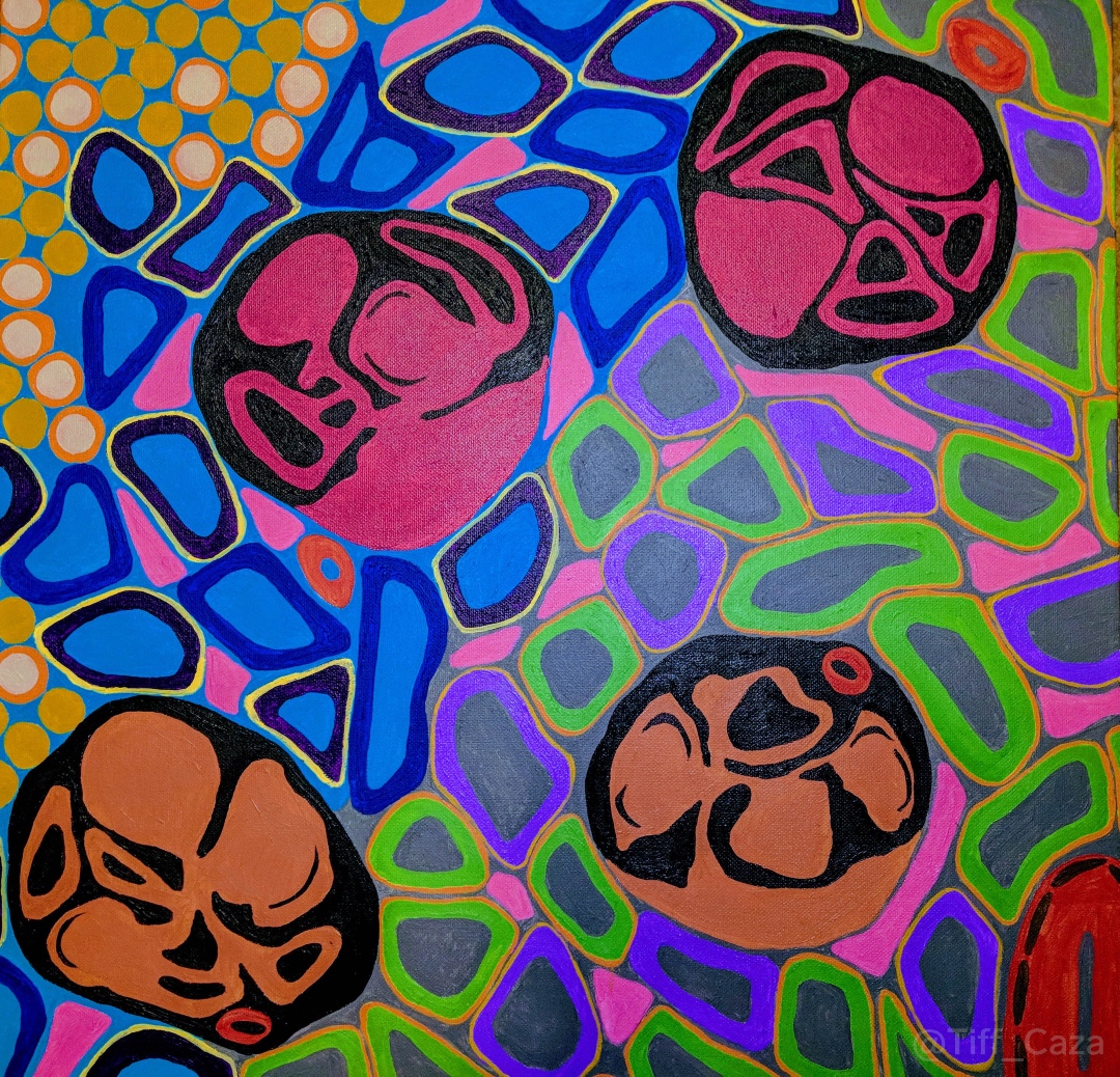
This painting shows glomeruli with nodular mesangial expansion and arterial hyalinosis, changes frequently seen in diabetic nephropathy. Diabetic nephropathy affects 30-40% of patients with diabetes mellitus, and is a major cause of end stage kidney disease in these patients.
Painting by Dr. @Tiff_Caza
#NationalKidneyMonth #ArtofMedicine #CKDAwareness #DiabeticNephropathy
Reminder that EM is still useful even when no gloms available. Example case of light chain deposition disease. IF highly suspicious with kappa restriction along TBMs; EM was helpful to confirm the diagnosis: powdery deposits on TBMs. #renalpath#nephrology#PathTwitter
Weekend paint project 🖌️🎨 . This is Alport syndrome by light microscopy. Overall, looks pretty normal except for some foci of interstitial foam cells. EM and genetics would tell us more.
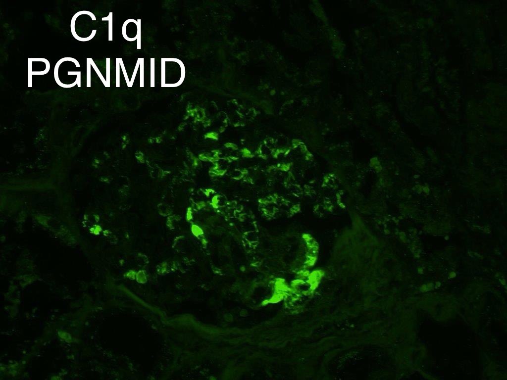
Teaching C1q in glomeruli on immunofluorescence microscopy.
My experience is if + it is real & is not non specific.
Positive:
1. Autoimmune disease=always +
2. Monoclonal Ig (PGNMID/MIDD)=often +
3. Infections= can be + or -
Negative:
1.IgA nephropathy
2.C3 glomerulopathy
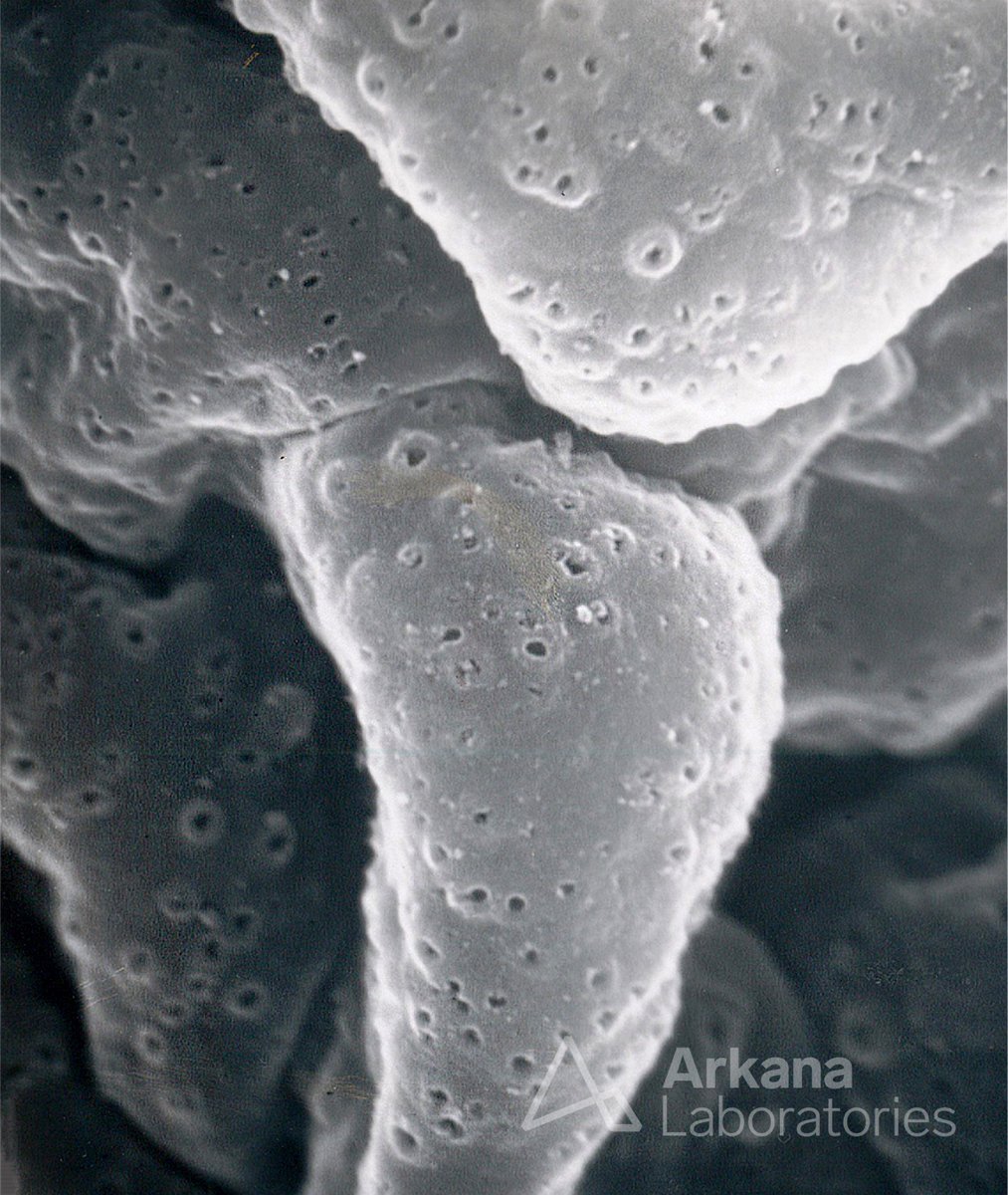
Today’s #eyeSCANdy shows an acellular scanning EM from a biopsy with membranous glomerulonephritis stage I, showing numerous small shallow depressions representing the site of immune complexes which were extracted.
Photo courtesy of Dr. Stephen Bonsib. #PathTwitter#PathX
Alport syndrome is a genetically heterogenous disorder thought to be affecting only males. Read these Clinical Insights into a 15-yr-old female w/a rare pathogenic variant in the COL4A5 gene & X-linked Alport syndrome. #OpenAccess
https://t.co/N4jEOxeFtQ
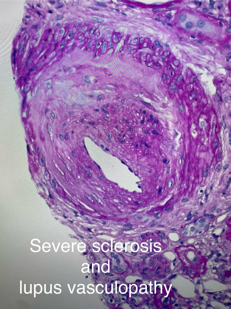
1. Focal necrotizing lupus arteritis.
Plus severe lupus vasculopathy.
Can be missed and is unfortunately not part of the lupus classification.
2. Glomeruli showed mesangial lupus nephritis, class II.
40-yr old woman with lupus & rise in serum creatinine,+ lupus anticoagulant.
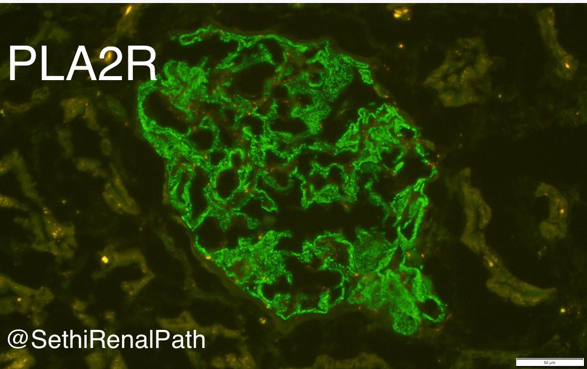
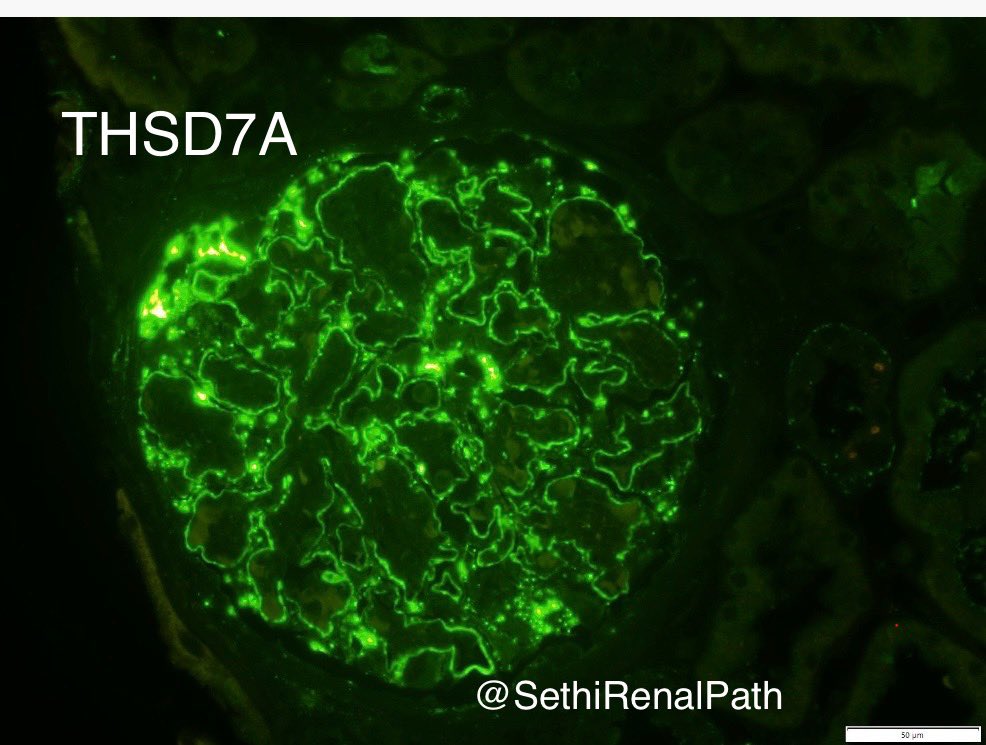
Nuances of Membranous antigen staining:
1. PLA2R: granular staining along GBM
2. THSD7A: lumpy-discrete, scattered deposits along GBM (linear staining=negative)
3. EXT: granular staining along GBM, can be intense
4. NELL1: granular, can be segmental,sometimes appears linear. 1/2