Nuclear Stars and Viral Auroras ✨
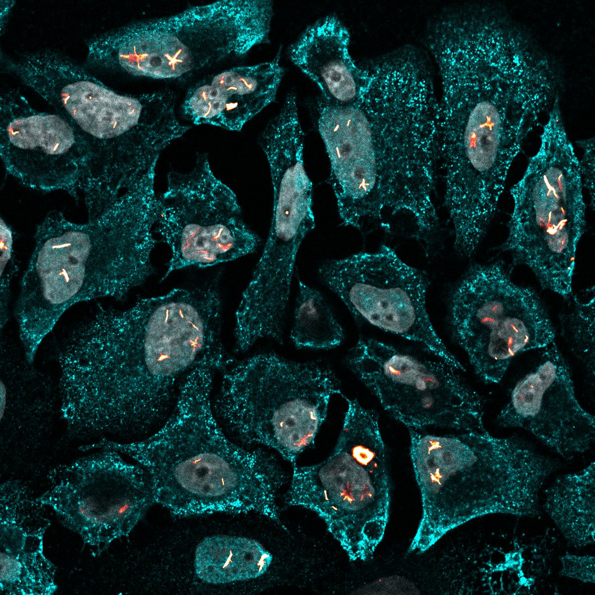
Red-hot nuclear constellations are frozen inside grey microcosmic vaults, while a bright viral aurora washes across the cytoplasm
🔴|🟠: Mx1-L7A
🔵: Influenza A virus nucleoprotein
⚪: Nuclei
🔬: Joe McKellar
#SciArt#MicroscopyMonday
Wild vs. Domesticated 🐯🐱
Immortalized cell lines live a cozy life, but these rugged primary mouse ear skin fibroblasts vibe differently:
🩷: Actin
🩵: DNA
How they are climbing over each other shows they haven't forgotten where they came from!
🔬: Joe McKellar
Welcome to The Grid 🕹️⚡
Mouse cells playing TRON 8 hours post-infection with VSV.
🟧 VSV-G: Membranes
🟦 GFP: Cytoplasm
⬜ DAPI: Cores
The viral program has rewritten the host code.
Orange circuits queuing at the perimeter.
Next sector loading...
🔬: Joe McKellar
#SciArt
#FluorescentFriday
As we celebrate the amazing images being submitted to our Microscopy Image Competition, we’re also highlighting the importance of antibody validation.
Have an image to share? Entries for the Microscopy Image Competition close 5 Oct.
https://t.co/qf1GFqelhS
Cellular Wormhole 🌀
The images I've been processing recently have me in a major sci-fi mood!
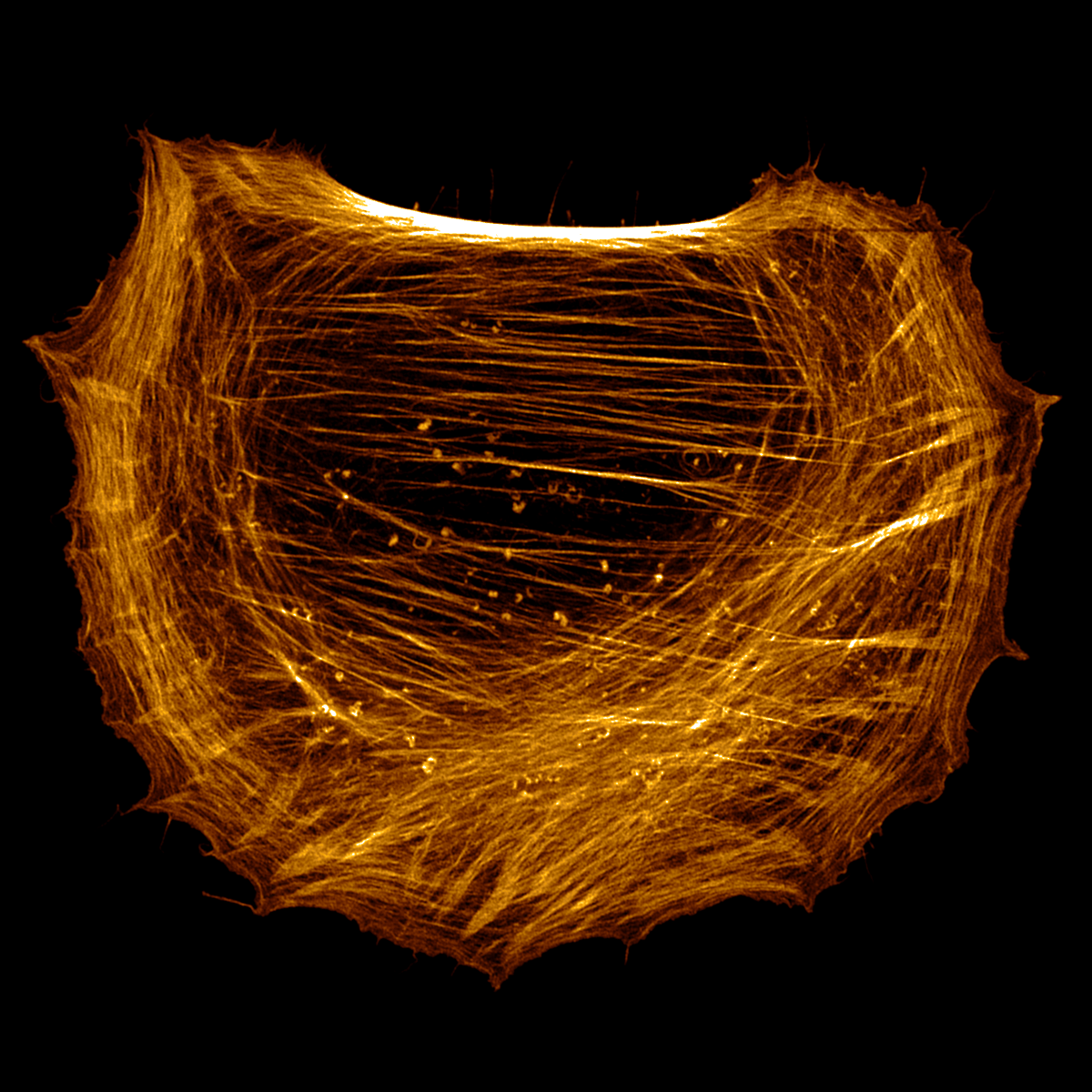
A glowing microscopic wormhole:
🟢: Actin
🟣: Tubulin
The microtubule web pulling toward the dark core gives event horizon vibes 👀
Where does it lead? 👇
🔬: Joe McKellar
Cellular Uniqueness 🧬✨
Clonal lines look uniform until you fire up the lasers.
💜 Magenta: Membrane
💛 Yellow: DNA
🩵 Cyan: Microtubules
See the two distinct microtubule exclusion zones (top & bottom right)? Organelle displacement or cell tension?
🔬: @_Viroscope
Want to catch a judge’s eye in this year's Proteintech Microscopy Image Competition? 👀
As guest judge, I teamed up with @Proteintech on a cheat sheet for winning SciArt.
Don't let your best shots slumber, show us what you've got!👇
🔗: https://t.co/5wF5xCItBG
End of a Microscopic Galaxy 🌌🔥
This high-density neuronal culture screams cosmic web collapsing into stellar fire.
🔥Fiery Orange: Actin
❄️Cool Blue: DNA
Sometimes, looking down through a microscope allows us to look up at the cosmos.
🔬: Joe McKellar
Viral Takeover ☣️
A microscopic siege frozen in time, as cellular life gives way to viral progeny.
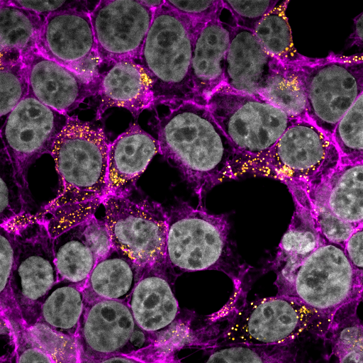
HEK293T cells infected with VSV:
🔴 Viral glycoprotein
🔵 DNA
🔬: Joe McKellar, @_Viroscope
Cellular Ballet 💃
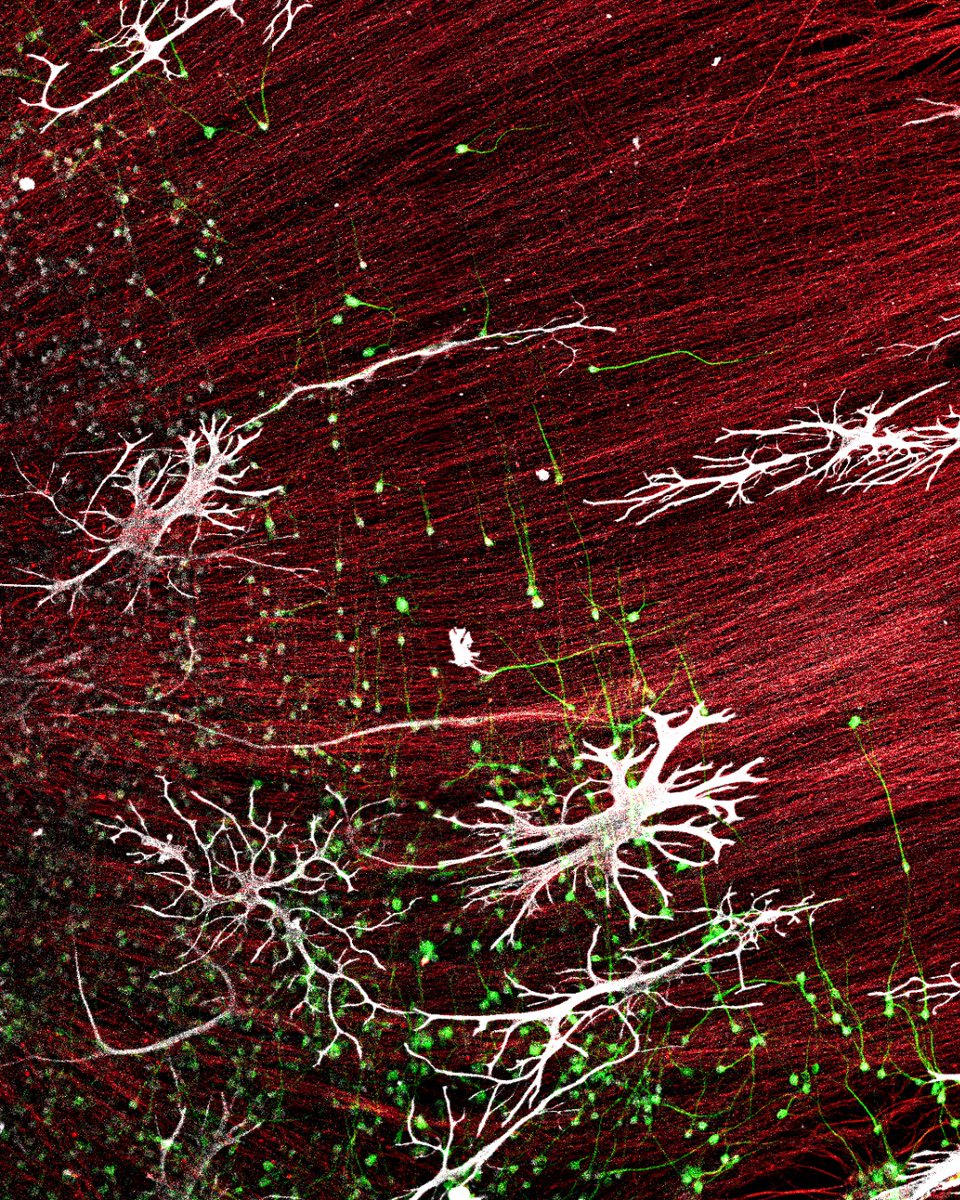
Two boa constrictor brain cells reaching through the dark to perform a microscopic ballet.
🟢 Membranes
🟡 DNA
⚪ Actin
The dance of life is always a spectacle to behold.
🔬: Joe McKellar
The final part of Ask the Expert!
Joe McKellar PhD (@_Viroscope) closes the series with one tip for every researcher just starting out in IF. Simple. Essential.
Thank you, Joe 🔬
Antibodies by Proteintech. Imaged using @zeiss_micro LSM880.
#AskTheExpert#Immunofluorescence
Entry Spotlight 🔬
A closer look at 5 incredible entries from our #Microscopy Image Competition.
Want to see your image featured next?
The 2026 Microscopy Image Competition is open for entries until 5 Oct.
Enter here: https://t.co/NcjN9vLxKa
#Immunofluorescence
Reconstituting mitosis ✨
I recreated a full cell division sequence using 9 separate cells I found from old files!
🟠 DNA
⚪ Tubulin (look the different structures!)
🟣 Actin
Which stage is your favorite? 👇
🔬: Joe McKellar
Does the colour scheme you choose for a multiplex IF image matter scientifically?
Joe McKellar PhD (@_Viroscope) breaks it down in Part 7 of Ask the Expert.
🟡 KIF5B (21632-1-AP)
🔵 IAV nucleoprotein
🟣 DNA
Imaged on @zeiss_micro LSM880 Airyscan.
#Immunofluorescence
How do you design a multiplex IF experiment with zero bleed-through?
Joe McKellar PhD (@_Viroscope) breaks it down in Part 6 of Ask the Expert.
🟡 KIF5B (21632-1-AP)
🔵 IAV nucleoprotein
🟣 DNA
Imaged on @zeiss_micro LSM880 Airyscan.
#AskTheExpert#Immunofluorescence
Cosmic horror at the sub-cellular scale 🌠🐙
Mutant mouse Mx1 (green) forming tentacle-like structures crawling through a genomic portal (pink; DNA).
Like Cthulhu breaching the barriers between realms...
Years later we still don't know why.
🔬: Joe McKellar