@MatthiasNahrend Deeply grateful to Prof. Nahrendorf for this thoughtful Preview. Honored to have our work discussed in the context of cardiac regeneration evolving from cardiomyocyte proliferation toward regenerative ecosystems. Much to learn and very inspired.
Very honored and grateful to see our work discussed by Prof. Nahrendorf in JACC. This perspective beautifully captures an emerging concept: cardiac regeneration is not just about cardiomyocyte proliferation, but about rebuilding regenerative ecosystems.
I’m truly honored to host Professor Lily Jan and Professor Yuh-Nung Jan in one of their three @AFAYale invited talks. As first-gen immigrants, Lily and Yuh-Nung have run a highly successful joint laboratory for over forty years, maintaining world-class scientific output and both becoming HHMI investigators and National Academy of Sciences members. They have over 100 former trainees who have become independent scientists leading their own research labs. In this talk titled "Scientific Excellence, Mentorship and Equity", Lily and Yuh-Nung discussed their insights and observations on these topics, including a “Bamboo Ceiling” and underrepresentation of Asian biomedical scientists in the U.S. in research prizes and in school leadership positions.
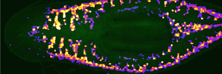
Excited to share our paper now published in GigaScience: “4D single-cell spatial transcriptomics reveals dynamic morphogenetic gradients and regenerative domains in planarians” We study how planarians rebuild body structure after injury. https://t.co/yYUFHJLben
Our paper is now live in @PNASNews
"A single brief cue leaves a day-long internal state imprint in planarians"
A tiny cue can echo through a whole day. Even in planarians.
https://t.co/xd5mutKr3c
Huge thanks to all co-authors, especially Drs. Yue Chen, Kai Han, Guangyi Fan, and collaborators, and colleagues who contributed to this long journey. We hope this dataset will be useful for exploring the principles of tissue patterning, regeneration, and spatial organization.
This work was inspired by many earlier research on gradient genes in the field. We deeply appreciate the groundwork done by the planarian community, and we invite everyone to explore the dataset at: https://t.co/BsmtLON6pe. We welcome your valuable comments and suggestions.
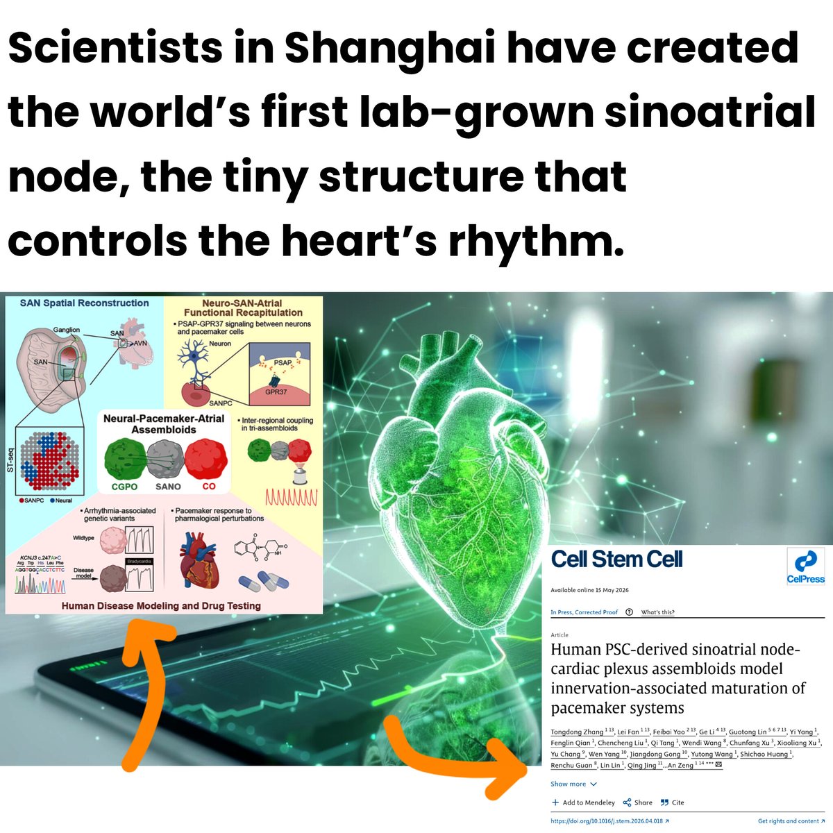
NEW BREAKTHROUGH: Chinese researchers have successfully created a lab grown biological pacemaker using human stem cells.
They engineered a functional sinoatrial node organoid, the exact region of the heart responsible for generating the electrical signals that control every heartbeat.
But the real breakthrough came when scientists connected the pacemaker tissue with neuron-rich organoids, allowing nerve cells to directly regulate the heartbeat rhythm just like inside a real human heart 👀
The lab-grown tissue could spontaneously generate rhythmic electrical impulses, speed up or slow down its beating, and transmit pacing signals into surrounding heart tissue.
Researchers also introduced disease-causing mutations linked to sinoatrial node dysfunction and successfully recreated abnormal slow heartbeat conditions in the organoid.
This is important because today’s electronic pacemakers still depend on batteries, implanted wires, repeated surgeries, and artificial hardware.
A future biological pacemaker could potentially replace electronic implants with living tissue grown from a patient’s own cells, allowing the heart to respond naturally to exercise, stress, and nervous system signals.
China's lab-grown heart organoid could offer alternative to pacemakers | Bojan Stojkovski, Interesting Engineering
Scientists in Shanghai have created the world’s first lab-grown sinoatrial node, the tiny structure that controls the heart’s rhythm.
Deep within the heart’s right atrium lies the sinoatrial node, a tiny cluster of cells responsible for controlling the body’s heartbeat. Often described as the heart’s natural pacemaker, it generates electrical impulses that keep the organ beating in a steady and coordinated rhythm.
Guided by signals from the nervous system, these impulses tell the heart’s upper and lower chambers when to contract, allowing blood to circulate properly throughout the body. When the sinoatrial node malfunctions, the heart can beat too slowly or even stop momentarily, disrupting blood flow and creating serious health risks.
In the most severe cases, failure of this critical control center can become life-threatening and require artificial pacemakers or emergency medical intervention.
Beating heart organoid linked to artificial nerve network
A team of scientists in Shanghai has developed a lab-grown biological pacemaker designed to mimic the heart’s natural rhythm control system. By working with human pluripotent stem cells, which can transform into many different types of tissue, the researchers created a three-dimensional sinoatrial node organoid capable of generating electrical impulses, the South China Morning Post reported.
To make the system more lifelike, the team linked the organoid to an artificial cardiac plexus, a network of nerves located near the base of the heart that helps regulate heartbeat activity. The achievement allowed researchers to recreate how the nervous system communicates with the heart, opening potential new paths for studying irregular heart rhythms and developing future treatments that could reduce reliance on electronic pacemakers.
The research, published in the journal Cell Stem Cell involved scientists from the Chinese Academy of Sciences and Fudan University. The team focused on the sinoatrial node, the tiny part of the heart responsible for controlling its rhythm. Although it plays a critical role in keeping the heart beating properly, the structure has been difficult for scientists to study because of its small size and hard-to-reach location inside the heart.
Positioned near the upper right chamber and close to one of the body’s largest veins, the sinoatrial node is rarely easy to access in human tissue samples, limiting research into how it works and how related heart conditions develop.
Scientists recreate human heart rhythm tissue after animal models fall short
Animal studies, especially those involving mice, have struggled to fully replicate how the human heart’s natural pacemaker works. Because of these limitations, scientists have increasingly turned to lab-grown models of the sinoatrial node to better understand heart rhythm disorders and explore new treatment options.
A 2024 study from SUNY Downstate Health Sciences University highlighted the potential of such models for studying disease and developing biological pacemakers. Building on that goal, researchers in Shanghai used human pluripotent stem cells to create a three-dimensional sinoatrial node organoid by recreating signals normally seen during early embryo development. The lab-grown tissue was able to produce stable and spontaneous beating, closely resembling the activity of the heart’s natural pacemaker.
The breakthrough allowed scientists to recreate, for the first time in a laboratory setting, the complete process through which the heart generates and carries electrical signals that control its rhythm. Researchers found that the lab-grown tissue closely matched human embryonic sinoatrial node cells in terms of gene activity and also reacted correctly to medications used to control heart rate.
The findings could help pave the way for future biological pacemakers based on transplanted cells or organoids, potentially offering an alternative to traditional electronic devices. Conventional cardiac pacemakers, which use electrical pulses to regulate the heartbeat, have been widely used in medicine for more than 50 years and remain one of the most common treatments for patients suffering from dangerous heart rhythm disorders or irregular heartbeats.
https://t.co/XyBWM76eVq