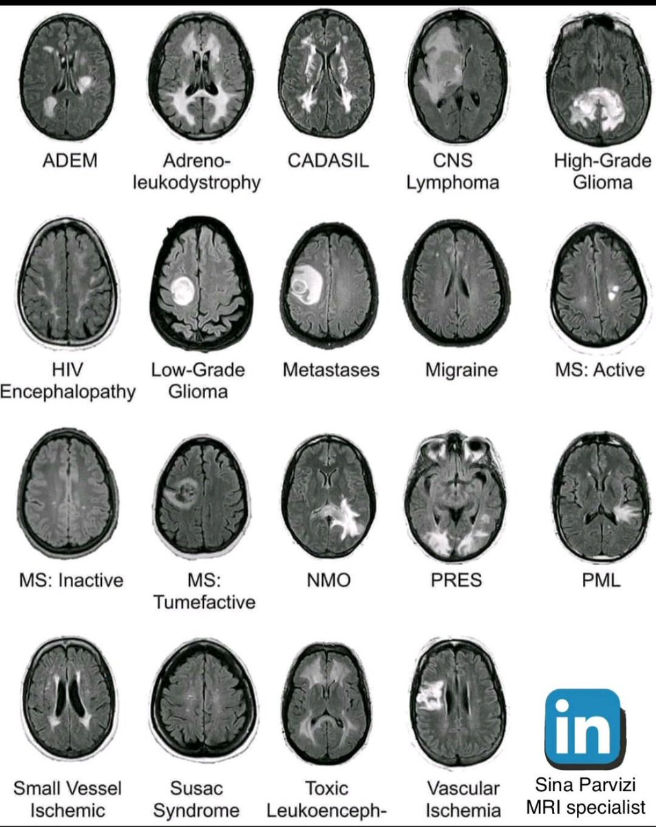
Common lesions (including tumors) on FLAIR MRI.
A time saver I wish I knew earlier(for residents and medical students)
Press like so everyone sees this ❤️. #MedX
20/So now you know the basic MRI sequences and what they are used for!
So hopefully now, the radiologist won’t sound like they are speaking a different language when they talk to you—they will just be nerdy and socially awkward when they do!
19/But other metals will be black too.
Notably, calcium, which is in our bones and in many other lesions
So remember, just all that glitters is not gold, not all that is black on gradient is blood—other metals are black too
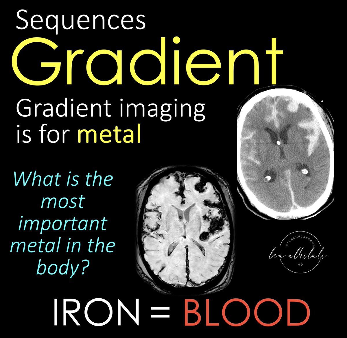
18/Blood is black on gradient
I remember this bc gradient is for metal—and when I think of metal, I think of blacksmiths forging metal products
So BLACKsmith=metal is BLACK on gradient.
17/Last but not least is gradient imaging
Gradient imaging is sensitive to metals
And what’s the most important metal in body? Iron!! Because iron is in blood!
So gradient is our blood sensitive sequence
15/Here’s an example
There's a ring enhancing lesion w/a lot of edema on T2
Centrally, there is restricted diffusion, meaning that there is something gooey or thick or dense centrally
Bc this central stuff doesn’t enhance, we know it’s not a mass. This is pus in an abscess!
14/Pus is also bright on diffusion
As a radiologist I don’t often see pus, but as a mom, I sure do!
It is thick and gooey and you can just imagine how difficult it is for water to travel through that gelantinous blob of pus.
13/Here's an example
The hemorrhage is bright on CT bc it is clotted, and thus more dense than the brain and CSF, which are closer in density to water.
For this same reason, the hemorrhage is bright on diffusion—bc the dense clot traps the water.
12/Hematomas are also bright on DWI
In normal blood, water flows happy & free—but once the clotting cascade starts & fibrin & thrombin & whatever stuff I don’t remember as a radiologist clumps everything together, things get tight—water is trapped in the clot interstices!
11/Here's an example
Here is a mass that is as bright as stroke on diffusion bc of its densely packed cells
On contrast images, we see it avidly enhance, as we would expect for a mass
On CT, the tumor is very dense bc of the densely packed cells.
10/But other things can make it crowded and difficult for water to move
For example, tightly packed cells in aggressive tumors will also fill spaces & make it difficult for water to move—it's trapped between the tumor cells!
So highly cellular tumors are often bright on DWI
9/So classically, it's from a stroke
When cells run out of ATP, the Na/K pump stops working & immediately water rushes in from osmotic pressure & cells swell
These swollen cells fill the interstitium & restrict the movement of water. This is why strokes are bright on DWI!
8/This is bc all diffusion does is detect how difficult it is for water to move
Anything that makes the space around water crowded and difficult to move will be bright on diffusion imaging
6/To review:
T1 is for anatomy and contrast
T2 (and FLAIR, which is a type of T2) is for water—which is bright on T2.
I remember this bc H20 has a 2 in it—T2 is for H20!
5/Now to T2!
T2 sequences are water sensitive sequences
What is pathologic water in the brain? Edema!
My attending once said, “Everything bad in this world is trying to turn you back into what you came from—water"
So T2 shows you edema—but this edema can be from many things
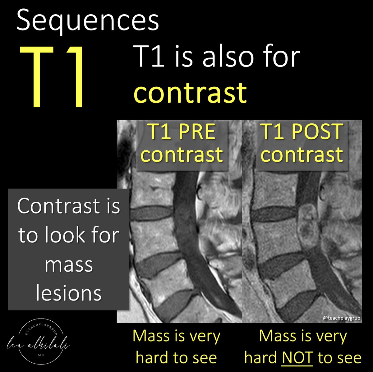
3/T1 is also for contrast
Contrast material helps us to see masses
Contrast can’t get into normal brain & spine bc of the blood brain barrier—but masses don’t have a blood brain barrier, so when you give contrast, masses will take it up & light up, making them easier to see.
2/Let’s start w/T1—it is #1 after all! T1 is for anatomy
Since it’s anatomic, brain structures will reflect the same color as real life
So gray matter is gray on T1 & white matter is white on T1
So if you see an image where gray is gray & white is white—you know it’s a T1
1/Do radiologists sound like they are speaking a different language when they talk about MRI?
T1 shortening what? T2 prolongation who?
Here’s a translation w/an introductory thread to MRI.