🧠🫀 Síncope: Evaluación y Manejo
📋Historia Clínica y Exploración
🩸Presión Ortostática
⚡Electrocardiograma y Arritmias
🚨Estratificación de Riesgo
💊Tratamiento Según el Mecanismo
🔰📚New England Journal of Medicine 2026
Artículo Completo👇🏻✅🆓
https://t.co/aR5SFK2xfj
Guarda esta imagen, luego me agradeces.
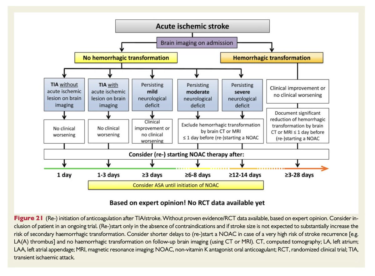
Si un paciente con FA presenta un ACVi mientras recibe anticoagulación, el riesgo de transformación hemorrágica obliga a suspenderla.
La pregunta de siempre es: ¿cuándo reiniciarla?
Esta gráfica ayuda a responderlo y, en general, favorece el reinicio durante la primera semana especialmente si no existe transformación hemorrágica o no es extensa la isquemia.
De los más relevante en Medicina de las últimas semanas. Dejo los links debajo.
1) TRIUMPH-1 Trial (Retatrutida, agonista triple para obesidad) en NEJM 2026
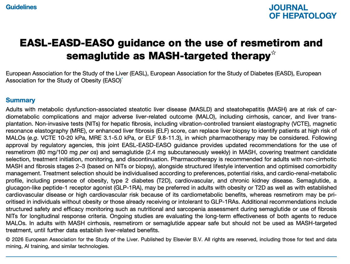
2) Guías 2026 EASL-EASD-EASO para uso de Resmetirom y Semaglutide en MASH y MASLD (alias “hígado graso”)
3) Consenso Español VI de tratamiento para Helicobacter pylori
4) Meta-análisis de antidepresivos y eventos cardiovasculares y prolongación del QT
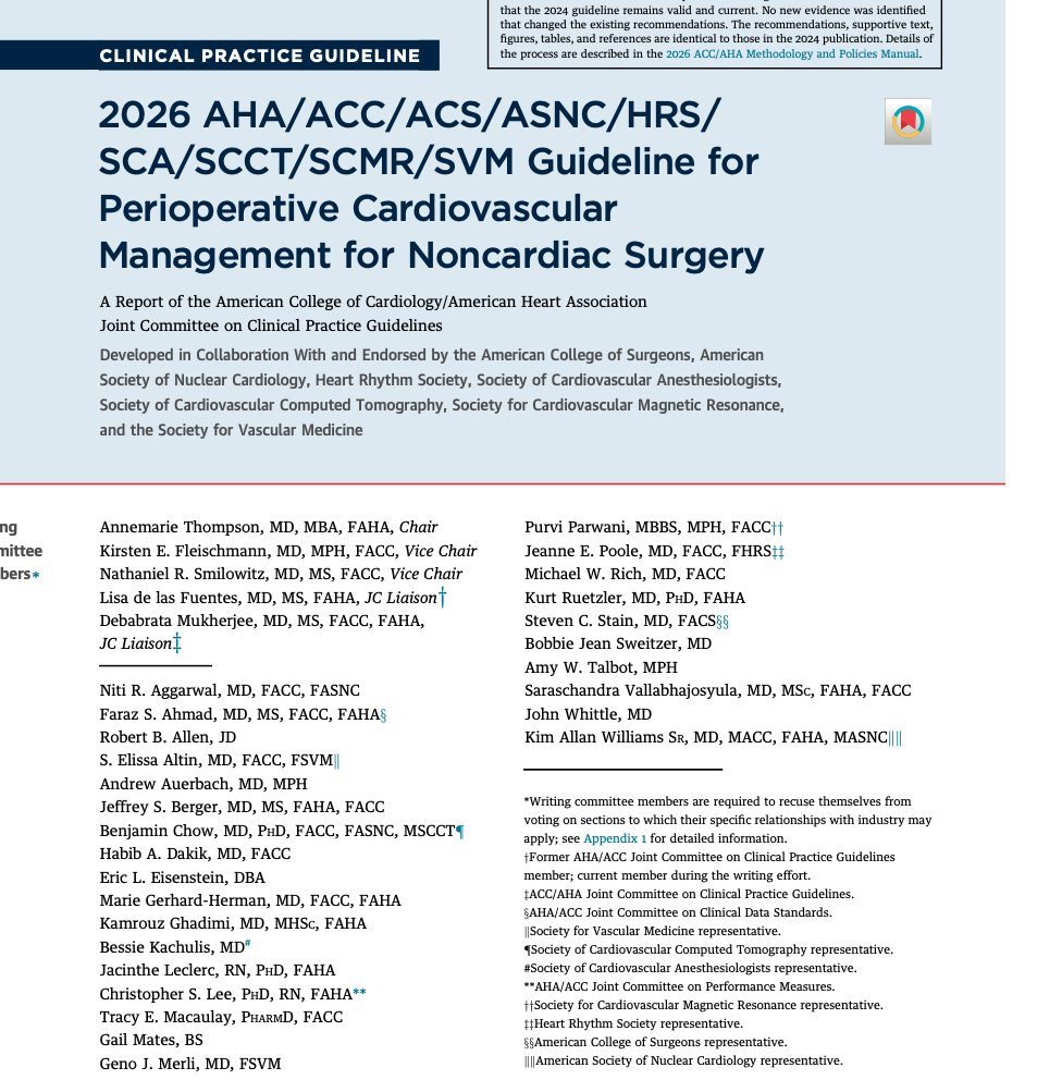
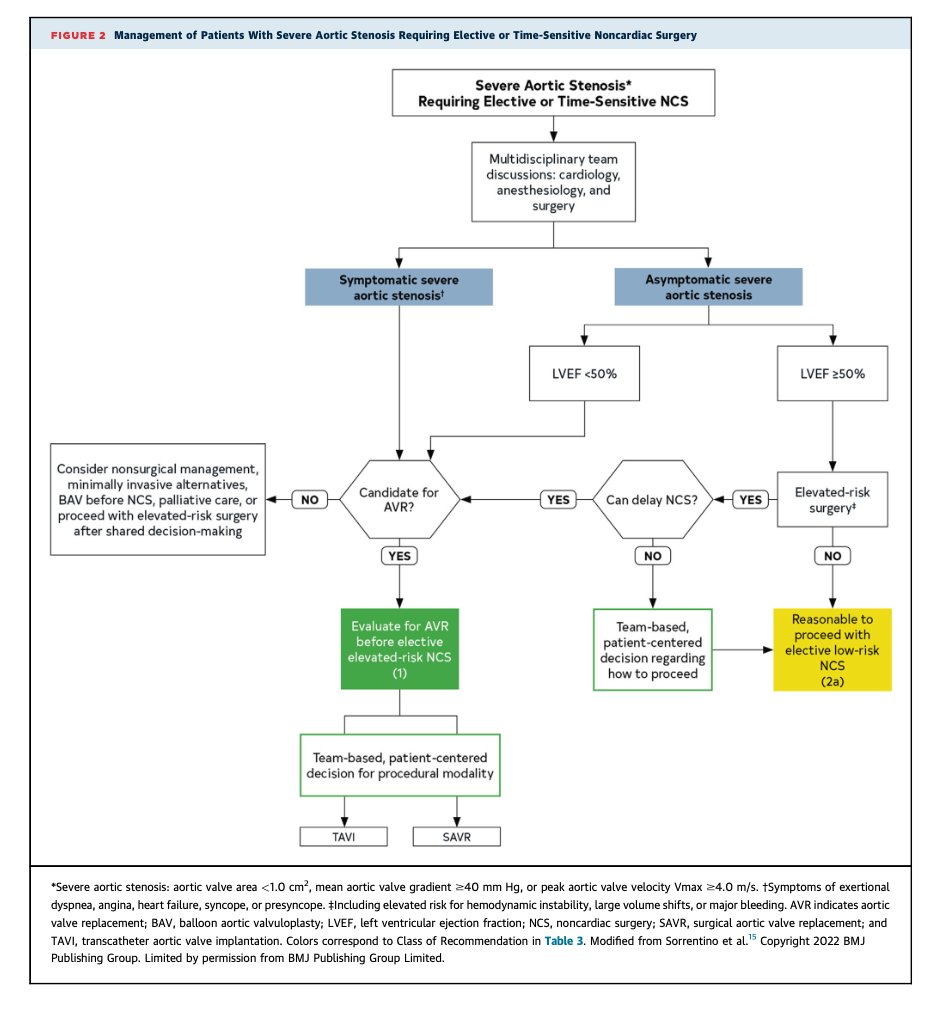
5) Guía perioperatoria AHA/ACC 2026
6) EloraTZP Trial (eloralintida + tirzepatida) en DM2 con obesidad
7) Guía AGA sobre enfermedad hepática metabólica asociada al alcohol (metALD)
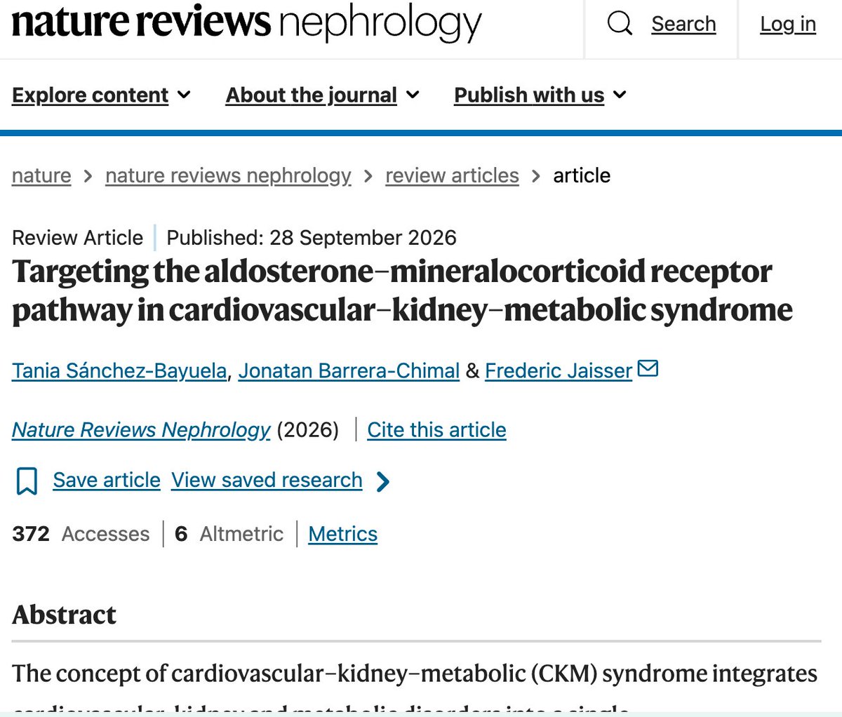
8) Eje aldosterona–receptor mineralocorticoide en el síndrome cardiovascular-renal-metabólico - Nature (2026)
9) Guía ADA y EASD 2026 para manejo de diabetes tipo 2 - Diabetología 2026
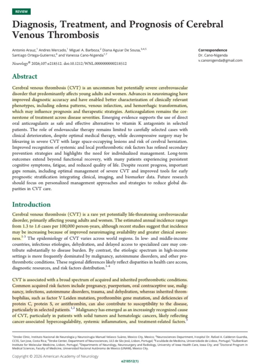
🧠🩸Abordaje y Manejo de la Trombosis Venosa Cerebral
🤕Cefalea y Déficit Neurológico
🧲RM y Angio-TC
💉Anticoagulación
💊DOACs
🧠Trombectomía-Craniectomía
🔰📚Neurology 2026
Artículo Completo👇🏻✅🆓
https://t.co/aR5SFK2xfj
Nefritis Lúpica 🤓
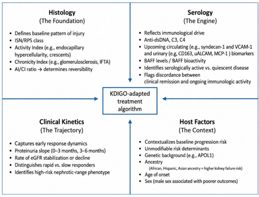
✔️Tratamiento: KDIGO propone basarse en 4 dominios para estratificar y decidir ✍️🏽👇🏼
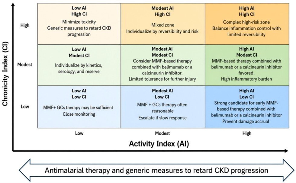
✔️Importante para decidir el objetivo de tratamiento los índices de actividad y cronicidad 🔝👇🏼
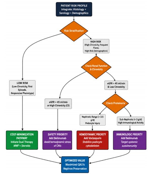
✔️Algoritmo propuesto por autores👀👇🏼
Sciascia et al. CKJ. 2026
#NephX#MedEd
Calculadora de perfusiones de noradrenalina, dobutamina, levosimendán y otros vasoactivos: de µg/kg/min a mL/h
CardioTeca publica una calculadora de perfusiones intravenosas continuas de noradrenalina, adrenalina, dopamina, dobutamina, milrinona, levosimendán y nitroglicerina en adultos. Convierte la dosis en velocidad de la bomba (mL/h) y al revés, y parte de las diluciones de la ficha técnica española.
https://t.co/CdboYnNo5q
🧮 Convierte µg/kg/min en mL/h y, a la inversa, la velocidad de la bomba en la dosis que recibe el paciente.
🧪 La dilución de partida es la de la ficha técnica española y se puede cambiar por la de la unidad; la adrenalina y la milrinona no tienen dilución para perfusión en su ficha técnica.
🚦 El aviso es verde dentro del intervalo de dosis, ámbar fuera de él y rojo por encima del máximo.
📋 Da la cuenta escrita para comprobarla a mano, una tabla de velocidades y un texto para pegar en el informe clínico.
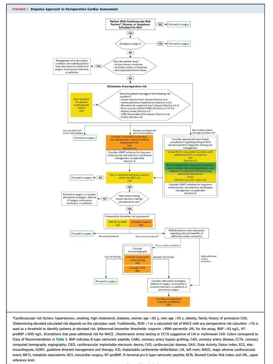
Guías 2026 de la AHA de manejo preoperatorio en cirugía no cardíaca (alias valoración preoperatoria). Recién salida del horno ayer. Puntos importantes:
🔴 Los iSGLT2 se suspenden 3 días antes por riesgo de cetoacidosis euglucémica (nota: la evidencia nueva apunta a que tal vez no se requiera).
🔴 Los aGLP-1 semanales se suspenden más de 1 semana antes (por broncoaspiración). Metformina: continuar.
🔴 Betabloqueador: continuar. Si apenas lo vas a iniciar: empezar 7 días antes.
🔴 IECA/ARA-II pueden omitirse 24 horas en hipertensión controlada + cirugía de alto riesgo, pero se continúan en insuficiencia cardíaca.
🔴 Prueba de estrés y angioTC coronaria: solo con riesgo alto (como <4 METs por DASI y otra indicación. CAC de 0 en los últimos 2 años: pasa a cirugía sin más.
🔴 El ECG en asintomáticos con cirugía de bajo riesgo: no aporta, no es necesario.
🔴 En cirugía de alto riesgo, >65 años, o sintomáticos: considera medir BNP o NT-proBNP). En general no se necesitan seriados.
🔴 Anticoagulación oral (con ACOD) sin puente en general, sin heparina. Puente solo si: AVK y riesgo trombótico muy alto: válvula mecánica mitral, EVC/AIT en los últimos 3 meses, trombofilia o CHA₂DS₂-VASc ≥7.
🔴 Tiempos e infarto. Después de un stent farmacoactivo: esperar 6-12 meses si cirugía electiva. Después de un EVC/AIT se esperan ≥3 meses.
ECG From Scratch #33: Left Atrial Abnormality
If right atrial abnormality tends to make the P wave taller, left atrial abnormality tends to make it wider.
(Why)
Because the left atrium normally depolarizes after the right atrium. Delayed or prolonged left atrial activation can therefore increase the total duration of atrial depolarization.
The classic ECG clue
P-wave duration ≥0.12 s (120 ms)
That is at least 3 small boxes.
The P wave may also become:
Broad and notched or humped
The second hump reflects delayed left atrial activation.
This pattern was historically called P mitrale, particularly because it was associated with mitral valve disease. The broader term left atrial abnormality (LAA) is preferable because a broad P wave does not necessarily prove anatomical left atrial enlargement.
Look at V1
🔵Lead V1 can provide another important clue.
A normal P wave in V1 is often biphasic:
small positive component ➡️ negative component
With LAA, the terminal negative component may become:
≥1 mm deep
and/or
>40 ms wide
Why?
The right atrium lies more anteriorly, while the left atrium is positioned posteriorly.
So the initial positive component reflects right atrial activation, while the prominent terminal negative component reflects delayed left atrial activation directed posteriorly, away from V1.
Where can LAA appear?
Common settings include:
- Mitral stenosis or regurgitation
- Aortic stenosis or regurgitation
- Hypertensive heart disease
- Dilated, hypertrophic, or restrictive cardiomyopathy
- Coronary artery disease
An important caveat
A broad or notched P wave does not prove anatomical left atrial enlargement.
A patient may have a broad P wave because of delayed atrial conduction even when imaging does not show an enlarged left atrium.
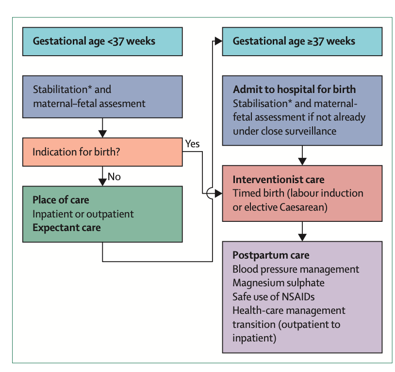
Preeclampsia, la última revisión en The Lancet (2026). Puntos importantes:
🔴 La proteinuria dejó de ser obligatoria para el diagnóstico. Preeclampsia = hipertensión después de las 20 SDG + disfunción de órgano blanco (neurológica, renal, hepática, hematológica) o uteroplacentaria.
🔴 Olviden la hipertensión permisiva: trata toda hipertensión del embarazo, meta <140/90 (diastólica 85), CHIPS y CHAP Trial. Labetalol, nifedipino o metildopa. IECA/ARA contraindicados por fetotoxicidad.
🔴Prevención con Aspirina 150 mg por la noche, antes de las 16 SDG, y se suspende a las 36. El calcio ya no se recomienda.
🔴 El único tratamiento sigue siendo el parto. El sulfato de magnesio no es profilaxis universal — es para preeclampsia con datos de gravedad y eclampsia.
🔴 En el puerperio, la presión arterial alcanza su pico los días 3 a 6 (monitorea en casa). Asociado a más: hipertensión, enfermedad renal crónica, diabetes, riesgo cardiovascular. Revisión médica anual recomendada.
Ya actualizamos las apps.
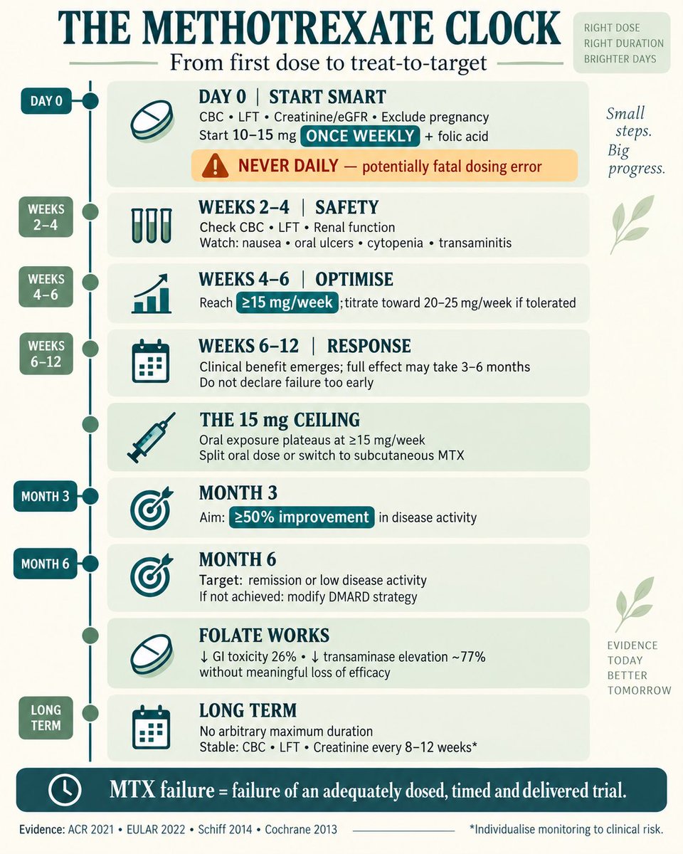
💊 THE METHOTREXATE CLOCK: FROM FIRST DOSE TO TREAT-TO-TARGET
DAY 0 | Start smart
CBC • LFT • creatinine/eGFR • pregnancy exclusion ± hepatitis screening
Start 10–15 mg ONCE WEEKLY + folic acid
⚠️ Never daily—potentially fatal error
WEEK 2–4 | Safety first
Check CBC/LFT/renal function.
Early nausea, fatigue, oral ulcers or transaminitis may occur.
WEEK 4–6 | Optimise early
Escalate to ≥15 mg/week, usually toward 20–25 mg/week, if tolerated.
✅ ACR recommends reaching ≥15 mg/week within 4–6 weeks.
WEEK 6–12 | Response window
Clinical benefit usually emerges; full effect may take 3–6 months.
❌ Do not label MTX failure after only 2–4 weeks.
THE 15-mg CEILING!
Oral MTX exposure tends to plateau at ≥15 mg/week. If response or tolerance is inadequate:
➡️ Split the oral dose over 24 h
➡️ Switch to subcutaneous MTX—greater, more predictable exposure
➡️ Optimise folate before abandoning MTX
MONTH 3 | First checkpoint
Aim for ≥50% improvement in disease activity.
If absent: check adherence, dose, route and diagnosis—then modify treatment.
MONTH 6 | Target checkpoint
Aim for remission or low disease activity.
Persistent active disease despite optimised MTX → add/switch DMARD therapy.
WHY FOLATE MATTERS
Folic/folinic acid reduces:
• GI toxicity by 26% relative
• Elevated transaminases by ~77% relative
• MTX discontinuation by ~61% relative
✅ Without meaningful loss of efficacy
LONG TERM
No arbitrary maximum duration—continue while effective and tolerated.
Once stable, monitor CBC/LFT/creatinine approximately every 8–12 weeks, individualised by risk.
🚨 STOP & ASSESS: cytopenia • persistent transaminitis • severe mucositis • significant infection • new cough/dyspnoea
📌 MTX failure should mean failure of adequately dosed, adequately timed and properly delivered MTX—not merely failure of a low oral dose.
Evidence:
ACR RA guideline: doi:10.1002/art.41752
EULAR recommendations: doi:10.1136/ard-2022-223356
Oral vs SC pharmacokinetics: doi:10.1002/acr.22212
Folate Cochrane review: doi:10.1002/14651858.CD000951.pub2
#Methotrexate #Rheumatology #RheumatoidArthritis #TreatToTarget #DMARDs #MedEd
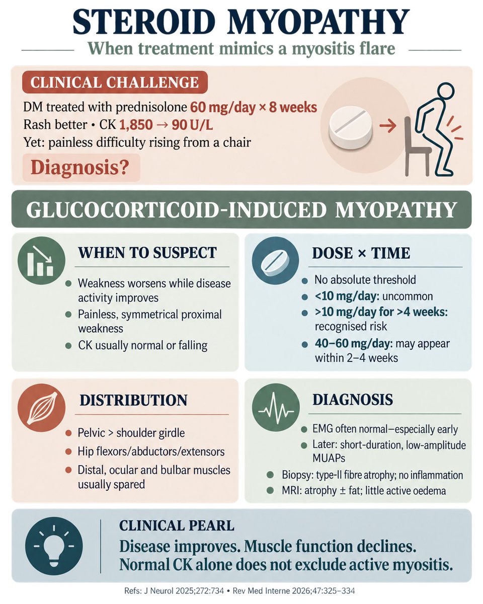
🧩 THE MYOSITIS PARADOX
The disease improves—but the patient gets weaker.
A 38-year-old woman with dermatomyositis receives IV methylprednisolone followed by prednisolone 60 mg/day. Eight weeks later:
Rash and dysphagia improved
CK: 1,850 → 90 U/L
MRI oedema markedly reduced
Yet she struggles to rise from a chair
Painless, symmetrical hip weakness
EMG essentially normal
❓Flare, deconditioning—or steroid-induced myopathy?
✅ Answer: GLUCOCORTICOID-INDUCED MYOPATHY
🔎 When to suspect?
New/progressive weakness despite control of the underlying disease
Insidious, painless proximal weakness ± visible wasting
Lower-limb function affected first: chair-rise, stairs, getting off the floor
CK remains normal or falls with treatment
No parallel worsening of rash, dysphagia or systemic inflammation
💊 Dose × duration
There is no validated diagnostic threshold—risk is cumulative and dose-dependent.
<10 mg/day prednisolone-equivalent: uncommon
>10 mg/day for >4 weeks: recognised risk
40–60 mg/day: weakness may appear within 2–4 weeks
Very high-dose IV steroids may contribute to an acute, diffuse ICU/critical-illness myopathy
Risk increases with older age, cancer, malnutrition, sepsis, inactivity and concurrent neuromuscular blockers. Fluorinated steroids—dexamethasone, betamethasone and triamcinolone—appear more myotoxic.
🎯 Distribution
Symmetrical and proximal
Pelvic girdle > shoulder girdle
Hip flexors/abductors/extensors prominently affected
Distal, facial, ocular and sphincter muscles generally spared
Respiratory weakness is unusual in chronic disease
⚡ EMG pearl
Often normal, especially early
Steroids predominantly injure type-II fibres, which routine needle EMG may inadequately detect
Later: low-amplitude, short-duration MUAPs with early recruitment
Prominent fibrillations/positive sharp waves favour active inflammatory or necrotising myopathy—although they are not mandatory for a flare
🧪 Diagnosis: no single confirmatory test
CK/aldolase/LDH: usually normal in chronic steroid myopathy
MRI: atrophy ± fatty replacement; absence of new oedema supports steroid myopathy
Biopsy, if uncertainty persists: selective type-II fibre atrophy without inflammation
Improvement after glucocorticoid reduction supports the diagnosis
⚠️ Normal CK or absent MRI oedema alone does not exclude active dermatomyositis—interpret the entire phenotype.
🛠️ Management
Use the lowest effective glucocorticoid dose, optimise steroid-sparing therapy, correct nutritional/electrolyte factors and begin supervised resistance plus aerobic exercise. Strength may start improving within 3–4 weeks, but complete recovery can take months.
📌 Steroid myopathy is a diagnosis of clinicobiological discordance: disease activity improves while muscle function declines.
References: Géfard-Gontier et al., 2025 • Solé et al., 2026 • Minetto et al., 2018 • Gupta & Gupta, 2013
#SteroidMyopathy #Myositis #Dermatomyositis #Rheumatology #ClinicalPearls
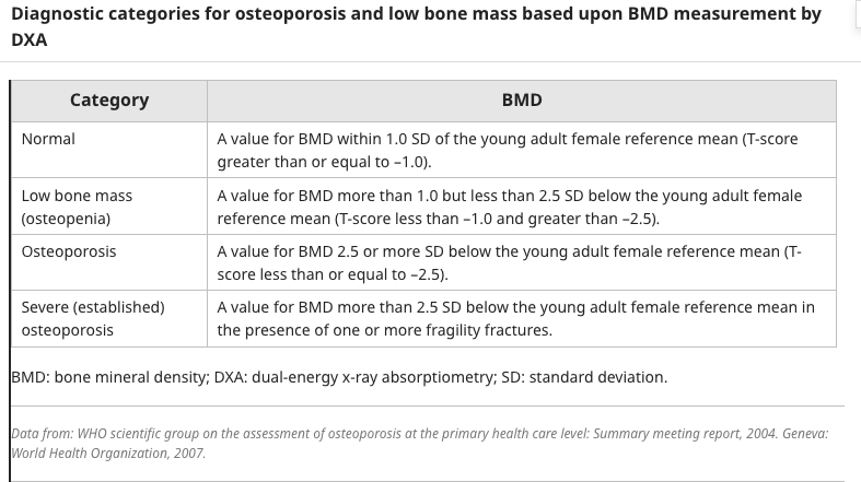
Estos son los criterios DENSITOMETRICOS para diagnosticar osteoporosis.
Recuerda que el T-Score se usa en pacientes post-menopasuia y en hombres que ya tengan mas de 50 años.
En mujeres con edad fertil u hombres <50 años se deberá enfocar con Z-Score + Clinica.
Anticoagulation reversal: know the antidote.
When major or life-threatening bleeding occurs, reversal depends on the anticoagulant involved:
🟥UFH: Protamine
🟦Dabigatran: Idarucizumab
⬜Apixaban/rivaroxaban:Andexanet alfa
🟩Warfarin: 4-factor PCC + vitamin K
For LMWH, protamine provides only partial reversal, while fondaparinux has no specific antidote.
The key is not simply knowing the drug.
Know its mechanism, half-life, renal clearance and reversal strategy.
Current evidence also emphasizes that reversal agents carry thrombotic risks and should be reserved for appropriate major/life-threatening bleeding or urgent situations.
Which anticoagulant reversal pair is most important to remember?
Disfunción en Cateter de Hemodiálisis 🫤
✔️Causa importante de tratamientos sub-optimos y problema común en unidades de HD
✔️Temprana < 7 dias luego de inserción
✔️Tardía > 1 semana luego inserción
✔️Causas 👀👇🏼
Lazarus et al. JASN. 2025
#NephX#MedEd#MedTwitter