Hi everyone! My name is Alex and I will be applying to cardiology fellowships this season. I come from Landmark Medical Center in Rhode Island where I’m a third year internal medicine resident. I look forward to meeting you all! My passion is electrophysiology!
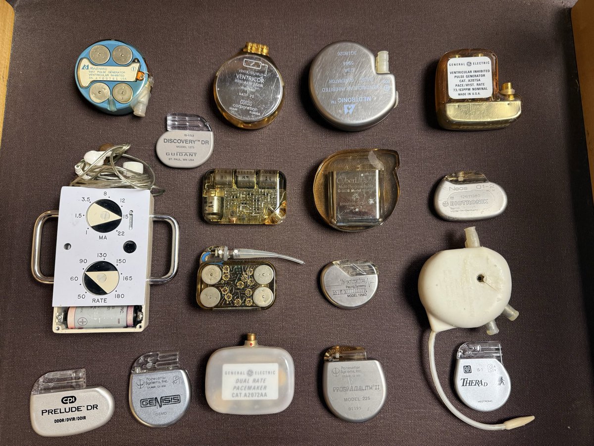
Fellow on HCM rotation asked what’s in the case. Here’s my dad’s collection of history of pacemakers.
From 1950s bulky devices to leadless wonders, they’ve come a long way.
Decades later, they still do what they do best: keep hearts (& history) ticking
Which is the earliest?
@MiguelP23970914 If irregular Cannon = VT
If irregular V waves = AF.
This was MVP severe MR, PH, TR with AF and fast ventricular rate. During rounds while looking at JVP, I also first thought irregular cannon waves and looked at the monitor which showed AF
Coronary CT will become the non-invasive standard to evaluate patients with stents
It will also help in understanding the root cause of stent restenosis (mechanical vs. neoatherosclerosis) and guide treatment
@djc795@ziadalinyc@Heart_SCCT@SCAI@crfheart
https://t.co/7nwapXoDs0
Our structural heart team performed their first transcather tricuspid valve replacement (TTVR) using a new FDA approved device, designed to treat severe tricuspid regurgitation without open-heart surgery. @RaniKHasan@frahman480@MatthewCzarnyMD@HopkinsACCMCard
Critical isthmus of VT is confined to small area despite extensive substrate!
👇
Epicardial map of ARVC demonstrating figure-of-8- reentry colocalized with DZ of ILAM. #EPeeps
Safety of pulsed field ablation of atrial fibrillation in patient with Left Ventricular Assist Device and heparin-induced thrombocytopenia
https://t.co/jaNBI4NVGy
This pt on DDD pacemaker had a few episodes of “giddiness” for which she was admitted and Holter was connected.same day we asked the pacemaker tech to have a look at the parameters.He told all parameters were normal.X-ray shows normal leads.Holter shows a strip of CHB. What to do
Today in EPlab:56 yo man with 41% PVC density and PVC induced cardiomyopathy. RVOT map with #ORION showed EGM preceding QRS by 22 ms. RF at this site was unsuccessful. Mapping sinuses of Valsalva showed in the RCC an early EGM.RF ablation in this point led to disappearence of PVC
Fascinating VT ablation case: basal inferolateral SUBENDOCARDIAL infarction. Voltages mostly preserved (bipolar & unipolar). No LAVA or late potentials on sinus rhythm/S1. S3 PROTOCOL unmasked deceleration zones, matching channel areas seen with ADAS @JuditMasC_
In the SUPPRESS-AF subanalysis, LVA ablation added to PVI was effective only in patients with enlarged left atrium (LAD >44mm), while no benefit was seen in smaller atria. Larger LA patients may benefit most #AHAJournals#Epeeps
https://t.co/cPgpvdOJHu