Because of our supporters, groundbreaking equine research is happening right now.
Grayson has funded hundreds of projects at leading veterinary institutions — improving prevention, diagnosis, and treatment.
Join the donors making a difference.
https://t.co/AnwZRNyzQI
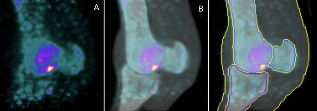
Equine PET can now be analyzed with AI
"Deep learning cascade networks for segmentation of F18-NaF PET scans of equine metacarpo- and metatarsophalangeal joints outperform atlas-based method"
https://t.co/CUq8g20RyM
@alienbytesoft@Sergey_ABSS@MathieuSpriet
10 years ago today, we performed the first equine PET @ucdavisvetmed It was academic curiosity at the time, but now with 12 active scanners, thousands of horses have been imaged and more to come. What a great adventure! @equinepet@Grayson_JC@ucdavis_ceh
Grayson has a proven record of funding research which has benefited, and continues to benefit, the horse.
Horses strive to be our champions, we must ALWAYS be theirs.
Donate now https://t.co/AnwZRNyzQI #EquineResearch
We are a couple of days late with this announcement, but it's huge. Wonderful innovation! Made in America and now available on Heard Island and McDonald Islands. Like and repost!
Don't miss the online @VetPD presentations/panel discussion "Positron Emission Tomography (PET): Clinical Applications & Case Discussions"
April 1, 1pm:4pm US Eastern
@MathieuSpriet, @ucdavisvetmed
Dr. Kyla Ortved, @pennvet
Dr. Natasha Werpy, Ocala Equine Hospital
Dr. Carter Judy, @ucdavisvetmed
Register here:
https://t.co/0jGbKNmKy4
Sponsored by: @equinepet@alienbytesoft
@TheHorse Advanced Imaging in Horses: Where, Why, When? by Lucile Vigouroux
"Paired with motion-correcting software, PET can tolerate minor movement, making the modality ideal for use under standing sedation, rather than anesthesia."
https://t.co/SI5O3UDI7C
"Diagnosing Equine Hock Joint Problems", in @thehorse by Dr.Stacey Oke, discusses common pathologies of the hock, as well as diagnostic options, including diagnostic analgesia, X-rays, CT, and PET, with Dr.Sue Dyson and Dr.Pablo Espinosa-Mur.
https://t.co/KJzSAGKTor
https://t.co/k8mRapHSgG
A very interesting case on the use of F18-NaF PET and MRI in equine septic hock published in @EVJltd Equine Veterinary Education by Dr.Arndt @tuftsvet and @ucdavisvetmed team
"Use of positron emission tomography and magnetic resonance imaging to identify central tarsal bone necrosis"
https://t.co/CNDYlSAhQa
@Grayson_JC @BEVAPresident @BEVA_news
From Discussion: "The marked 18F-NaF [PET] uptake in the central and third tarsal bones, combined with the clinical history, was strongly suggestive of osteomyelitis, but the large photopenic area at the dorsal aspect of the central tarsal bone was an unexpected finding."
“A specific spot on the sesamoid bone will light up on PET scans in 5-10% of (unsound or underperforming) horses we scanned. And we’ve learned from necropsies that this spot is commonly involved in breakdowns. So we can confidently say that horses with this finding on PET are at greater risk of breakdown.”
"We once looked at imaging like a PET scan as good for a short period of time. But as we've done these sequential studies, if you have a clean PET scan, you can feel very confident for the next four, six, eight weeks that you're not going to have a problem."
Dr.Ryan Carpenter
https://t.co/xSEpmb6Nqr
It all starts with research. Take a minute and read the research spotlight Development of PET Technology for the Equine Athlete. https://t.co/uYyKb2P6Kv
Donate now https://t.co/AnwZRNz7Gg #EquineResearch


![equinepet's tweet photo. From Discussion: "The marked 18F-NaF [PET] uptake in the central and third tarsal bones, combined with the clinical history, was strongly suggestive of osteomyelitis, but the large photopenic area at the dorsal aspect of the central tarsal bone was an unexpected finding." https://t.co/t9OU6OpFR2](https://pbs.twimg.com/media/GkqVJmNW0AA78eq.jpg)


