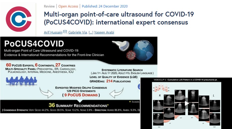
Lung #ultrasound based approach for early diagnosis of #COVID19 pneumonia:
➡️20 US/European hospitals
➡️1462 pts
Combining #LUS patterns of probability + clinical characteristics allow to rapidly rule in/out #SARSCoV2 pneumonia at bedside w high accuracy!
https://t.co/GqsOZoIdgc
Diuresis baffled me when I first got out of school, but it's pure bread and butter for critical care. When should you start? How do you dose furosemide? What if it's not working? An overview of these basics, and more... #medtwitter#FOAMcc#pulmcc https://t.co/UTpkp956Mc
Loculated pericardial effusion causing clinical tamponade. LV is being collapsed in diastole (not the RV). Went for a pericardial window right after the POCUS was performed. #echofirst@HGHED
Adhikari's Image of the Week: Lemierre Syndrome. Characterized by the spread infection into lateral pharyngeal space followed by septic thrombophlebitis of internal jugular vein. Look for: Noncompressible vein, thickened wall, +/- visible thrombus in lumen #POCUS
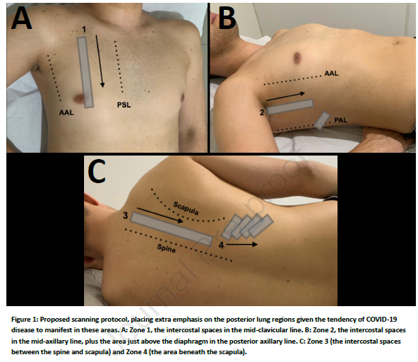
New Article: How I do it – Lung Ultrasound for Patients with COVID-19 Pulmonary Disease. Scott J. Millington, MD, Seth Koenig, MD, Paul Mayo, MD, Giovanni Volipcelli, MD https://t.co/RlcTZgOoHR More Info: https://t.co/Va3648O5ln
@POCUSbot@IM_POCUS@emergencypdx@NephroP