μsam is live in Nature Methods!🥳
Huge thanks to @cppape for being the absolute best PI, guiding me through every step in this journey, @rita_strack for amazing handling of our paper & all reviewers for helping us strengthen μsam.
Go check our tool and paper right away!😉🧵
After a long journey, Segment Anything for Microscopy is now published in Nature Methods! We significantly improve SAM for interactive and automatic segmentation in light and electron microscopy and build a user-friendly tool. https://t.co/HL19SGMdNt
How can we use foundation models such as (micro)SAM to improve electron microscopy segmentation? Check out our preprint! We found substantial improvements for nucleus, mito, and neurite-segmentation using initialization and semi-supervised learning with foundation models.
There are exciting opportunities and foreseeable clinical applications of vision foundation models for biomedical image analysis! The MIDL Community did seem excited, and so are we! (some amazing follow-up applications coming soon, keep an eye open 😉)
We presented our latest work on "PathoSAM" and "Late PEFT" last week at #MIDL2025 (Salt Lake City)!
The community is growing and MIDL is becoming the venue-to-go for high quality research discussion!🧵
And both of our efforts and these beautiful presentations at #MIDL2025 have been successful due to immense efforts from our amazing lab members, Carolin Teuber and Titus Griebel!
And of-course @cppape being the absolute best PI, as always! (I can't say this enough tbh)!
Are you looking for an exciting position at the intersection of super-resolution microscopy and AI? Then check out the PhD and PostDoc position we offer for a joint project with the Group of Stephan Hell at MPI Göttingen. Please RT, read on for links and details.
A nice advance for imaging-based spatially resolved transcriptomics from the Weigert and La Manno labs. Spotiflow uses deep learning for subpixel-accurate spot detection in diverse 2D and 3D images. https://t.co/qVGlT7MXYC
Announcing the new release v1.4.0 of microSAM. The main changes are:
1. Simplified installation on windows.
2. Preliminary support for automatic tracking.
3. Improved interface for model selection.
Read on for a quick summary of the changes.
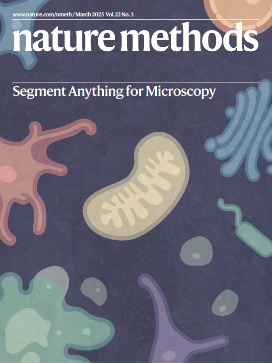
Another feather for Segment Anything for Microscopy. We made it to the cover for @naturemethods!
And all thanks to our amazing Sebastian for this! <3
Our March issue is now live! 🥳
https://t.co/ODK1Eqq2bJ
The cover represents the process of cell and organelle segmentation by Segment Anything for Microscopy.
Paper here: https://t.co/eFVi5XB4Ar
Cover by Sebastian von Haaren.
nnU-Net v2.6 released!
Most important change: Rework of the preprocessed data format 🚀 We now use Blosc2 for partially reading compressed files! Faster IO with less disk space required. Many thanks to Jan Sellner from @DKFZ_IMSY_lab and the @Blosc2 team for their amazing help!
That's a wrap for #EMBLDeepLearning 🌠
A huge thank you to everyone who attended this advanced course 🙌 We hope the hands-on element of applying deep learning-based methods to your own data and image analysis problems was useful. Until next time!
Because we have seen these improvements and due to popular demand, we have decided to start a call for community data submission to further improve our models: https://t.co/A0J08125QN. Looking forward to any feedback!
Our first deposition of synaptic vesicles segmentations is now in the Cryo ET Portal! We segmented vesicles in over 50 tomograms to enable analysis of membrane proteins and more.
https://t.co/O9AbcoAVhe
After a long journey, Segment Anything for Microscopy is now published in Nature Methods! We significantly improve SAM for interactive and automatic segmentation in light and electron microscopy and build a user-friendly tool. https://t.co/HL19SGMdNt
Two absolutely fantastic bioimage analysis papers out today offering exceptional, generalizable tools for segmentation--Cellpose3 and Segment Anything for Microscopy. (1/3)
Segment Anything for Microscopy (μSAM) is based on Segment Anything, the vision transformer model for image segmentation, and offers generalist models for light and electron microscopy segmentation tasks. @cppape@AnwaiArchit@DataNerdery@Sushmita_Nair
https://t.co/eFVi5XB4Ar
A huge thanks to all co-authors and especially
@AnwaiArchit who led this project! Also thanks to
@rita_strack or the excellent handling of our paper. Stay tuned for more updates on micro_sam in the next few days!