Olfactory nerve damage after a motor vehicle collision may be permanent. Most patients are never told this — because most patients are never tested.
Olfactory receptor neurons are among the only neurons in the adult brain with regenerative capacity. That sounds optimistic. The outcome data is not: approximately one-third of patients with post-traumatic olfactory loss do not fully recover baseline function. Their sense of smell does not return. Their flavor perception does not come back to pre-collision levels.
That is a permanent injury from a collision that may have shown nothing on standard imaging.
It compounds: impaired smell affects nutrition — people with anosmia eat less varied diets and are at higher risk for weight loss and nutritional deficiency.
It affects safety — they cannot detect gas leaks, smoke, or spoiled food. It affects daily quality of life in ways that are measurable, specific, and compensable under AMA Guides.
The trajectory is set at the moment of injury. Documentation and impairment rating require only that someone tested CN I before the window for establishing causation closed.
The test is thirty seconds. The permanence of the injury is not.
The olfactory nerve enters the brain through perforations in the skull base. When the brain moves and the skull doesn't, those nerve fibers shear.
During a motor vehicle collision, the brain lags behind the skull — inertia keeps it moving while the skull has already changed direction. The olfactory fila pass through the cribriform plate and are in the exact path of that differential movement. Partial shearing produces measurable olfactory deficit — hyposmia, anosmia, or parosmia.
And this region is directly adjacent to the entorhinal cortex and hippocampal projections responsible for short-term memory. The olfactory nerve bypasses the thalamus entirely and synapses straight into the limbic system.
Which is why the clinical instruction is anatomically sound — when your patient reports memory problems after a crash, test their sense of smell. The proximity is not coincidental. It is structural.
@LoganARobison Looking back to when I was a kid, I think my family dentist was doing the same thing. I got drilled so many times I lost count. My teeth have been F'd ever since then.
"My sense of smell is probably fine — I don't notice anything." That is not a negative finding. That is an untested system.
Patients with olfactory nerve damage rarely present saying they can't smell. They say food doesn't taste right. They say their morning coffee doesn't smell the way it used to. They say they can tell something is burning but can't identify what it is. They've adapted. They attribute the change to a cold, to stress, to getting older.
Olfaction and flavor perception are neurologically coupled — approximately 80 percent of what we experience as "taste" is retronasal olfactory signal.
When CN I is damaged, the patient doesn't experience a smell problem. They experience a food problem. They stop enjoying meals without knowing why.
The provider who asks "Since the accident, has your sense of smell changed? Does food taste different?" is examining cranial nerve function.
The patient who answers yes has given a positive olfactory finding. That answer belongs in the record, with a specific ICD-10 code and a documented impairment pathway.
"I don't notice anything wrong" is not reassurance. It is a symptom of a system that has never been asked to perform.
@OldVetvp I had a teacher in College who had an arm that didn't work. From polio. It was atrophied, and he just kept that hand in his pocket while he lectured. It seemed kind of rubbery, and it wiggled when he walked around and when he wrote on the chalkboard.
The olfactory nerve is the most commonly injured cranial nerve in motor vehicle collisions. It almost never appears in the clinical record.
Standard post-collision neurological documentation reads "cranial nerves 2-12 grossly intact." CN I — the olfactory nerve — is not in that range. It was not tested. It was not documented. It was omitted.
The test that identifies it takes thirty seconds. A familiar scent — coffee, mint, citrus — presented to one nostril at a time. A patient who cannot identify it has a positive finding. That finding has its own ICD-10 code, qualifies for a 1-5% impairment rating under AMA Guides, and connects through anatomy to the memory and cognitive complaints the patient is already reporting.
Most providers don't test CN I because it wasn't emphasized in training. That's a correctable gap — and the thirty-second exam is the correction.
The patients who take longest to recover from a motor vehicle collision are often the ones with undocumented PTSD driving the physical plateau.
Untreated psychological trauma reduces physical therapy compliance, elevates pain sensitization, and keeps the nervous system in a chronic threat state that is physiologically incompatible with recovery.
The physical injury is real. The treatment plan is appropriate. The patient is not improving. No one has asked whether they can drive yet.
Early identification changes both outcomes: faster physical recovery and stronger case documentation. Coordinated psychological and physical care outperforms physical care alone — every time.
The question is not whether PTSD affects recovery. It does. The question is whether it was found in time to treat it.
https://t.co/O5da6uDV2r
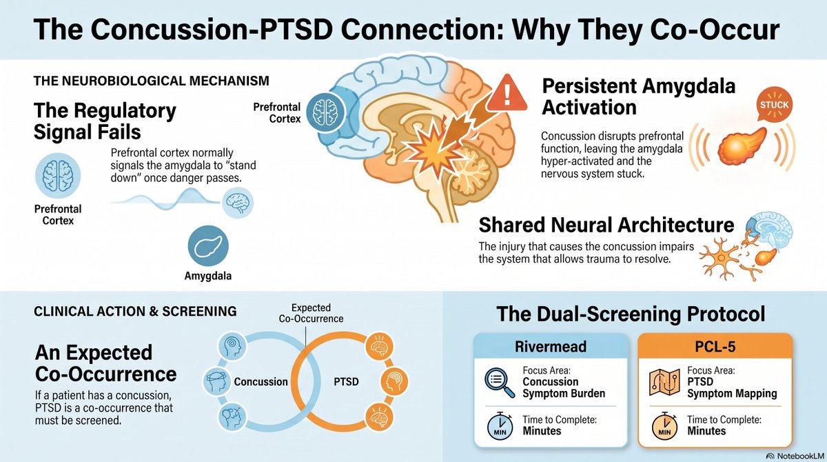
Concussion does not just injure the brain. It injures the system that prevents PTSD from taking hold.
The prefrontal cortex normally regulates the amygdala's threat response — it provides the signal that says "the danger is over, you can stand down." When a concussion disrupts prefrontal function, that regulatory signal fails. The amygdala stays activated. Threat responses persist. The nervous system cannot return to baseline.
This is why concussion and PTSD co-occur so predictably: the concussion impairs the exact neural architecture that allows psychological trauma to resolve. One injury amplifies the other through a shared mechanism.
If your patient has a concussion, PTSD is not a possibility to consider. It is a co-occurrence to screen for. The Rivermead captures one. The PCL-5 captures the other. Both take minutes. Neither is optional.
"You're coping well given what happened." That is not a PTSD screen. That is a social observation.
PTSD does not announce itself. Patients who are struggling to drive, who changed their route to avoid the crash location, who wake up reliving the impact, who tense up every time a car brakes near them — they often describe this as "stress" or "being nervous" or "just not feeling like myself." They are not going to self-diagnose. They are waiting to be asked.
The PCL-5 asks. Five minutes, 20 items, one cutoff score. A 32 or higher is a clinical finding, not a subjective impression.
It connects to a referral, a diagnosis, a treatment plan, and a documented injury that is as real and as compensable as the herniated disc in the same patient's neck.
"Coping well" is an observation. A PCL-5 score is evidence.
Between 25 and 33 percent of motor vehicle collision survivors develop PTSD within the first year. That is not a rare complication — it is the expected outcome for roughly one in three patients.
The screening tool that identifies it takes five minutes. The PCL-5 is a 20-item validated questionnaire developed by the Veterans Administration. A score of 32 or above indicates probable PTSD. It can be administered by a front-desk intake coordinator. Positive results go to a psychologist for confirmation.
The referral takes a phone call. The documentation takes two sentences.
One in three collision patients has PTSD. Most are never asked.
The patients who don't recover from seatbelt-related crashes are often the ones whose injuries were never found — because they were documented as "neck and back pain" while they had rib fractures, AC joint separations, and rotational disc injuries that went unimaged and unnamed.
Treatment without a diagnosis is educated guessing. A cervical manipulation protocol doesn't address a Grade II AC separation. A lumbar stretch routine doesn't heal a non-displaced posterior rib fracture.
When the pain doesn't respond to treatment, it's often because the treatment is targeting the complaint rather than the injury — and the injury was never identified because no one palpated the ribs, examined the shoulder, or wrote the causation statement that would have driven the right imaging order.
The seatbelt mechanism predicts the injury map. The injury map tells you what to look for. Who heals and who doesn't often traces back to whether anyone looked.
If you were wearing a driver-side shoulder belt when your vehicle crashed, your spine rotated. That is not a possibility — it is a mechanical certainty.
Here's the physics: the belt restrains your left shoulder. Your right shoulder continues forward. Your torso rotates around the restraint point. Your intervertebral discs, which tolerate compression and moderate flexion-extension reasonably well, experience torsional shear — the loading direction they are least equipped to resist.
Your facet capsules, already implicated in ~55% of chronic post-whiplash neck pain (Lord et al., Spine), take that same torsional load in the direction they have the least mechanical advantage to handle.
Seatbelt restraint doesn't just prevent ejection. It creates the rotation that loads your spine in a pattern that pure flexion-extension doesn't. That mechanism belongs in the clinical record — not as speculation, but as applied biomechanics derived directly from the crash geometry.
Most post-collision reports don't include it. They note the seatbelt was worn and move on
The ER told you everything was fine. That means you were not dying. It does not mean your ribs are intact.
Non-displaced rib fractures are notoriously difficult to identify on the plain chest X-ray the ER orders — the fracture line often doesn't appear radiographically until periosteal callus forms 7–10 days after injury.
CT is three to five times more sensitive. But CT isn't ordered unless someone suspects a rib fracture — which requires palpating the ribs, which requires knowing the shoulder belt creates rib fractures.
ER clearance rules out immediate life-threatening injury. It is not a mechanism-based post-collision evaluation. The wrong imaging was ordered. That's not the same as the fracture not being there.
Seatbelt bruising photographs change case value. Not because the bruise is the injury — because the bruise proves the forces.
Insurance valuation systems like Colossus weight objective, visible evidence more heavily than subjective symptom reports.
A photograph of diagonal chest bruising is not a pain report — it is a visual record of tissue hemorrhage sufficient to contuse soft tissue through clothing. Force magnitude, contact geometry, and injury location, all in one image.
The window is approximately two weeks. Bruising evolves from red to purple to green to yellow — and then it's gone permanently unless it was photographed.
Most patients don't present within the first 48 hours. Inspect for seatbelt bruising at every post-collision intake. The evidence expires whether or not anyone records it.
The patients who don't recover from concussion aren't the ones with the worst injuries. They're the ones who were never treated for the right thing.
Cervicogenic dizziness.
Convergence insufficiency.
Vestibular-visual mismatch.
These are not vague sequelae of a brain injury. They are specific, diagnosable, treatable conditions that persist in the background of post-concussive syndrome because no one looked for them.
Convergence insufficiency — the inability to sustain convergent eye position at near distance — is present in a documented proportion of concussed patients and is a direct cause of the reading difficulty and screen intolerance they report. It responds to vision therapy. It does not respond to rest.
The patients still symptomatic at three months aren't failing to recover. They're recovering from an injury that was never fully diagnosed.
The brain injury may have resolved. The cervical spine, the vestibular system, and the visual system are still waiting for treatment.
Adrenaline masks concussion. The injury and the masking happen at the same time.
At the moment of a collision or impact, the hypothalamic-pituitary-adrenal axis triggers a full stress response. Catecholamines flood the system. Heart rate rises. Attention sharpens. And the neurological disruption that just occurred — the axonal shear, the cytokine release beginning, the metabolic crisis initiating — is suppressed by the same biology that evolved to keep injured organisms functional long enough to survive.
This is why patients present as "mild" in the ER and are completely incapacitated by day three. The assessment happened during peak adrenaline. The injury is being assessed at its most masked.
By the time the catecholamines clear — 24 to 72 hours post-incident — the neuroinflammatory cascade is building toward its peak. Microglia are activated. Cytokines are signaling. The brain the patient goes home with is not the brain that will be presenting symptoms tomorrow. The ER file is already closed.
Prolonged rest after a concussion doesn't protect your brain. It trains your nervous system to stay broken.
Cocoon therapy — strict physical and cognitive rest until symptom resolution — was the standard concussion prescription for years.
A randomized controlled trial by Thomas et al. (2015) in Pediatrics tested it directly. The strict rest group did not recover faster. They reported higher rates of persistent symptoms.
Rest has a window: the first 24–48 hours. After that, graduated return to activity drives the cerebrovascular recovery and glymphatic clearance that passive rest cannot.
The glymphatic system — your brain's nighttime waste-clearing network — is partly dependent on cardiovascular dynamics to function. A body at rest clears less. A brain that isn't being challenged isn't being rebuilt.
The prescription that feels conservative is, past day two, actively delaying recovery.