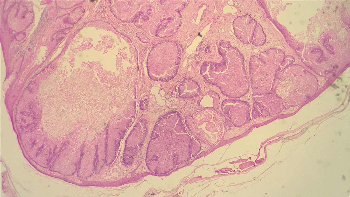
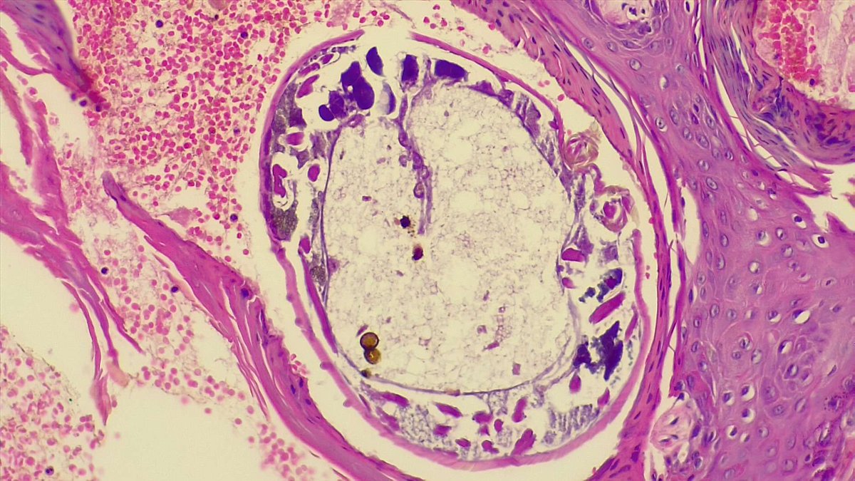
An incidental finding made by @christiestwit and @ChingLi31549786 😊 An ulcer was noted upon the gallbladder during a laparotomy for stab abdomen. The gallbladder was removed and here is the guilty culprit... What eagle-eyes we have in our department 🦅
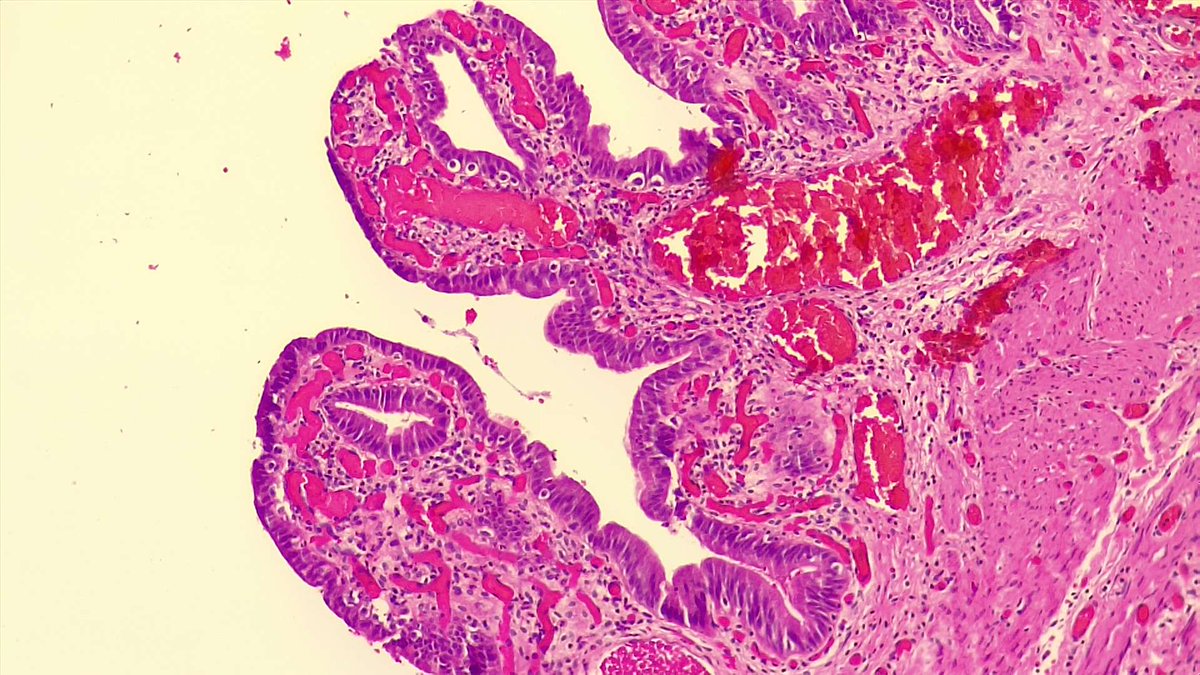
A cavernous hemangioma. A well circumscribed lesion with dilated vascular spaces, filled with blood and suspended in a fibrous stroma. Here is another example of a hemangioma in a bladder biopsy of a patient with haematuria and cervix carcinoma🩸thank you @christiestwit
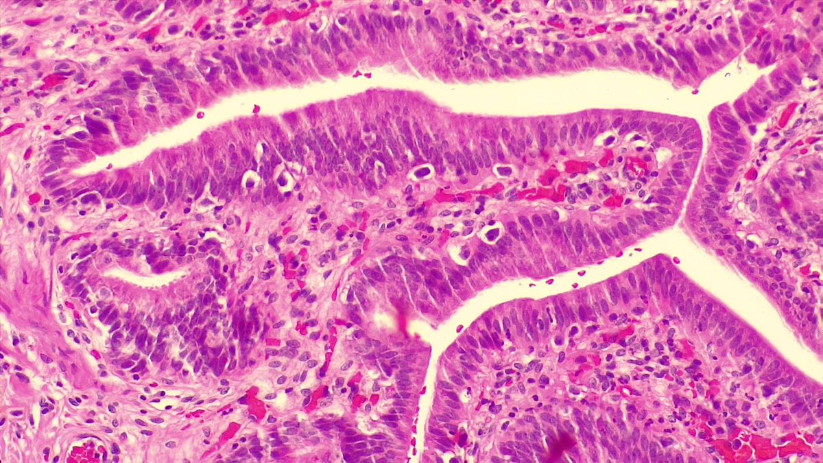
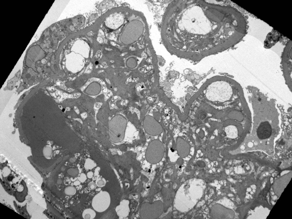
At first glance, the abundant foamy histiocytes can make you think this is just xanthogranulomatous cystitis. But on closer inspection, the Michaelis-Gutmann bodies give away the diagnosis: malakoplakia 🤓 von Kossa and PAS stains will highlight the MG bodies.
A retinoblastoma. Small round blue cell tumour seen in children. Flexner Wintersteiner rosettes: central lumen filled with mucopolysaccharide, surrounding tumour cells have nuclei located away from lumen. Homer Wright rosettes: do not have central lumina (less common than FW).
An example of the most common paediatric hepatic neoplasm: a hepatoblastoma. There are many histological variants (too many to describe in a 280 character Tweet 🙈). Notice the pink and blue components and the bone too. This is a mixed epithelial and mesenchymal hepatoblastoma 🤪