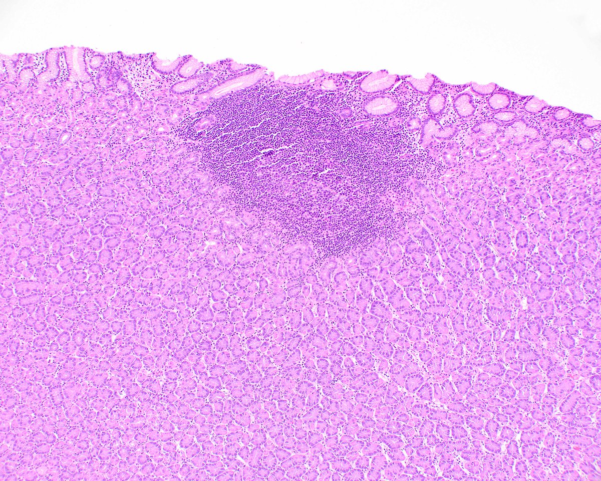
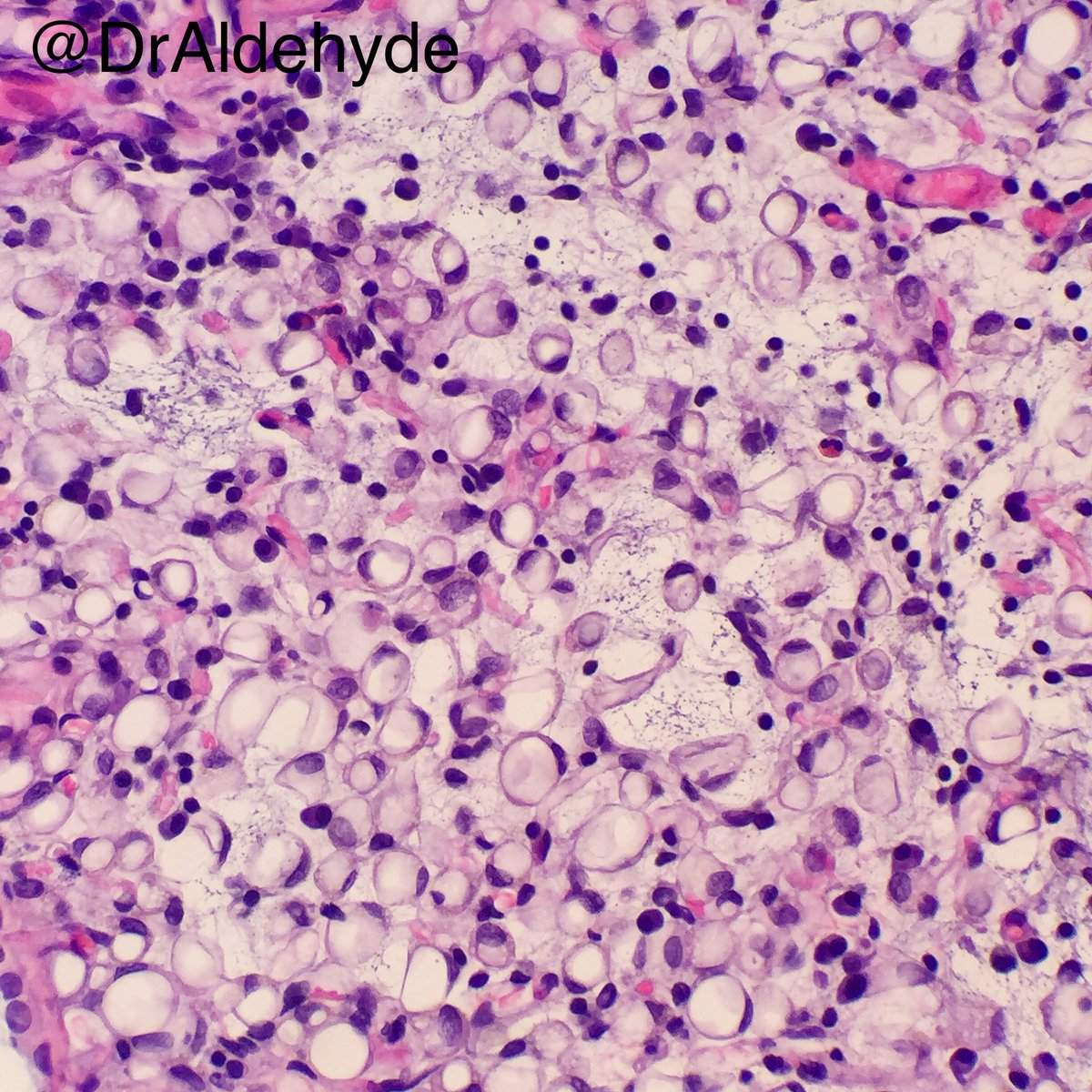
Clever Penn colleague Beth Furth pointed out a fascinating pattern of H. pylori ANTIGEN deposition (but not organisms) in germinal centers. Check out her article PMID: 32657781.
🔬 Bone and Soft Tissue #Pathology, day 2
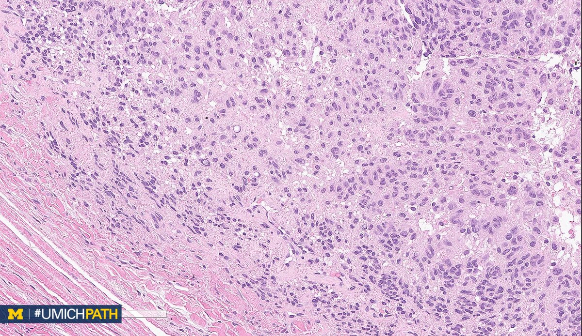
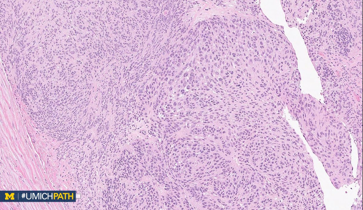
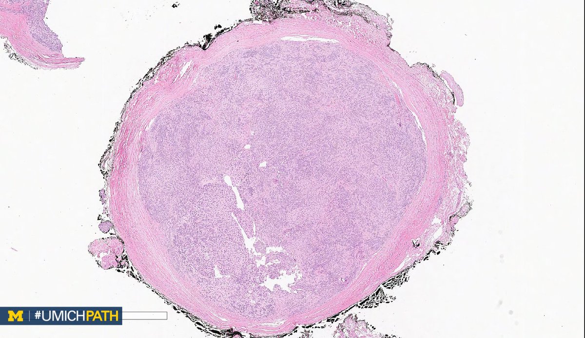
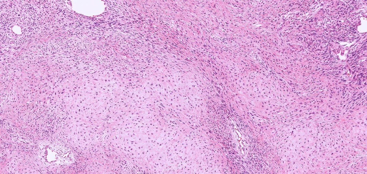
➡ What's your #DifferentialDiagnosis of this superficial soft tissue mass?
➡ What #DailyDx do you favor, and what confirmatory stain(s) would you order?
➡ We’ll tweet the answers and some quick facts tomorrow! #UMichPath#BSTPath
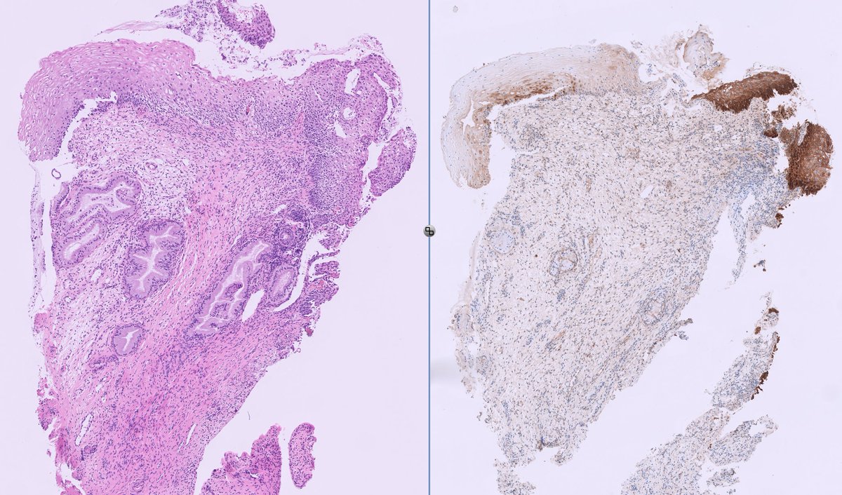
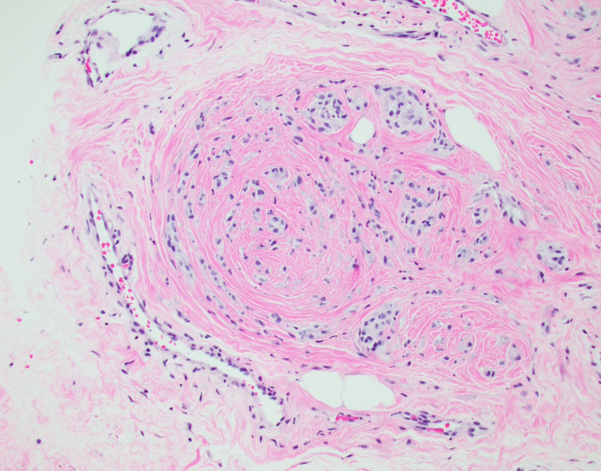
Prostate apex core needle biopsy.
Does this finding require a more careful apical dissection if a radical prostatectomy is performed ?
ie should we ever report? @slusagar
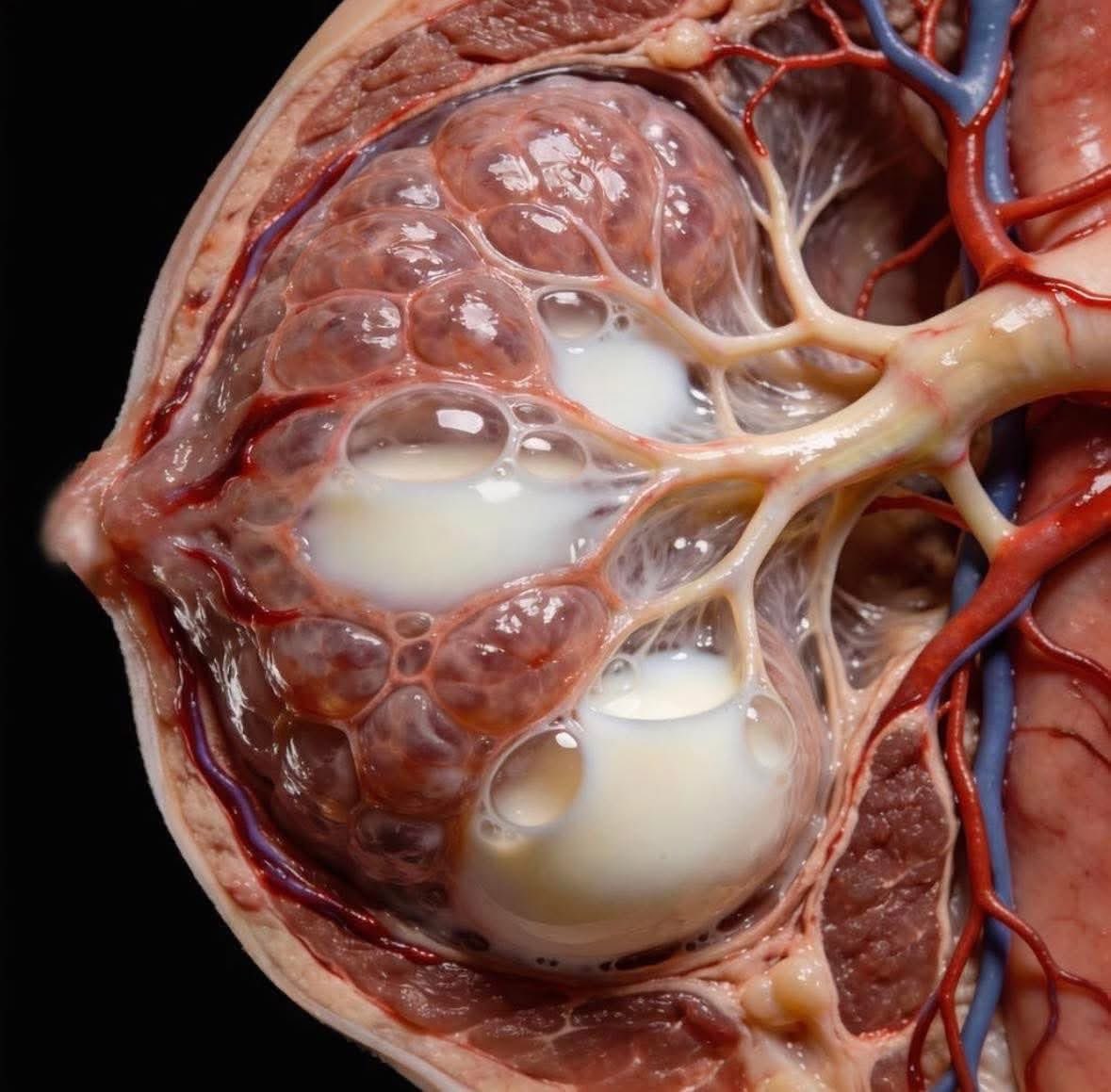
Most people picture breast milk as something simple.
White. Plain. Just food.
But this is what it looks like inside.
This is a glimpse of the inside of a breast while it is making milk.
Thousands of tiny milk making sacs, each one filling, flowing, responding in real time to a baby’s needs.
Every drop is alive.
Breast milk is not just nutrition.
It is a living, adaptive system.
It changes by the hour.
By the day.
By the age of the baby.
It adjusts for illness.
For growth spurts.
For comfort.
For survival.
Your body reads your baby’s saliva and responds with antibodies.
It knows when your baby is premature.
It knows when your baby is sick.
It knows when your baby just needs closeness.
No lab can recreate this.
No formula can copy this intelligence.
No machine can replace this connection.
This is biology at its most powerful.
This is love in liquid form.
This is the miracle happening quietly inside millions of women every single day.
And if no one has told you lately
Your body is incredible!
✨🙌🏾💫
Disclaimer: This image is AI just meant to represent what it may look like. Image by Salud Articular
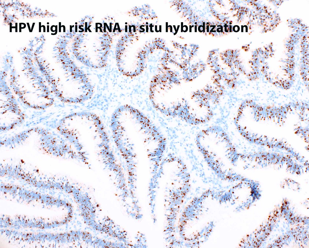
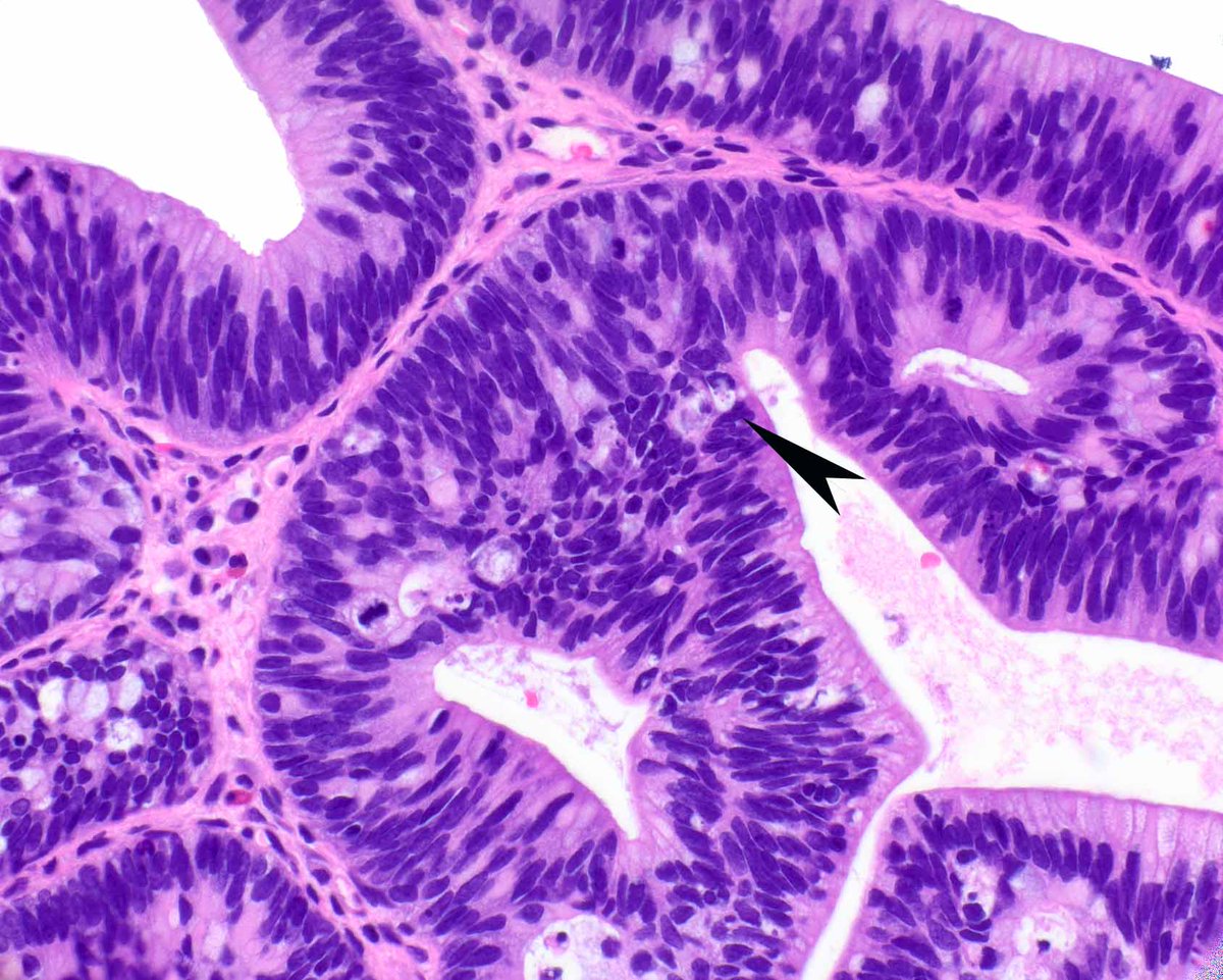
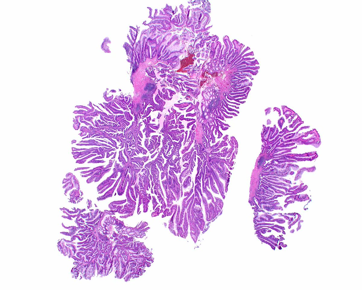
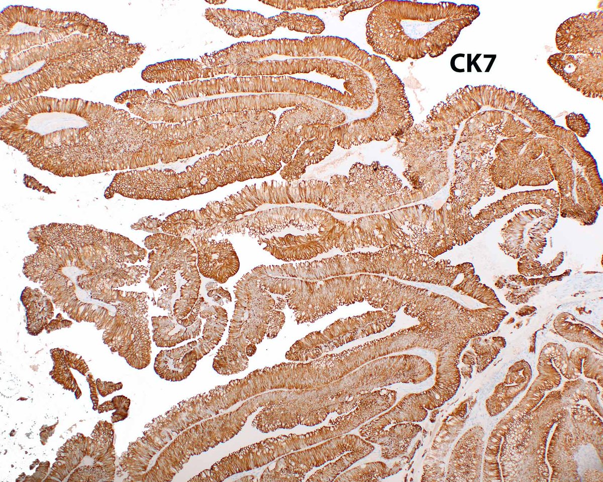
Distal rectal polyp - HPV-associated glandular neoplasms are rare and mimic tubulovillous adenomas. The mucin is peculiar and apical apoptotic bodies can be plentiful (arrow).
Voltaggio L, et al. Mod Pathol. 2020 May;33(5):944-952. PMID: 31857682.
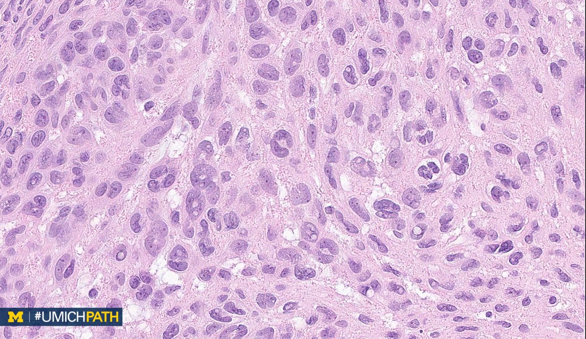
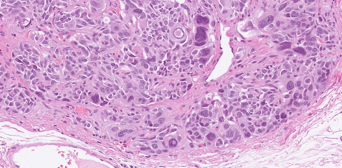
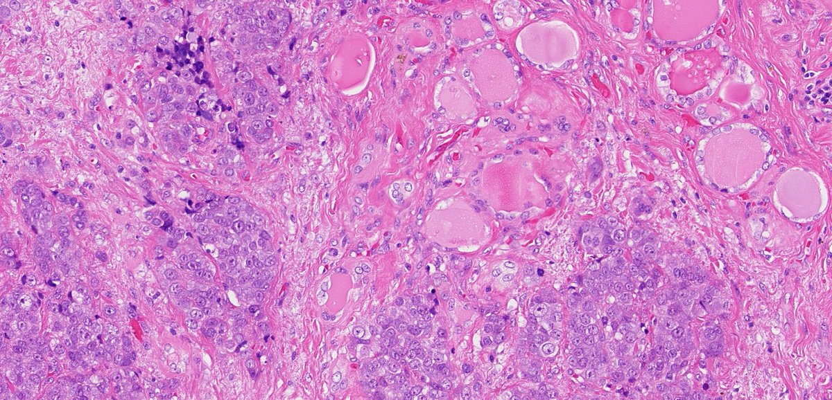
Medullary thyroid carcinoma (MTC) can show pleomorphic nuclei with bizarre shapes and inclusions. This low-grade MTC (Ki-67 <1%) demonstrates striking yet misleading features that might resemble anaplastic or papillary thyroid carcinoma. Morphology can be deceptive!
Spindle cell neuroendocrine tumor (NET) of the duodenum - an ultra-rare entity often mistaken for sarcoma, GIST, solitary fibrous tumor, or even paraganglioma. Accurate diagnosis requires careful histopathology and immunoprofiling! Some IHC images in the thread below...
Head & Neck & Endocrine Pathology Residents - Quiz Time!
An elderly woman presents with an expanding left thyroid mass invading strap muscles, the recurrent laryngeal nerve, and the esophagus. Lobectomy is performed. What’s your dx? (Follow the thread!)
This charming little hidradenoma papilliferum features oncocytic change and presented as an external hemorrhoid. Note the double layer of cells. Immunohistochemical stains can result in silly ideas.
TRPS1 Is Consistently Expressed in Hidradenoma Papilliferum. PMID: 38959400.
Monomorphic epitheliotropic intestinal T cell lymphoma (MEITL), formerly termed Type II enteropathy-associated T cell lymphoma is NOT associated with celiac disease. #UMiamiPath
They are usually easy to spot on H&E, but Modern Pathology takes a deep dive into EBV-associated gastric carcinomas.
Angerilli V, Gasparello J, Collesei A, Ceccon C, Bergamo F, Sabbadin M, Parente P, Vanoli A, Niero M, Luchini C, Gafà R, Dei Tos AP, Grillo F, Mastracci L, Lonardi S, Fassan M. Epstein-Barr Virus-Associated Gastric Cancer: A Histopathologic Study With Comprehensive Molecular Profiling. Mod Pathol. 2025 Nov;38(11):100881. PMID: 40912408.
Welcome back to Breast Consult Case of the Week 10 #BCCW10!
This one was tricky, and I apologize for leading you astray by not giving you the whole IHC panel, but this is exactly the kind of situation where serious misdiagnoses will happen.
The correct answer is myofibroblastoma (MFB) with epithelioid features.
The problem, of course, is that MFB is ER and PR positive and E-cadherin negative. Which means that if you do not suspect the diagnosis and don't include CK, CD34, and desmin at the very least, you will be led down the wrong diagnostic path.
Some of the useful morphologic features include the following:
1. Amphophilic cytoplasm that tends to contrast with eosinophilic collagen fibers/ropes
2. Open chromatin, no hyperchromasia
3. Despite the contrast between the cytoplasm and surrounding collagen, the cells appear to have a close relationship with the stroma, without clefting or evidence of desmoplasia
4. Often well-circumscribed radiologically
Naturally, none of these features is always present or enough on its own. The key here is to consider the possibility and let IHC resolve the issue.
One more thing to remember is that MFB is a great mimicker in its own right, with epithelioid, palisaded, myxoid, pleomorphic, and lipomatous variants. And though they're often well-circumscribed, they can show irregular and infiltrative edges on occasion, eliciting clinical concern.
Below are images of our case with ER and Desmin, followed by examples of morphologic variants of MFB.
Thank you all and see you next week!
@washu_pathology@washupathedu #breastpath #PathX #PathX