“Myocardial Strain Imaging: Theory, Current Practice, and the Future”
PDF at: https://t.co/a3lpsNClFk
1. Introduction to Myocardial Strain Imaging
• Definition: Myocardial strain quantifies heart muscle deformation, describing changes in length, thickness, and twisting motions.
• Clinical Relevance: This imaging technique, superior to ejection fraction (EF), detects mild systolic dysfunction, especially in patients with heart failure with preserved EF (HFpEF), valvular diseases, or chemotherapy-related myocardial dysfunction.
• Modality Variants: Includes echocardiography (e.g., speckle-tracking echocardiography, STE) and cardiac magnetic resonance (CMR) methods like feature tracking (FT).
2. Historical Development
• Early Techniques: Used implanted markers and ultrasonic dimension crystals.
• Modern Methods: Progressed from tissue Doppler to advanced CMR tagging and FT for better 3D and segmental analysis.
3. Technological and Methodological Insights
• CMR Techniques:
• Tagging: Applies magnetic grids on myocardium to monitor deformation.
• Feature Tracking (FT): Tracks anatomical landmarks for strain calculation; simpler and widely used clinically.
• Emerging Techniques: 3D CMR and strain-encoded imaging offer better spatial resolution.
• Echocardiography Techniques:
• STE dominates clinical use due to multidirectional tracking of speckles, providing reliable global and segmental strain metrics.
4. Clinical Applications
• Global Longitudinal Strain (GLS):
• More sensitive than EF in detecting early systolic dysfunction.
• Used in HFpEF, specific cardiomyopathies, and chemotherapy monitoring.
• Segmental Strains:
• Useful in identifying localized dysfunctions (e.g., ischemia, hypertrophic cardiomyopathy).
• Right and Left Atrial Strains:
• Indicate diastolic dysfunction and filling pressures.
• Twist and Torsion Analysis:
• Provides insight into ventricular mechanics but is less commonly applied due to technical challenges.
5. Advances and Challenges
• Advantages:
• Offers both global and regional myocardial function analysis.
• Extends to right ventricle (RV) and atria, expanding diagnostic capabilities.
• Challenges:
• Significant vendor variability, dependency on image quality, and complex standardization requirements.
• Limited adoption of advanced techniques like 3D strain due to resolution constraints and complexity.
6. Future Directions
• Layer-Specific Strain Analysis:
• Investigates strain gradients across myocardial layers; currently limited due to technical and reproducibility issues.
• Myocardial Work Index:
• Combines strain with non-invasive pressure estimates to evaluate energy efficiency.
• High Frame Rate Imaging:
• Enhances the detection of rapid cardiac events.
• Prospective Studies:
• Required to validate clinical outcomes and optimize guideline implementation.
7. Practical Considerations
• Strain imaging requires high-quality image acquisition, appropriate region-of-interest (ROI) placement, and software-dependent analysis. It demands awareness of confounding factors like loading conditions, chamber geometry, and patient-specific variables.
The review emphasizes myocardial strain imaging’s transformative potential in diagnosing and managing cardiovascular diseases, while recognizing the need for standardization, technological improvements, and further research. If you’d like, I can extract specific sections or elaborate further on any topic.
🫀 A deep dive into mitral annular disjunction (MAD) in MVP patients published this month in @JournalASEcho
🔍Pts with MVP were subclassified based on the mitral annulus phenotype in systole & diastole
📊True-MAD ➔ clear separation btw P2 & LV myocardium in both diastole & systole
⚖️Pseudo-MAD ➔ defined as the presence of only systolic apparent disjunction
⚠️ Pseudo-MAD linked to arrhythmic MVP features, including systolic curling and Pickelhaube appearance
📈 TTE proves is a reliable tool for identifying MAD phenotypes, with 89% accuracy & high intra-/interrater agreement
💡 Why it matters: Early recognition of Pseudo-MAD can help assess risk for arrhythmic MVP & guide treatment strategies, especially in advanced cases with severe MR
🔗Link ➔ https://t.co/zaTyVku9zj
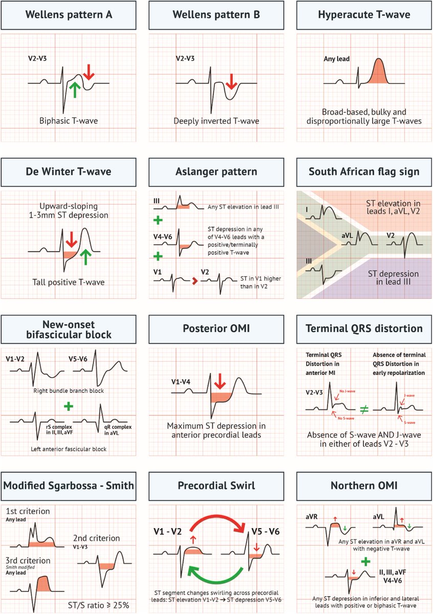
New #EKG patterns in acute myocardial infarction due to thrombotic plaque rupture:
❌ STEMI / NSTEMI
✅ State of the coronary artery
Great work by @fabrizioricci@martini_chia!
Understand the best practices for BP measurement for #hypertension patients with this infographic. 👇 Access the infographic, patient case quizzes & more 🆓 education w/ ACC's online course: https://t.co/BouB1rDXYk
#FusterCVS#ACCEd
Əfsus ki, bizlərdə insanlara doğru yol göstərildiyində yol göstərinin hansısa məqsədi olduğu düşünülür. Bu özəlliklə xəstələrə aid bir vəziyyətdir, ixtisaslaşmış həkimə yönləndirilmək anlayışı bizim millətin təfəkküründə heç vaxt yer etməyəcək.