Registration is now open for the next Multisite #Pediatric#Ultrasound Conference with @ChildrensLA@HaiThuyNguyenMD leading our discussion of midgut volvulus #imagingourfuture
Thursday, August 10th at 11am MT/1pm ET
Register at the following link: https://t.co/shtxbFQ3s2
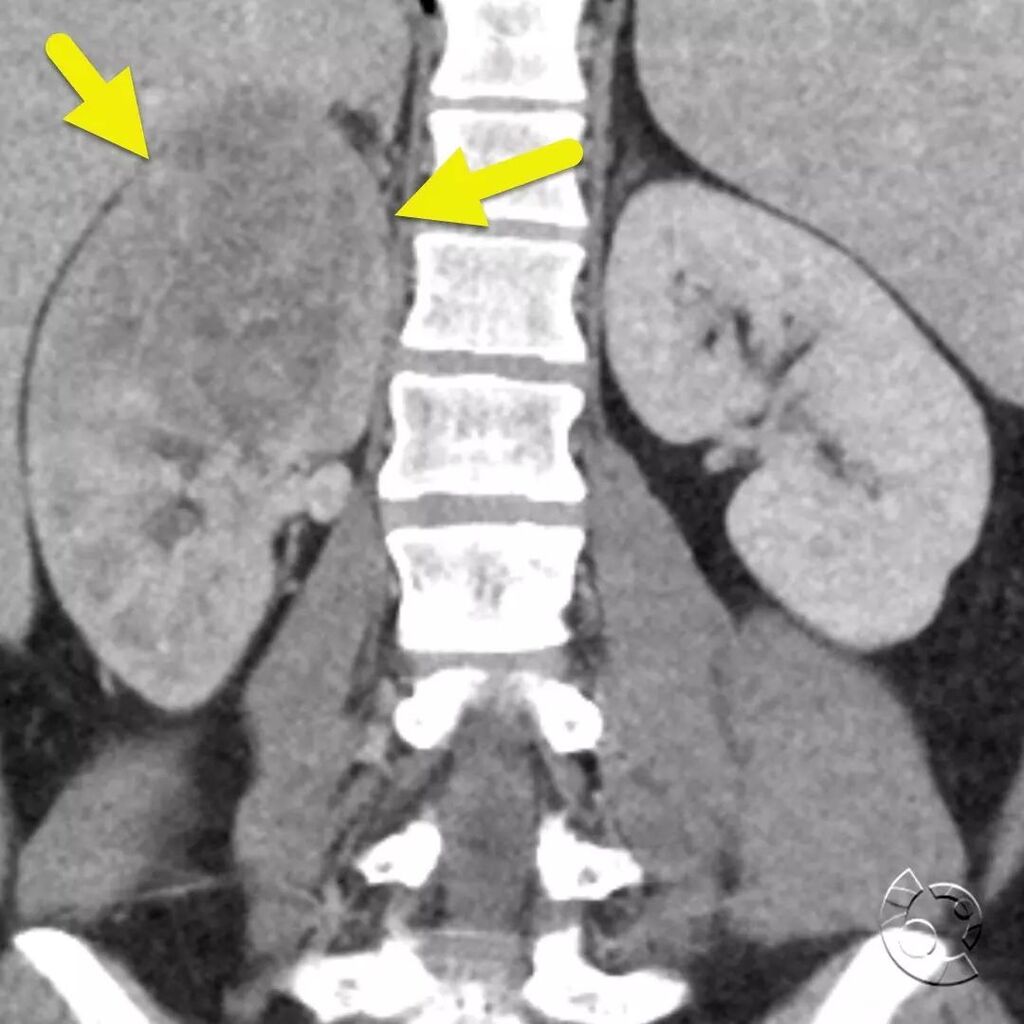
Coronal (image 1) and axial (image 2 and 3) CT of the kidneys shows a poorly defined mass (arrow) in the upper pole of the right kidney. The mass was ultimately diagnosed as an extraosseous Ewing sarcoma. Extraosseous Ewing sarcoma is a rare variant of E… https://t.co/4U0cJqS72v
Sagittal (image 1) and coronal (image 2) contrast-enhanced CT at the thoracoabdominal junction shows a congenital diaphragmatic hernia containing the right kidney. Congenital diaphragmatic hernias typically occur posteriorly and are more common on the le… https://t.co/9G8YAFe5yr
Frontal (image 1) and lateral (image 2) radiograph of the chest shows a soft tissue bulge (arrow) at the thoracoabdominal junction. What could be causing this appearance? https://t.co/u1KRRQ0sRr
Axial (image 1), coronal (image 2), and sagittal (image 3) contrast-enhanced CT shows a large heterogeneous mass (arrow) arising from the inferior margin of the liver. The mass is mostly solid but has large vascular spaces and a central nonenhancing comp… https://t.co/iibUx0GVJD
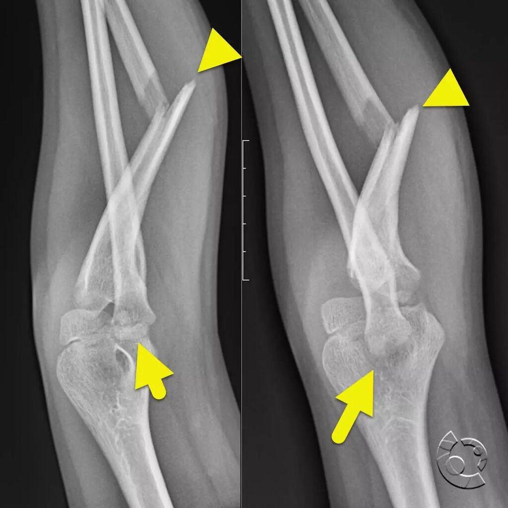
AP (image 1) and lateral (image 2) radiograph focused at the elbow shows a fracture of the mid ulna (arrowhead) and dislocation of the proximal radius (arrow) consistent with a Monteggia fracture. A Monteggia fracture is caused by a blow to the forearm w… https://t.co/pU4GyYK0Vm
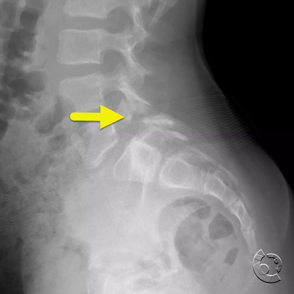
Lateral radiograph of the lumbar spine shows a pars defect (arrow) at the L5 level with mild anteriolisthesis of L5 on S1. Pars defects most commonly occur at the L5 level. They are relatively common occurring in approximately 1 and 20 patients. Most pat… https://t.co/ijjFS2T786
Coronal (image 1) and axial (image 2) images from fetal MRI show an abnormal appearance of the kidneys (arrows) with multiple cortical cysts and massive enlargement of the ureters (arrowheads). On postnatal voiding cystourethrogram (image 3) bladder rupt… https://t.co/v4f6whTB7H
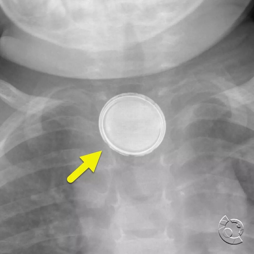
Coronel image from CT angiogram shows metallic residua (arrow) of the button battery. Ingested button batteries are a medical emergency. The battery can conduct a charge across the tissue leading to soft tissue burns. These burns can lead to esophageal i… https://t.co/y3SEdTDi1I
AP (image 1) and lateral (image 2) radiograph of the airway showing ingested button battery (button batter) with erosion of the inferior margin. The battery is causing significant edema. This is seen on the lateral view where there is widening of the spa… https://t.co/9QGoI3nPPQ
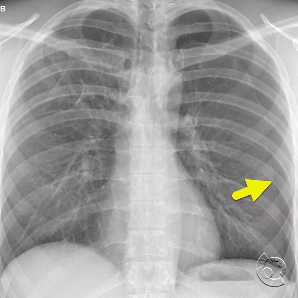
Frontal radiograph (image 1) of the chest shows a large left pneumothorax (arrow). Focused radiograph at the left lung apex (image 2) shows the pneumothorax, as well as a large pulmonary bleb (arrowhead). Coronal CT images (images 3 and 4) and 3-D reform… https://t.co/EOKQNbIRFo
A lateral radiograph of the thoracolumbar junction shows a focal kyphosis, anterior/posterior shortening of the vertebral body, and anterior inferior beaking (arrow) of the L1 vertebral body characteristic of Hurler's syndrome. Hurler syndrome is a mucop… https://t.co/Mg3vVdGURS
Oblique radiograph of the foot (image 1) and zoomed-in portion of the radiograph (image 2) show a fracture (arrow) of the proximal fifth metatarsal, a Jones fracture. The fracture is differentiated from the normal fifth metatarsal apophysis (arrowhead) b… https://t.co/87JgKnK6iC
Spinal Hematoma Visualized with Dual-energy CT-derived Electron Density Overlay Maps https://t.co/EIOThatt7C
(B) Dual-layer dual-energy CT reconstructions show the corresponding CT angiography scans of the chest. Electron density overlay images (left) clearly depict the hematoma (green; arrows) that is protruding in the spinal canal. Conventional images (right) in the sagittal and axial plane. The hematoma is almost entirely imperceivable (arrows).
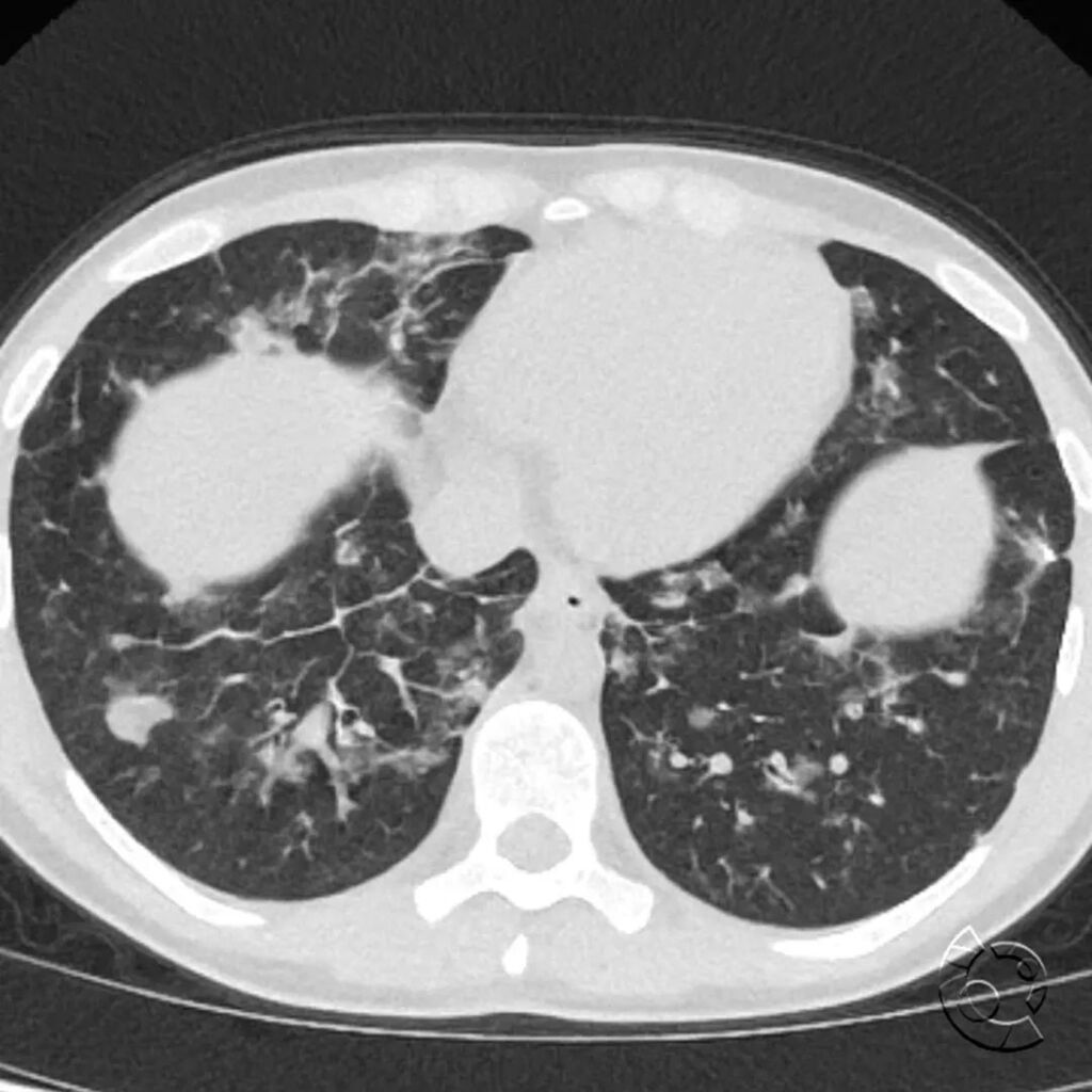
Axial CT images of the chest (images 1 and 2) show solid and ground-glass pulmonary nodules and interstitial thickening. There is mild associated bronchiectasis (arrow, image 2). Areas of ground-glass opacity are increased on expiratory images (image 3).… https://t.co/ZWiSGTV7sV
Axial CT of the chest shows multiple pulmonary nodules, interstitial thickening worse in the right lower lobe. The patient was diagnosed with granulomatous and lymphocytic interstitial lung disease. What underlying disorder is associated granulomatous an… https://t.co/Q6QKZkUn1l