MRI shows a complete transection of the median nerve → Sunderland grade V injury.
Key question: what should we look for on imaging in this scenario?
#MSKrad#Radiology#MRI#PeripheralNerve#MedianNerve
Back after a long break — teaching and work took over, but glad to be back sharing MSK cases.
📌 58-year-old male
📌 History: sharp laceration at the volar wrist
Presents with:
Loss of thumb opposition
Decreased sensation in median nerve territory
@DrMarwanAl_D @carlespedret Excellent case. A question about type 3: I’ve never seen a case like that, where only the free aponeurosis is affected. I usually see it associated with myoaponeurotic involvement. Have you ever seen a type 3 case?
@bhavinj One more thing: the patients I’ve seen with this type of tumor showed a reddish-bluish discoloration of the nail prior to undergoing ultrasound or MRI. Thanks for sharing!
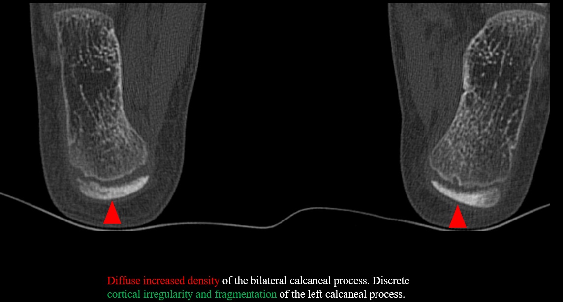
@northwoods1980 As I mentioned, the diagnosis is made clinically. In this case, it already came with suspicion. Likewise in children I always look at the density of the calcaneal processes to see normality. Additionally, the irregularity of the left calcaneus can be useful. Do not give up!😅
Sportsly active teenager with intense heel pain.
Tomographic findings of calcaneal apophysitis (Sever's disease).
Although the traumatologist diagnoses it clinically, imaging studies can help make a differential diagnosis (stress fracture, Achilles tendon).
#MSK#radiology
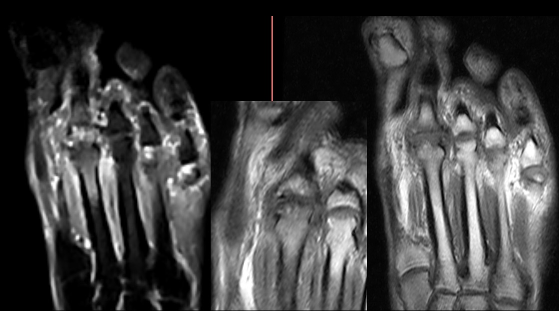
70-year-old, female with chronic metatarsalgia.
Various images of osteochondrosis in the head of the second metatarsal.
Note the deformity, cortical irregularity of the head of the second metatarsal, with subchondral bone edema on MRI.
#feet#MRI#MSK#radiology
@samrad77@WFUMB Put your hand in the back pocket of your pants! (I didn't know the name 😅). Extremely useful position in daily practice for those small tears that raise doubts with the classic position to assess the supraspinatus.
25-year-old male, with chronic pain, which increases in the post-match🏉🏉.
Example of imaging findings of athletic pubalgia and a short summary of secondary cleft.
#MRI#trauma#radiology#FOAMed
Female ⚽️ 20 y, chronic knee blockage, without trauma.
Example of ACL ganglion cyst, which when symptomatic must be resected.
Talking to the traumatologist, he is going to undergo arthroscopic cytoreduction (let's wait for the images).
#MSK#radiology#knee#ACL
@handoshera Mucoid degeneration usually keeps the ACL tract only thicker and hyperintense (celery stalk). Ganglions are focal images. However, it is believed that degeneration may predispose to the formation of cysts (elderly patients, which would not be the case☝️)
35 y, omalgia without trauma.
Distal osteolysis of the clavicle (DCO) without trauma occurs due to a repetitive stress mechanism. Exploring a little, the patient was a gym regular and the pain was aggravated when lifting weights🏋️♂️.
Interesting reading 🤓
#MSK#FOAMed#radiology
Example of subtle signs of aggressiveness of a chondral lesion: the size (> 5 cm), the bone disruption and remodeling, and the margins were signs that made me suspect that it was not a simple chondroblastoma.
Histology confirmed: Central atypical cartilaginous tumor
#bone#msk
Chronic pain in the anterior aspect of the knee in a young professional runner.🏃🏃
The findings described demonstrate the signs of jumper's knee or proximal enthesopathy of the patellar tendon, common in the context of repetitive forced extension.
#MSK#knee#trauma#radiology