We are a non profit organization promoting use of ultrasonography by Canadian rheumatologists to optimize clinical decision-making and therapeutic management
📣 @CrusSurc@lihi_eder Dr Lihi Eder Dr Sibel Aydin and Dr Gurjit Kaeley validation handheld us devices = moderate agreement - need for more validation 👇👇
https://t.co/3ZUSiafwyC
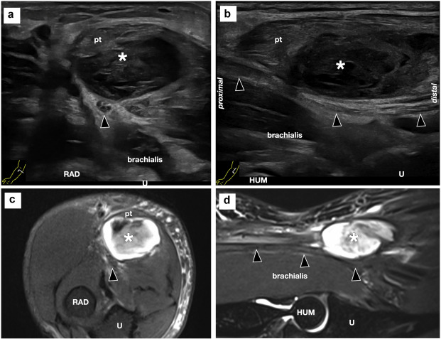
Tenosynovitis of the Tibialis Posterior in an anti-CCP-positive patient presenting with medial ankle pain.
Ultrasound features to look for:
1. Proliferative tenosynovitis -> hypoechoic thickening of the tendon sheath
2. Increased hyperemia -> Doppler signal at the tendon sheath thickening
Clinical Pearls:
* Tenosynovitis often occur in early rheumatoid arthritis (up to 80% in ACPA-positive early disease).
* High-yield anatomical sites: ECU and finger flexor tendons.
* The presence of tenosynovitis in an ACPA-positive patient predicts evolution to inflammatory arthritis (more superior than synovitis itself in a recent MRI-driven study):
Reference for a deeper dive:
1. Abacar K, Tabuchi Y, Matteo AD, Duquenne L, Rowbotham E, Nam J, Emery P, McGonagle D, Mankia K. Quantitative MRI tenosynovitis volume explains the association between tendon involvement and future development of clinical arthritis in anti-cyclic citrullinated peptide-positive at-risk individuals. Ann Rheum Dis. 2026 Mar 10:S0003-4967(25)04459-0. doi: 10.1016/j.ard.2025.10.020. PMID: 41813507.
2. Kleyer A, Krieter M, Oliveira I, Faustini F, Simon D, Kaemmerer N, Cavalcante A, Tabosa T, Rech J, Hueber A, Schett G. High prevalence of tenosynovial inflammation before onset of rheumatoid arthritis and its link to progression to RA-A combined MRI/CT study. Semin Arthritis Rheum. 2016 Oct;46(2):143-150. doi: 10.1016/j.semarthrit.2016.05.002. Epub 2016 May 18. PMID: 27342772.
#RheumUS #MKSUS
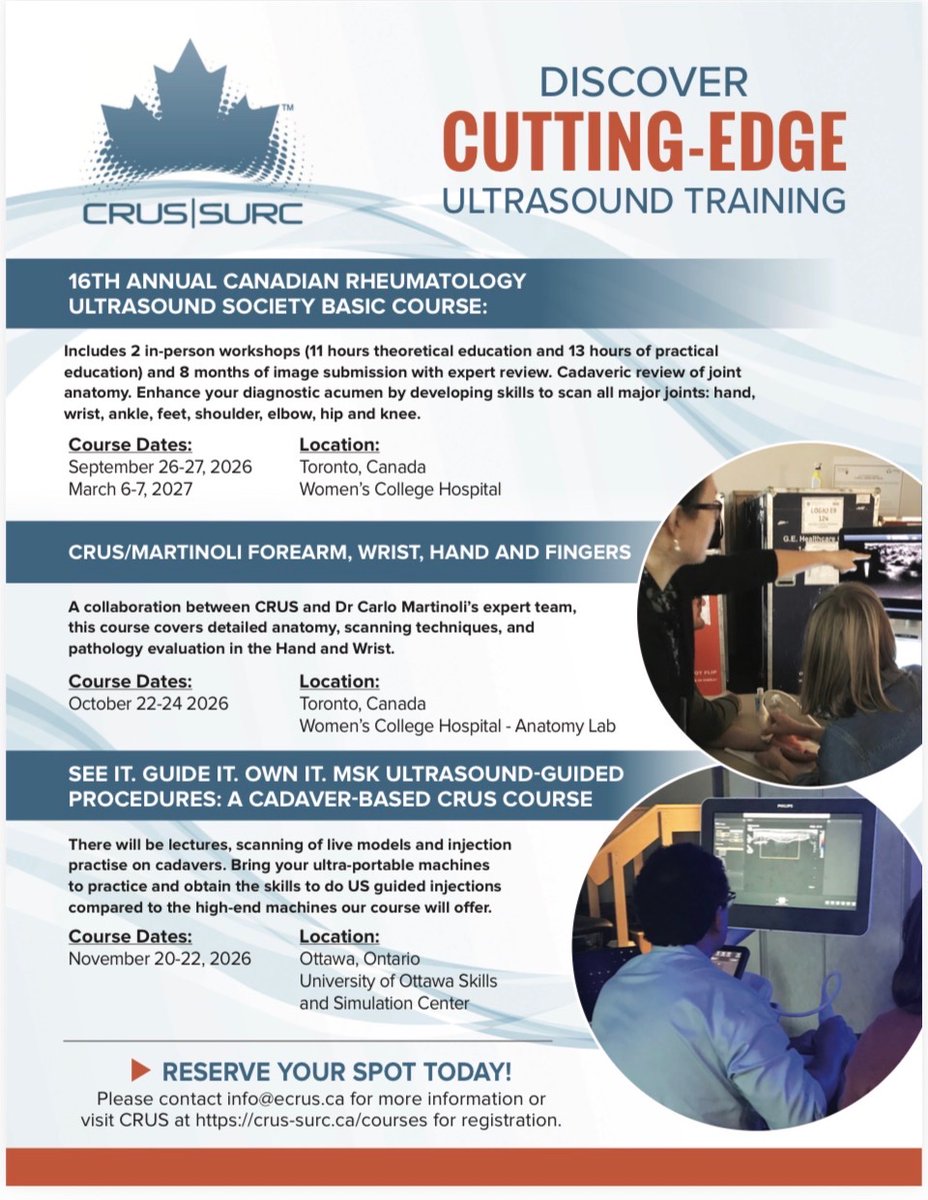
📣✅ ANNOUNCEMENT - @CrusSurc basic course and advanced courses now open for registration 👉👉👉 https://t.co/KfRtDZtDgL
Get your spot secured - we look forward to sharing with you in the joy of #mskus
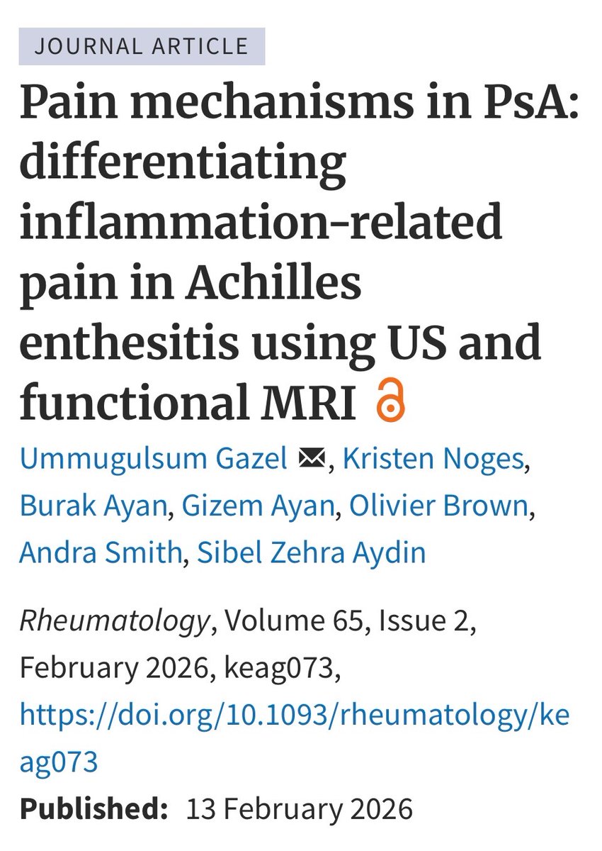
🔥hot topic - incoming @CrusSurc president Sibel Z Aydin studied enthesitis sites seen peripherally on mskus and central pain processing centres activated on functional MRI - not all enthesopathies are the same! https://t.co/zFxkhJ5uFa
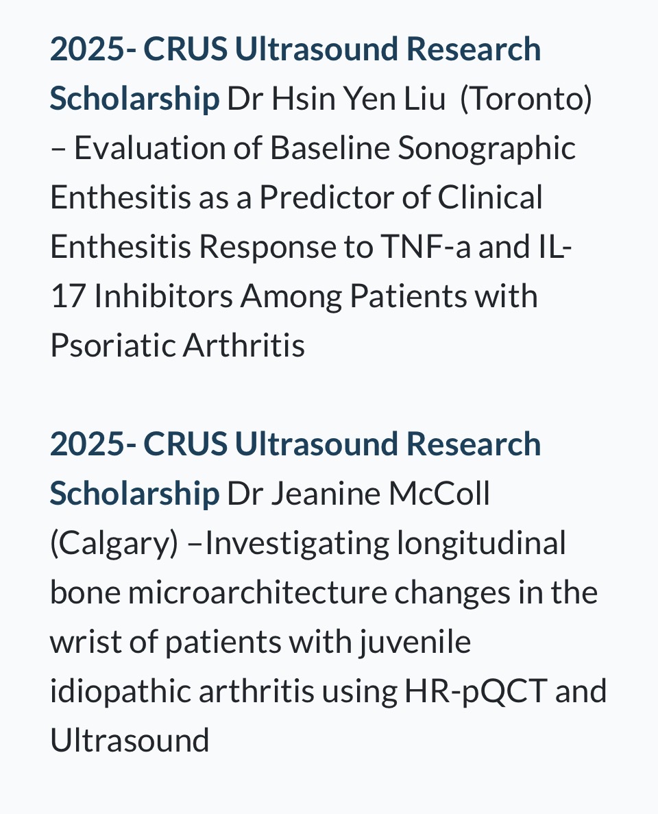
🤩🏆 @crussurc 2025 research award winners announced - congratulations to Dr. Alan Zhou, Dr. Samar Aboulenain, Dr. Hsin Yen Liu and Dr. Jeanine McColl 👇👇👇https://t.co/c3hu7rKH6f
Chondrocalcinosis of the knee 👇
Pearls:
- Fibrocartilage CPPD (in the meniscus) occurs at a younger age and is more prevalent than femoral hyaline cartilage CPPD.
- Chondrocalcinosis in both the fibrocartilage and hyaline cartilage is frequent in patients > 80 years old (47% and 23%, respectively).
What is your age cutoff to screen for secondary causes of CPPD?! --> hypophasatesia, hypomagnesemia, hyperparathyroidism and hemochromatosis
Reference for a deeper dive: Cipolletta E, Francioso F, Smerilli G, Di Battista J, Filippucci E. Ultrasound reveals a high prevalence of CPPD in consecutive patients with knee pain. Clin Rheumatol. 2024 Jan;43(1):435-441. doi: 10.1007/s10067-023-06805-3. Epub 2023 Nov 17. PMID: 37975949.
#MSKUS #RheumUS
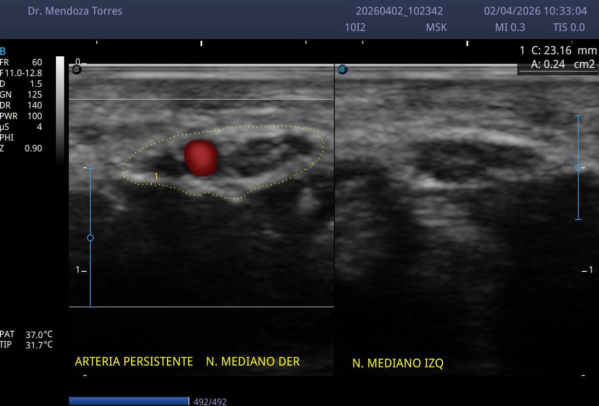
Síndrome de túnel del carpo secundario a nervio mediano bífido con arteria mediana persistente, variantes anatómicas que incrementan el riesgo por aumento significativo del área de sección transversal..
🔥Hot off the press @jrheum#mskus Dr. Filippucci et al. re DEUS initiative (enthesitis and SpA) ✅ Utility of higher Doppler scores near the entheseal site -> activity ✅ Complimentary role of #mskus & physical exam ✅proposal of a modified scoring system https://t.co/8cQ0ssZCuE
Recommended reading for a deeper dive:
Filippou G, Sirotti S, Cipolletta E, Filippucci E. Optimizing the Use of Ultrasound in Calcium Pyrophosphate Deposition (CPPD): A Review from the Ground Up. Gout, Urate, and Crystal Deposition Disease. 2024; 2(1):17-33. https://t.co/fPYtf74N48
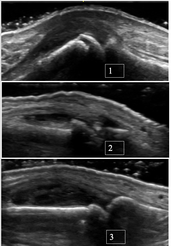
Quiz Time! 🧠
Which PIP joint ultrasound pattern matches each condition: rheumatoid arthritis, osteoarthritis, or psoriatic arthritis?
Take a close look and make your best guess 👀
Which PIP belongs to which diagnosis?
Drop your answers in the comments below 👇
#MSKUS #RheumUS
A case of chronic CPPD at the MCP & Wrist with Pseudo-Double Contour
Case Summary:
A 67-year-old female with seronegative rheumatoid arthritis referred for diagnostic clarification, as she has not responded to multiple csDMARDs or biologics, before escalating therapy. On exam, there were multiple tender and swollen joints, including MCP 1 and 3 bilaterally, as well as bilateral anterior ankle tenosynovial effusions.
Ultrasound Findings:
Pseudo-double contour over hyaline cartilage at the ulnar head and MCP joints, as well as focal intracapsular calcifications and chondrocalcinosis in the TFCC.
Key Learning Point:
*CPPD can represent a pseudo‑double contour (pseudo-DC) sign.
*Unlike the classic DC sign of gout, which moves with the subchondral bone, the pseudo-DC in CPPD moves in the opposite direction during dynamic assessment. This is thought to be due to the CPP crystals depositing in capsules or ligaments, not directly on cartilage like mono-sodium urate crystals usually do.
*Dynamic US evaluation is crucial: static images alone may mimic gout DC sign in CPPD (~10% of cases).
Diagnostic Confirmation:
Ankle aspiration from the extensor digitorum longus tenosynovial effusion confirmed CPP crystals, supporting the pseudo-DC interpretation.
References:
Di Matteo A, Grassi W, Filippucci E. Dynamic behaviour of the double contour sign in gout vs CPPD. RMD Open 2023;9:e002940.
PIP joint synovitis/tenosynovitis in rheumatoid arthritis:
Key elemental findings:
• Synovial hypertrophy → hypoechoic/isoechoic synovial thickening
• Doppler signal → active inflammation (when present where hypertrophy = proliferative synovitis AKA pannus). If this is present at the site of detected erosions -> high risk of radiographic progression
• Tenosynovitis → thickened tendon sheath with fluid ± Doppler signal
PEARL:
In RA, tenosynovitis usually shows up along with synovitis in the same finger. In contrast, half of SLE patients with tenosynovitis have no concomitant synovitis.
Reference for a deeper dive:
Ogura T, Hirata A, Hayashi N, et al. Comparison of ultrasonographic joint and tendon findings in hands between early, treatment-naïve patients with SLE and RA. Lupus. 2017;26(7):707–714.
#RheumUS #MSKUS
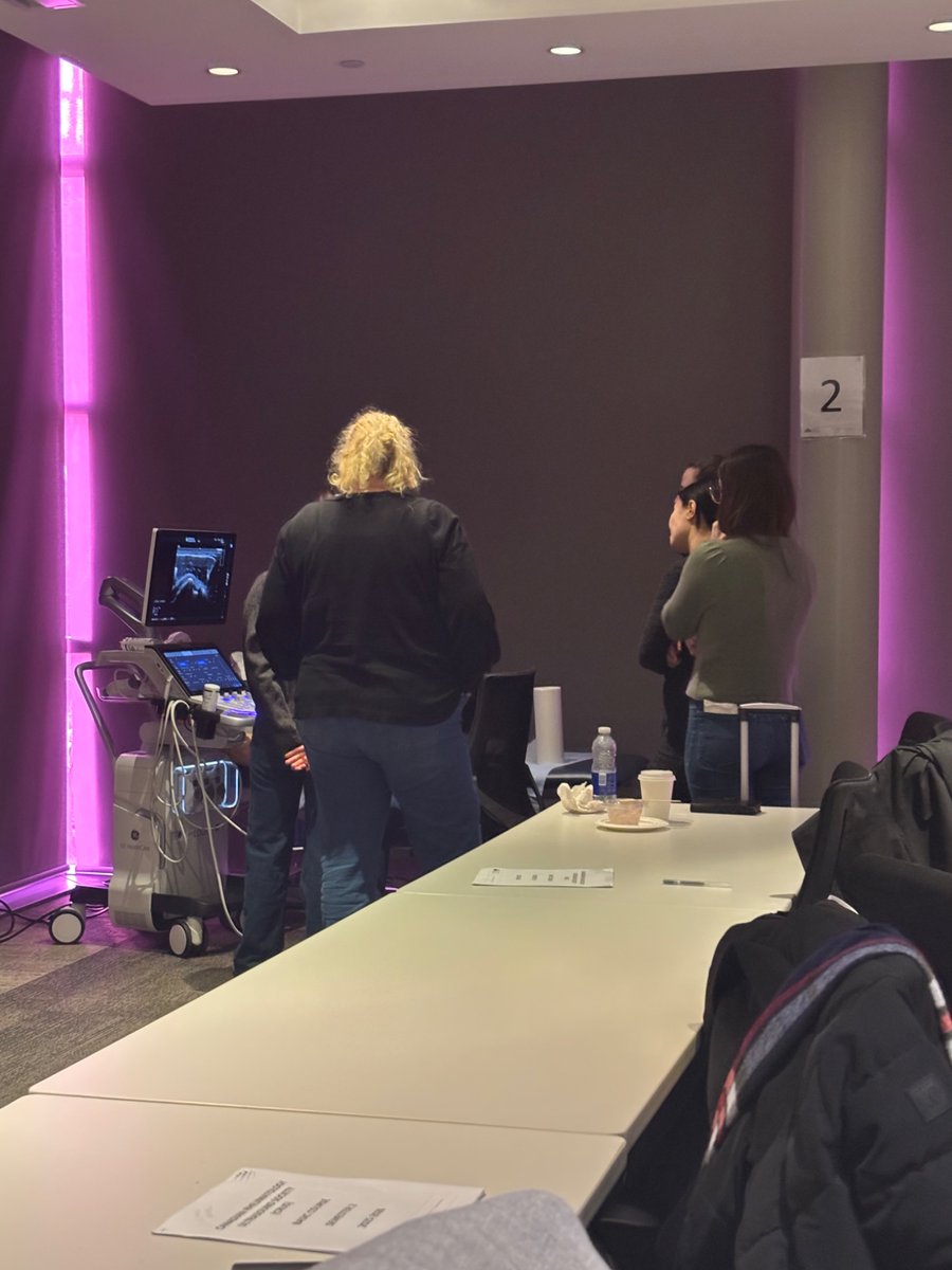
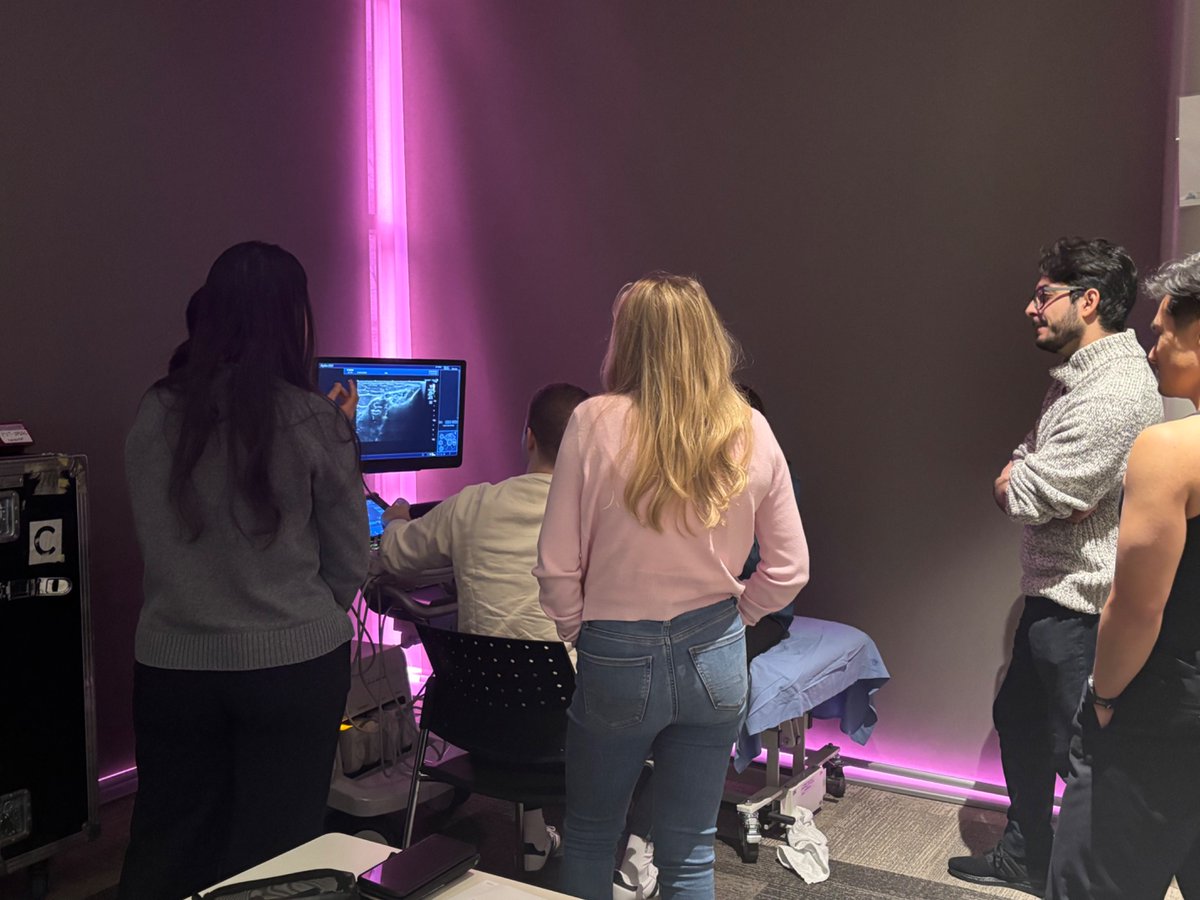
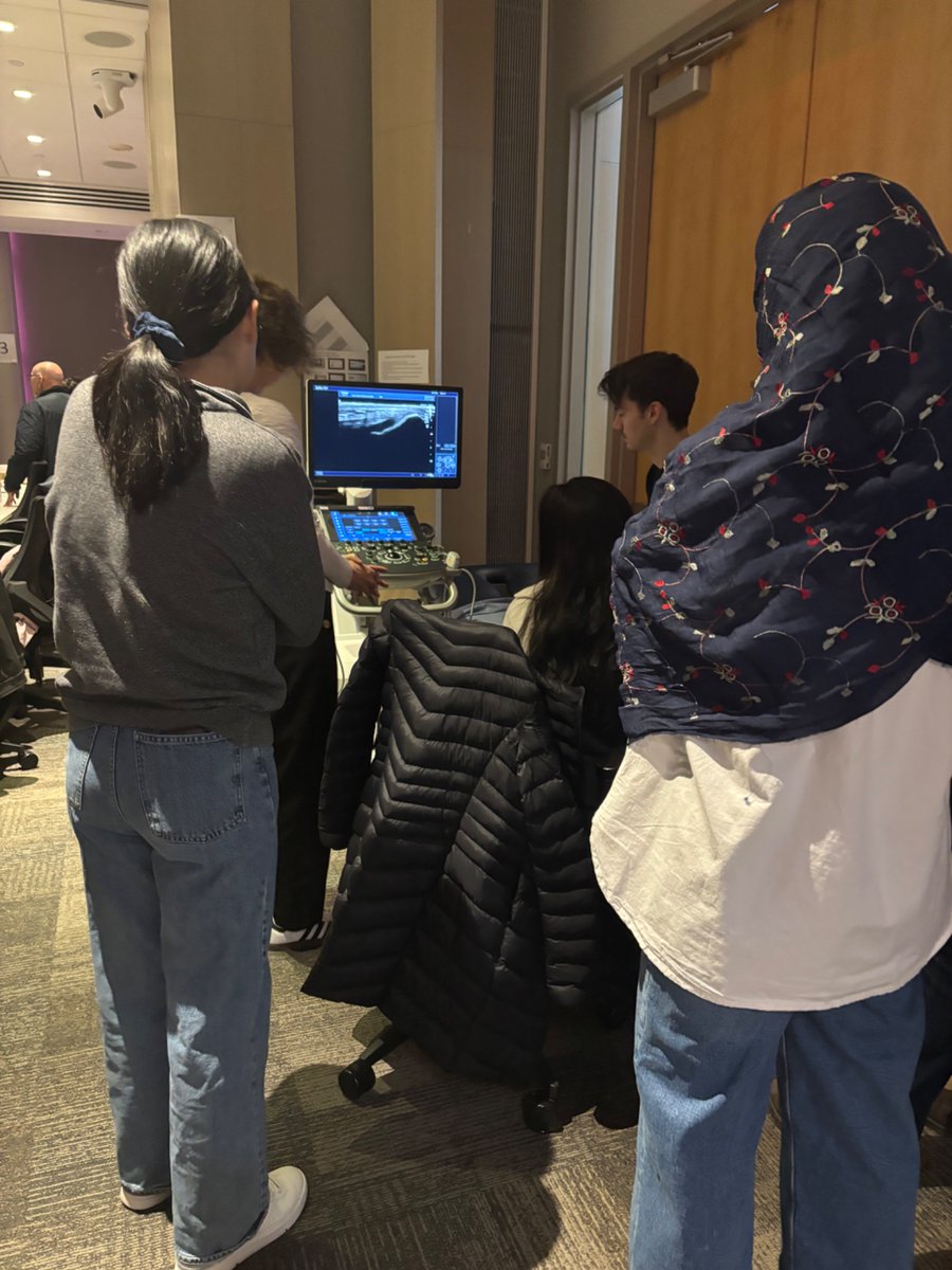
💕Day 2 pink cube Crus Surc basic course Women's College Hospital - hands on practice scanning - note the small groups - one of the benefits of our course - come see for yourself 😉 - we love sharing with you all in the joy of MSKUS!
✅ More @crussurc Basic course pearls - dynamo Dr. Aydin presents on MSKUS shoulder rotator cuff - partial tears, bursal surface vs articular surface and intrasubstance tears. Pearl- don’t mistake anistropy for tears ✅
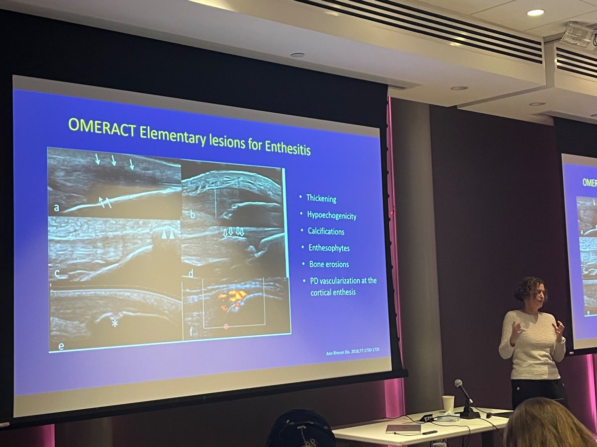
✅ More @CrusSurc pearls Day 2 basic course - the fabulous Dr. Eder presenting on enthesitis - pearls defined by enthesophytes, erosions, hypoechogenicity, calcifications, PD vascular Doppler. Make sure to correlate clinically to avoid biomechanical confounders