New from our lab: FidlTrack—structure-aware single-particle tracking benchmarks/boost SPT fidelity
It resolves in organelles e.g. BACE1 amyloidogenic APP cleavage #Alzheimers, ER exit, map nanobody binding in realtime in ER/organelles 🔬🧠#SingleMolecule
https://t.co/7hhAu5Z0qV
A xeno-free protocol for rapid differentiation of human iPSC-derived microglia from the KOLF2.1J reference line. https://t.co/Bf4ILVGpy7 #biorxiv_cellbio
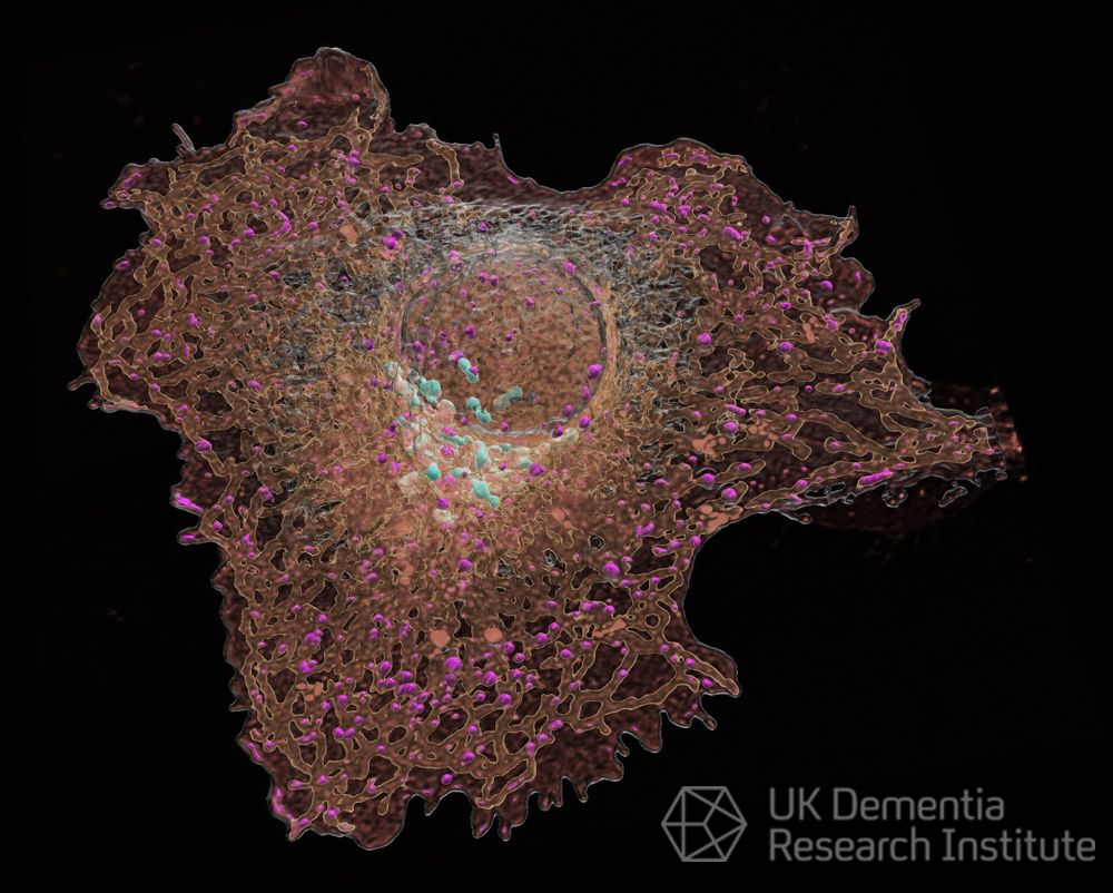
Image 5 of 5: AstrocytER
The endoplasmic reticulum (ER), a network of membranes through which proteins and other molecules move, is stained within astrocytes, the star shaped supporting brain cells.
7/7
Image 4 of 5: Amyloid Precursor Protein (APP) - an intracellular journey
APP produces the toxic amyloid beta protein that forms amyloid plaques in Alzheimer's disease. Researchers found that APP's location in the cell is important for how much amyloid is produced.
6/7
📰Our method for probing protein aggregation in cells with Halotag/FLIM now published, by @karnika0696 & Daniel Maddison @D6Mad 🔬
https://t.co/hjh7KjqwZk
Tech we used to visualise stress-induced disaggregation in https://t.co/Gipx1LuMB2
New paper by @EdwardAvezov@AvezovL in @CellReports reveals crucial insight into the role of the endoplasmic reticulum (ER) in suppressing neuronal regeneration in the central nervous system, offering promising treatment strategies for neurodegeneration👉https://t.co/tG8ZH8Sncz
#FluorescenceFriday#cellbiology#bioart@wonderofscience 4D organelle remodeling in dividing HeLa cells. Please expand to see greater detail.
Left: 3D volumes of chromosomes, mitochondria, and the endoplasmic reticulum (ER) in whole cells.
Middle: Inner details revealed in successive 2 micron thick slabs computationally cut though the cells.
Right: Expanded view in one slab. As the cells progress through division, mitos fragment, ER forms sheets, and mito fragments nestle into ER pockets as both organelles partition into the two daughter cells.
As seen by 4D lattice light sheet microscopy. https://t.co/vpdwTXqrdm
Our new paper modelling #mitochondrial disease in Drosophila is now available. UPR and #ATF4 activation in the brain depends on the severity of mitochondrial complex I loss. https://t.co/n6FgPpBx5Q
📢Job alert - Postdoc position📢 Are you excited about using iPSC-derived microglia to understand endocytic mechanisms underlying Alzheimer's? Fancy developing some cool new imaging methods? Join us! Please RT & share @UKDRI@dem_researcher@ARUKscientist https://t.co/tJ45L6p4tT
💡Closing soon: opportunity for 2x Research Assistant/Associates to join @AvezovL!
Lead an exciting project exploring pharmacological approaches to antagonise amyloidogenesis through enhancing cell proteostasis👉https://t.co/U70SAxM5fM
🔬 Join our lab @Cambridge_Uni@UKDRI 🧠 and lead an exciting project exploring pharmacological approaches to antagonise amyloidogenesis through enhancing cell proteostasis.
https://t.co/aHcEFjKzMx
Go check out my recent manuscript that we uploaded to @biorxiv_neursci.
We found that the protective variant of PLCG2 expressed by microglia in the brain preserves synaptic integrity independently from amyloid burden in an amyloidosis model of AD. @UKDRI
https://t.co/MI71sN7cYG
Delighted out lastest work has been published at Cell Death Discovery! We investigated the unusual role of Cdk12 in neurons. @UKDRI@SpringerNature
https://t.co/MNrn1X9ap7