Postdoc at UCLA, Department of Radiological Sciences | PhD from The Univ of Tokyo, Chemical Biology & Biotechnology | Researching Alzheimer’s Disease & Beyond
Real-time tissue-clearing imaging: A groundbreaking paper overcoming a major challenge in the field! Personally curious about the impact of azoreductase—how compatible is it with biological processes or chemical tools involving azoreductase enzyme activity?https://t.co/CsTXTo2g7O
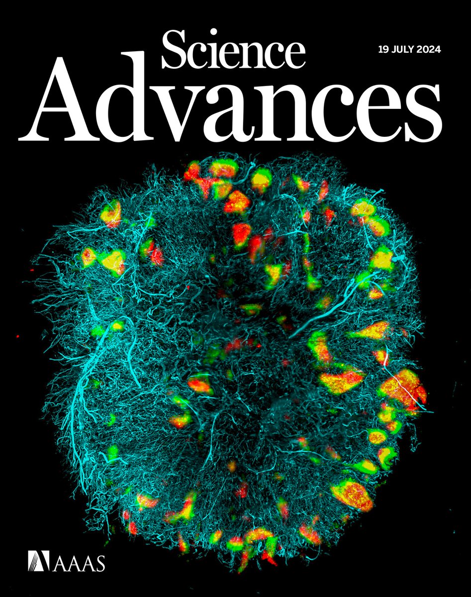
Researchers have developed Click3D, a method for thoroughly staining whole organs using click chemistry.
Learn more in this week’s issue of Science Advances: https://t.co/NEubW1JLky
This research was primarily conducted by Dr. Iori Tamura, under the supervision of Prof Shinsuke Sando (https://t.co/NV14yC8Hum). I am honored to have supported and introduced this work.
Click3D, a whole-organ 3D imaging method utilizing click chemistry, is now hot off the press in @ScienceAdvances! Congratulations to my former colleague Dr. Iori!
"Click3D: click reaction across deep tissues for whole-organ 3D fluorescence imaging"
https://t.co/ODFb6YiHDL
To those developing clickable chemical probes or working with tissues containing targets with a click handle, why not consider using Click3D? Unexpected discoveries might await you!
And what I especially want to say to chemists who are developing covalent molecular probes or anchorable molecular probes with fixatives is this: You might step into unexpected chemical biology when you clear your tissue samples.
One thing to pay attention to here is that the fluorescent labeling must be covalently anchored within the tissue. Otherwise, the fluorescent labeling may be washed out during the clearing process.
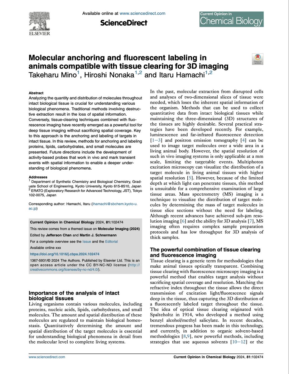
A spot-on review paper on the latest techniques for labeling and anchoring biological targets within tissue samples for tissue-clearing imaging, written by my friend @TMino12 and behind-the-scenes Assoc Prof Nonaka from an esteemed chemical biology lab in Japan @HamachiLabKyoto
Excited to share our new review: “Molecular anchoring and fluorescent labeling in animals compatible with tissue clearing for 3D imaging.” 🔬 This review explores anchoring and labeling various molecules in intact tissues, key for deep-tissue imaging. ��https://t.co/UQ8zbfhp7M
In the process of tissue clearing, tissue samples are typically highly permeabilized. This enables the effective penetration of various clearing reagents and labeling agents into the tissue, washing out unwanted substances for observation, and resulting in high-quality imaging.
We have developed a Pimonidazole-alkyne conjugate named Pimo-yne, enabling sensitive detection of in vivo hypoxia. Congratulations to my colleague Iori! The true potential of Pimo-yne will be revealed soon.
https://t.co/ClJsX38Quh
To address these issues, we have developed a Pimonidazole-alkyne conjugate named Pimo-yne. Pimo-yne can be detected by a click reaction with a reduced background signal, and its capability for hypoxia detection is comparable to that of Pimonidazole.