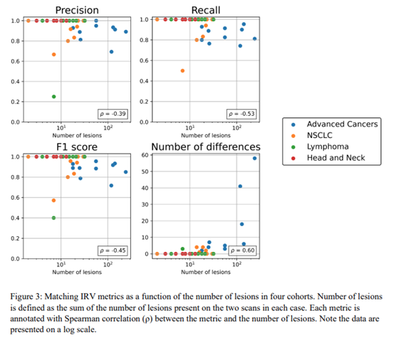
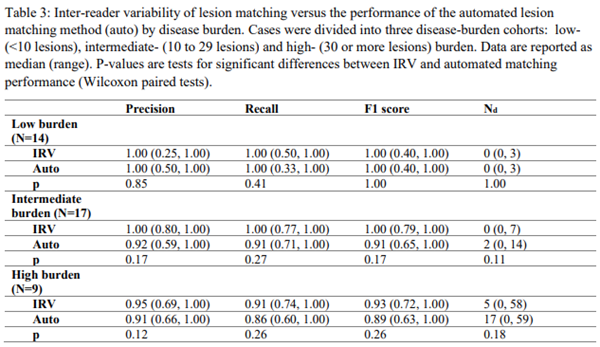
5/ Lesion matching is a harder task (for both 👩⚕️and 🤖) in subjects with higher disease burden. Here are IRV metrics as a function of lesion count – plots look similar if you compare automated to physician consensus.
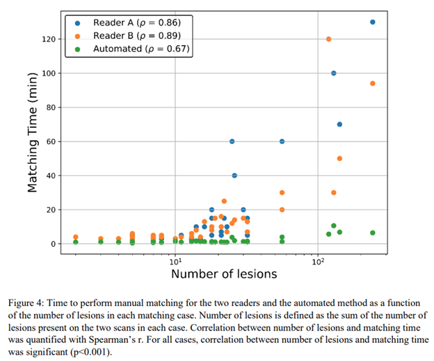
6/ The big win for automated 🤖matching is in time savings. Automated matching took up to 11 minutes (on a typical desktop workstation – no NASA supercomputer here!), while clinicians took up to 130 minutes (⌛️!!) for the highest burden cases.
4/ Automated matching met the benchmark of IRV in all study cohorts – meaning the variability between 🤖 and 👩⚕️ was similar to variability between 👨⚕️ and 👩⚕️
3/ In our study, two #nucmed physicians matched lesions between pairs of #PETCT images of cancer pts. Then, we used an automated 🤖approach to do the same. We assess differences between clinicians 👨⚕️ and differences between physician consensus 👨⚕️👩⚕️ and automated🤖
2/ Inter-reader variability (IRV) has been widely studied in image segmentation contexts, but another task clinicians 👩⚕️👨⚕️ perform is longitudinal comparison of multiple images to track lesions in time and determine change.
This task is critical in determining tx. response!
1/ Excited to share our latest paper! 🧵
Performance of an automated registration-based method for longitudinal lesion matching and comparison to inter-reader variability https://t.co/xWvQsHNmxs via @ioppublishing
Join us next Monday at #RSNA22. AIQ's Eric Horler will give a presentation sponsored by @OneMedNet. We hope to see you there, and please contact us if you'd like to connect in Chicago.
This month has been pretty cool - I defended my PhD on imaging #immunotherapy response using #PET from @wiscmedphys, and am now starting as a research scientist at @aiq_solutions. Excited for new challenges! 😄
Are you a @UWMadGSEd student or @Badgerdocs / @UWPostdocOffice researcher interested in science careers? Join us at @DiscoveryBldg May 19 to chat with scientists who have careers in the communications space. Registration requested. https://t.co/8NNZbT5073
4/ Early detection+monitoring of toxicity is critical for keeping pts on #immunotherapy! Our work suggests #PETimaging can play a role here, but big limitation is when scans are acquired after tx start.
1/ A belated xmas present - our latest article detecting #immunotherapy toxicity on FDG PET was published in #EJNMMI. Elevated uptake in thyroid, lung, and bowel can indicate toxicity in those organs!
Read the deets at: https://t.co/JqhqrDVXyf
3/ Zooming out to all pts, things get messier - but general trend is elevated organ uptake at time of clinical toxicity dx (or sometimes before!).
Worth noting that organ PET quantification here is automatic thanks to #AI-based organ segmentation.