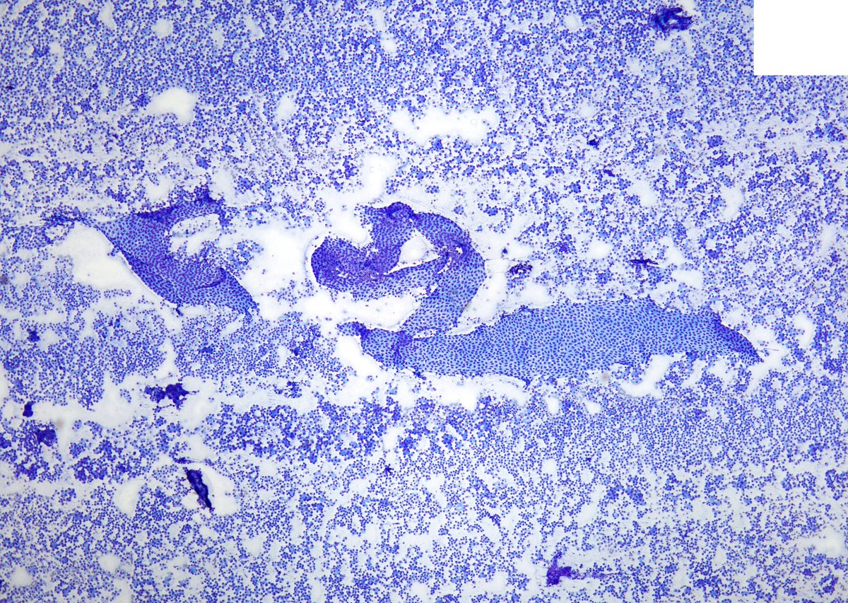
Collision of good versus bad in this pelvic wash: sheet of benign mesothelial cells versus metastatic high-grade serous carcinoma
#CytoPath#PathTwitter
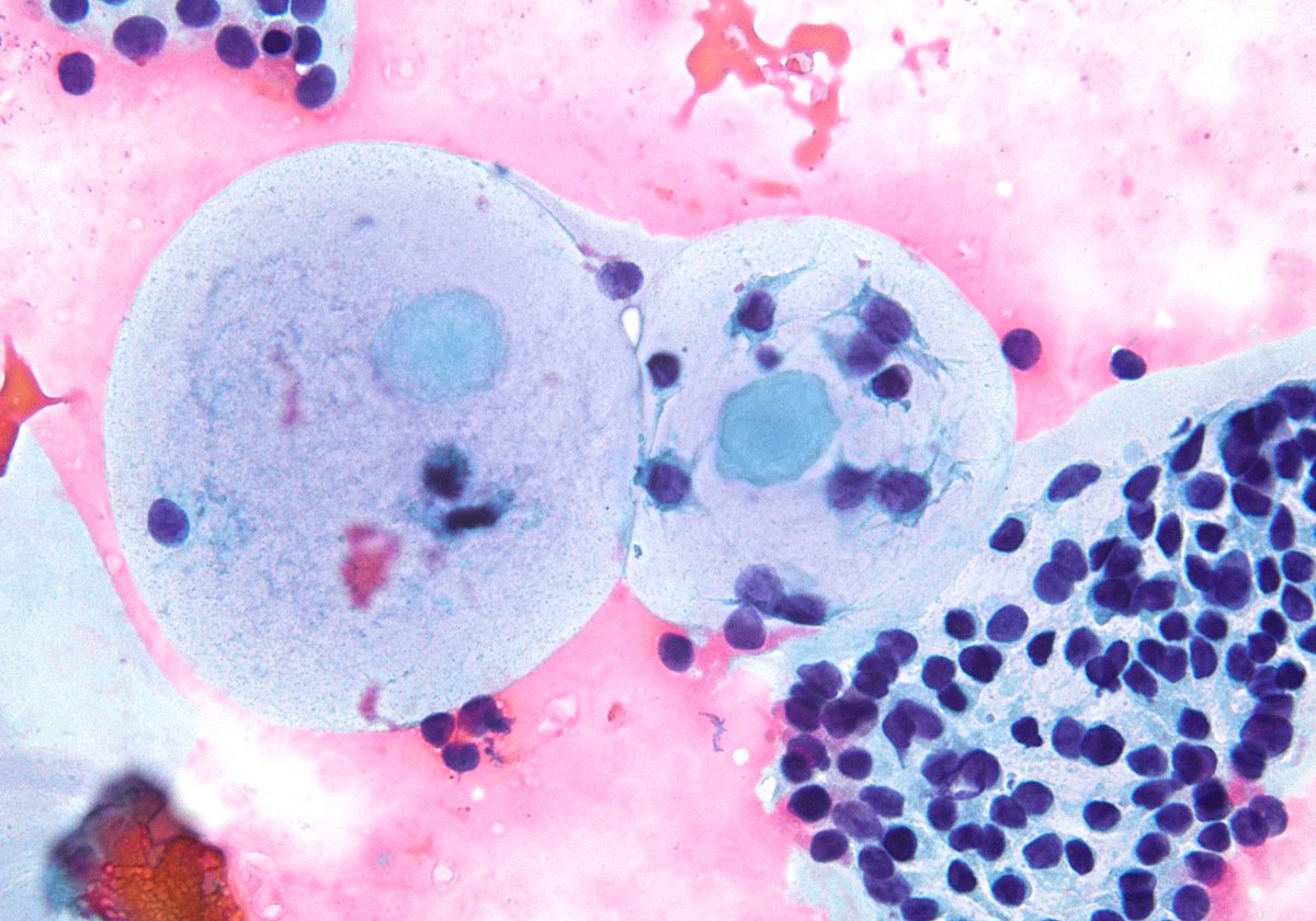
The so-called “circle artifact” has been traditionally defined with GYN malignancies on a ThinPrep PAP test as a result of tumor diathesis and less often with extensive blood or inflammation. This voided urine had extensive acute inflammation with candidiasis.
Adenocarcinoma, presenting a sharp contrast with the juxtaposed benign squamous cells in a lower esophagus brushing. Prominent solitary macronucleoli, lack of adhered PMNs and a clean slide background with absence of inflammatory debris rules out reactive condition. (ThinPrep)
Couldn't be more excited to announce the launch of https://t.co/iJqFUSnnfj, the virtual slide site I wish had existed during my pathology residency. You can:
- Order special stains to work up cases
- See annotated histologic features
- Prepare for exams with board-style questions
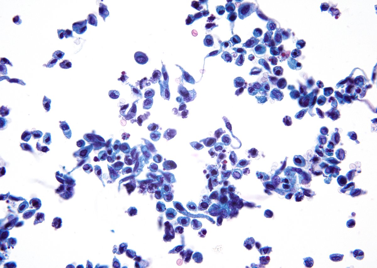
Glands, Acini or Rosettes? Occasionally, the vague "tigroid" smear background helps. This pancreatic FNA with IHC help was diagnosed as "Acinar Cell Carcinoma". (EUS-FNA)
"NEVER forget the path of the traversing FNA needle". Liver mass in a 50/M with history of "Pancreatic Neuroendocrine Tumor (PanNET)". The flat sheets of mesothelial cells present a beautiful and eye-catching contrast with the background metastatic PanNET.
NOT benign osteoblasts on FNA from a sclerotic spine lesion. Despite the innocuous appearance, these cells represent bone metastasis from the patient's known "Lobular Breast CA". (FNA)
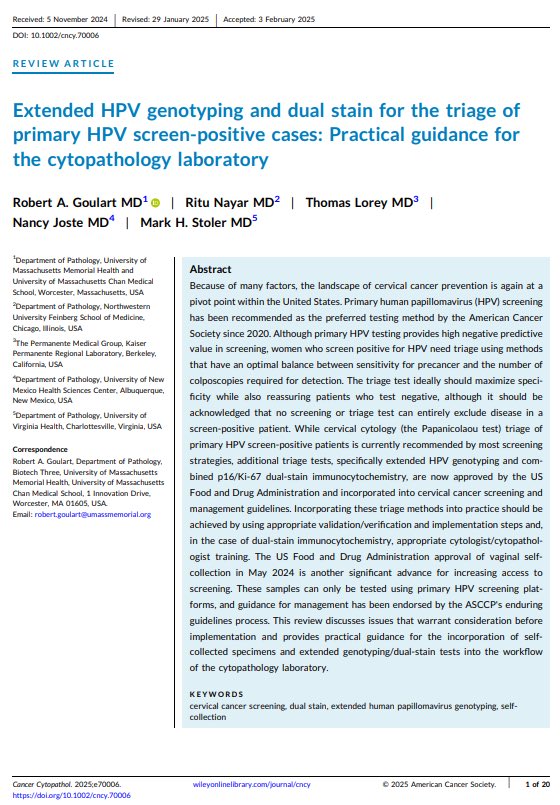
Extended HPV Genotyping and Dual Stain for triage of primary HPV screen-positive cases: Practical guidance for the cytopathology laboratory. Work done by the ACS Cervical Cancer Roundtable’s Laboratory Infrastructure Workgroup. @CancerCytopath@cytopathology@cervicalRT
"Micropapillary Adenocarcinoma" of the lung involving pleural effusion. Impressive tumor cytoarchitecture with psammoma bodies. (cytospin and cell block)