Diagnosis: Fordyce spots (ectopic sebaceous glands).
Diagnostic justification: Multiple asymptomatic, yellow-white papules on the labial mucosa are the hallmark clinical feature of Fordyce spots. The first panel demonstrates numerous discrete yellow-white papules on the inner upper lip. The second panel shows similar papules distributed along the labial mucosa. The third panel highlights the characteristic small, yellowish papules against otherwise intact mucosa.
Management: Reassurance and patient education; treatment is unnecessary for asymptomatic lesions. #dentistzone
Tooth discoloration isn't always just a surface stain.
Changes in tooth color can originate inside the tooth, and the appearance may provide an important clue to the underlying cause. Tooth discoloration is broadly classified as extrinsic, intrinsic, or a combination of both.
This visual highlights four examples:
🔸Calcification - changes in the internal hard tissues can alter the tooth's normal appearance.
🔸Internal resorption may sometimes produce a characteristic pinkish discoloration, often called a “pink tooth."
🔸Pulp necrosis breakdown or necrosis of the pulp can cause intrinsic darkening or discoloration.
🔸Tetracycline staining exposure during tooth development can produce intrinsic discoloration, often with characteristic banding.
Because similar looking discoloration can have different causes, clinical and radiographic examination is important before deciding on treatment. #dentistzone
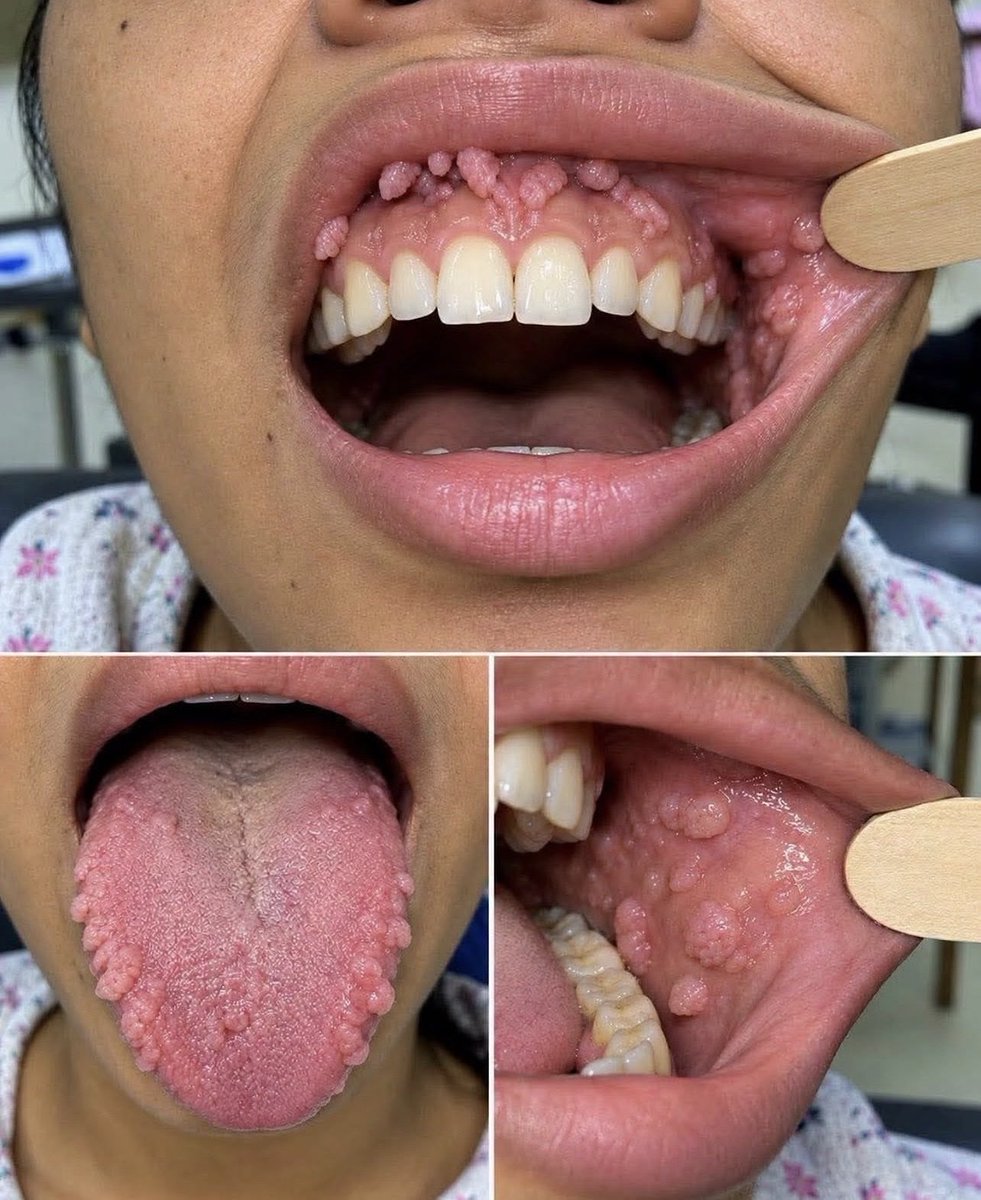
These growths are oral papillomas or warts caused by human papillomavirus (HPV). Certain low-risk HPV types infect the oral mucosa through direct contact, including oral sex, producing these clusters of pink cauliflower-like lesions on the gums, tongue, and cheeks. #dentistzone
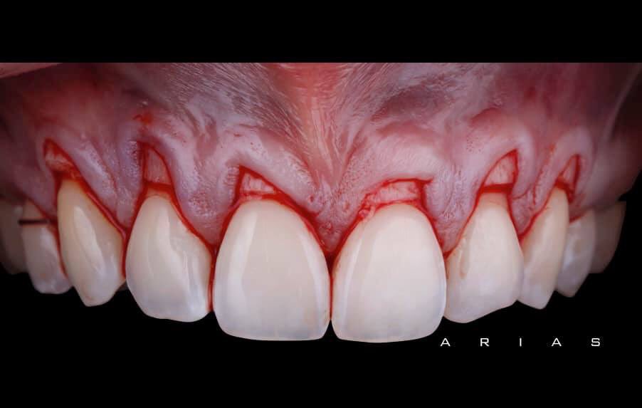
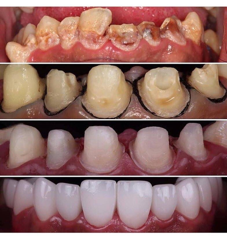
GINGIVOPLASTY is the surgical reshaping of gum tissue around the teeth. It is often is done simply to make gums look better. They may have an unusual shape or may not be formed normally. The causes can include a person's genes, disease or trauma.#dentistzone