Are you right when it’s bright?
Bright cortical signal on diffusion images is classically associated w/hypoxic-ischemic injury.
But there are many mimics!
Do you know how to recognize the different patterns of cortical restricted diffusion? Here’s a figure & some pearls to help!
Cortical restricted diffusion many seen in a variety of conditions—many with VERY different pathophysiology & prognosis.
There are 6 main patterns:
➡️Cortex & deep gray
➡️Diffuse Cortex
➡️Focal Cortex
➡️Limbic
➡️Deep gray
➡️White matter
Here are some steps to help differentiate:
🔸Is the presentation acute or chronic?
🔸If it’s acute, toxic-metabolic & hypoxic-ischemic events account for the majority of cases.
🔸If it's acute, seizure & encephalitis are less frequent but should be considered if more focal
🔸 In the chronic setting, isolated cortical involvement is almost diagnostic of CJD
Hopefully this will help clear up any confusion about the diffusion!!
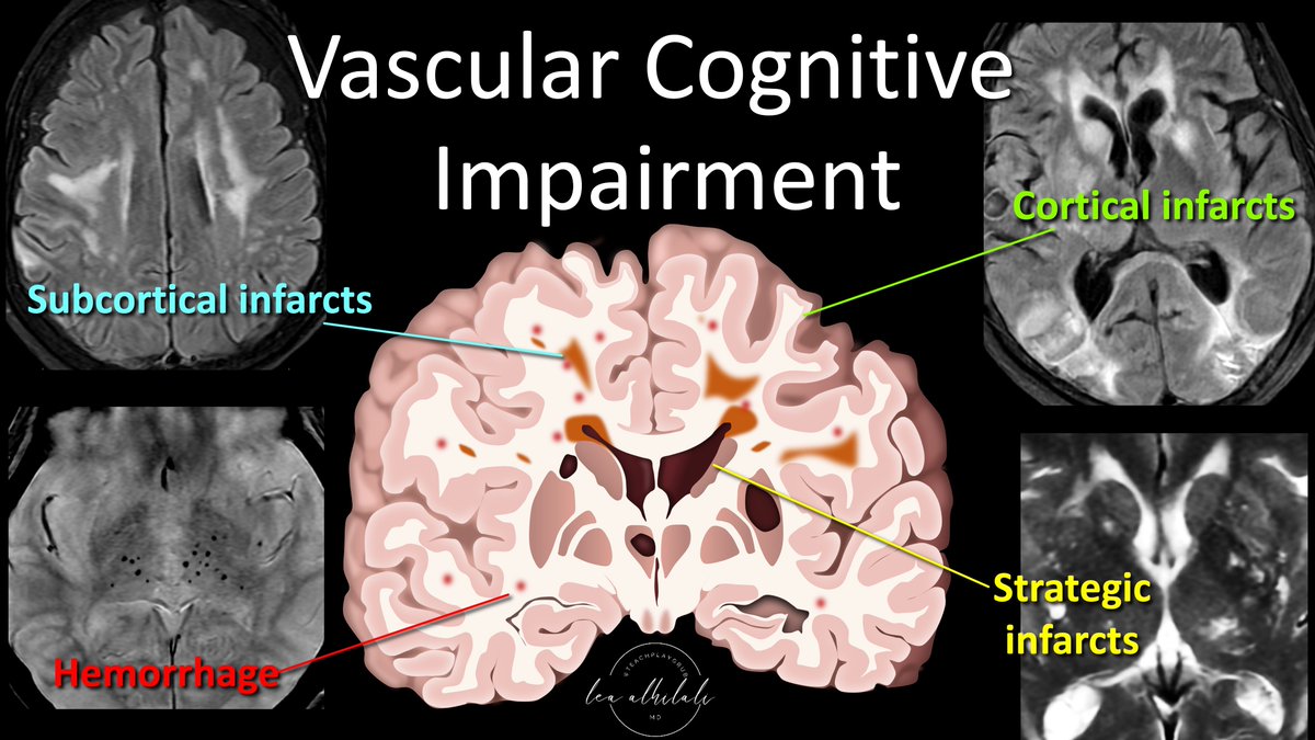
1/Having trouble remembering what you should look for in vascular dementia on imaging?
Almost everyone worked up for dementia has infarcts. Which ones are important?
Here’s a thread on the key findings in vascular dementia!
1/Correlate clinically!
It’s harder than you think in THALAMUS—where its size is small & but the clinical symptoms are large.
Here’s a thread to help you remember the main thalamic syndromes & their locations!
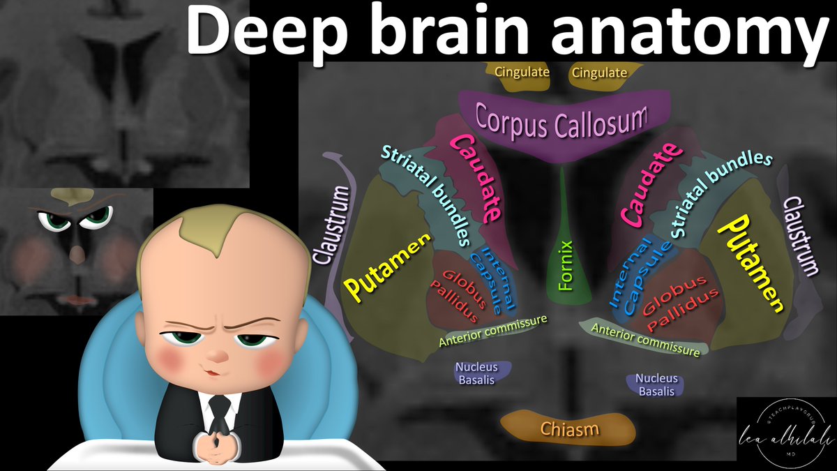
Trying to cover your bases when it comes basal ganglia region anatomy?
Need some core knowledge of the corpus striatum?
Want to be a boss when it comes to anatomy here?
Can you see the Boss Baby w/the puffy cheeks here?
Here's how this baby can help you w/this anatomy...
Deep brain anatomy in the coronal plane looks like a baby with puffy cheeks. This can help you remember the anatomy & function!
Here’s how:
➡️Cingulate gyrus:
🔸Looks like the baby’s hair. It’s a SINGLE tuft of hair for the CINGulate gyrus
🔸Cingulate gyrus is involved in processing painful emotions
🔸Remember: CINGulate is for things that SINGED you
➡️Corpus callosum:
🔸Looks like the evil boss baby eye brows
🔸It’s the crossing fibers so it’s what makes the baby look cross!
➡️Fornix:
🔸Looks like the baby’s nose
🔸Involved in memory
🔸Remember the NOSE KNOWS things and FORnix never FORgets
➡️Corpus striatum:
🔸Looks like the baby puffy cheeks
🔸Made up of many different structures
▶️Putamen: important for motor skills. PUT-amen help you PUT-one foot in front of the other
▶️Striatal bundles: important for learning actions w/a reward. STRIatal bundles make you want STRIVE for better
▶️Caudate: involved in associated learning. CAUdate helps you learn the CAUSES
➡️Claustrum:
🔸Looks like the baby’s ears
🔸Inhibits motor function, esp during sleep
🔸Remember Santa CLAUStrum only comes when we sleep
Hopefully now you have a deep knowledge of these deep brain structures!!
1/ I always say, "Anyone can see the bright spot on diffusion images—what sets you apart is if you can tell them why it’s there!”
If you don't why a stroke happened, you can't prevent the next one!
Can YOU tell a stroke’s etiology from an MRI?
Here’s a thread to show you how!
1/ “Now listen carefully!”
Everyone has so much fear about the anatomy where they hear!
Do you dread temporal bone anatomy?
Do find the understanding ossicles impossible?
Do you know the ice cream cone sign on CT & then nada?
Then you need this thread on ossicular anatomy!
1/Have disagreements between radiologists on the degree of cervical canal stenosis become a pain in the neck?!
Worried about sticking your neck out and calling severe cervical stenosis?
Here’s a thread on cervical stenosis grading that’s easy, reproducible & evidence based!
Do you know why they call it the "sella turica"? Although technically speaking the term "sella turcica" refers to the floor of the pituitary fossa, it is often used as a synonym for the pituitary fossa. #RadEd#FOAMed#neurorad
@OkromelidzeLela If you truly want the motherload of all links/resources and shocked no one has said this yet.
https://t.co/g371OC9rDj
Also don't forget about
https://t.co/3VCfOCID9L free!
Knowing spinal arterial anatomy is a given.
Knowing spinal venous anatomy is next level
Vertebral venous plexus can be both good & evil--good for collaterals, bad for tumor spread, infxn, & emboli...and spinal CSF fistulas!
Here’s the anatomy you MUST know if you are doing spine
3 rings: internal, anterior & posterior plexus
➡️Internal is the epidural plexus. It drains the basivertebral veins from the vertebral body
➡️Anterior drains the basivertebral veins and foraminal veins.
➡️Posterior drains the veins from the deep paraspinous musculature.
🌟Internal plexus connects the anterior & posterior plexuses via foraminal veins along the nerve root
These foraminal veins or foraminal plexus are site of CSF venous fistulas & the target for transvenous embolization!
So it's important to learn this anatomy, no matter how draining it can be!
1/Time to go with the flow!
Hoping no one notices you don’t know the anatomy of internal carotid (ICA)?
Do you say “carotid siphon” & hope no one asks for more detail?
Here’s a thread to help you siphon off some information about ICA anatomy!
Is trying to remember toxic encephalopathy patterns making you toxic?
Does memorizing metabolic encephalopathy patterns put you into a metabolic coma?
Don't become encephalopathic!
Here is the cheat sheet of the encephalopathy patterns you NEED to know & how to remember them!
➡️Corpus Callosum:
🔸Lesions here are called cytotoxic lesions of the corpus callosum or CLOCC.
🔸Usually transient from cytotoxicity, especially viral infections.
🔸Remember they are commonly viral & transient bc CLOCC lesions are on the CLOCK
➡️Central Pons:
🔸Usually osmotic demyelination
🔸Remember this bc the T2 hyperintensity is in the shape of O for Osmotic
➡️Dentate:
🔸Usually flagyl toxicity
🔸Remember this bc the wavy appearance of the dentate looks like a FLAG for FLAGyl
➡️Corticospinal tract:
🔸Usually from cobalamin deficienty
🔸Remember cortiCOspinal for CObalamin
➡️Symmetric Gray Matter:
🔸Differential is EXTENSIVE
🔸Remember the differential list for deep gray is deep!
➡️Cortical Gray Matter:
🔸Typically hypoglycemia & hyperammonemia
🔸Remember it looks like the brain has been coated in shimmering sugar = related to glucose
🔸Hyperammonemia has about 1000 Ms in it & the undulated cortex looks like an M
➡️Symmetric White Matter:
🔸Commonly from chasing the dragon (heroin) & methotrexate
🔸Remember that the puffy white matter signal looks like the smoke from a dragon
🔸Remember methotrexate as methoTRACTSate = affects many white matter TRACTS
➡️Posterior subcortical edema:
🔸This is PRES or posterior reversible encephalopathy
🔸So if it’s all POSTERIOR, think POSTERIOR reversible encephalopathy
➡️Asymmetric White Matter:
🔸This is usually demyelinating from chemotherapy
🔸Demyelinating lesions have a C shaped pattern of enhancement
🔸So if you see a C enhancing think of C-hemotherapy
Now you know all the toxic/metabolic encephalopathy patterns.
So hopefully now remembering the patterns of toxic/metabolic won’t seem so diabolic!
✅Cortical vein sign: cortical veins located adjacent to the inner table. In BESSI/cerebral atrophy
❗️Displaced cortical vein sign: cortical veins running separated from the inner table. In subdural collections.
⬆️Relevant in pediatrics. See what´s necessary & what´s superfluous
Reversible Cerebral Vasoconstriction Syndrome (RCVS) in this 40 y/o F with acute onset headache
🔷Thought to be caused by loss of control of cerebrovascular tone leading to vasospasm
Imaging and learning points in thread 🧠
#radres#Neurology#MedEd#neurosurgery#medicine
Even if you’re in it for the LONG haul, sometimes it’s good to come up SHORT!
If you can’t summarize, you’ll never memorize!
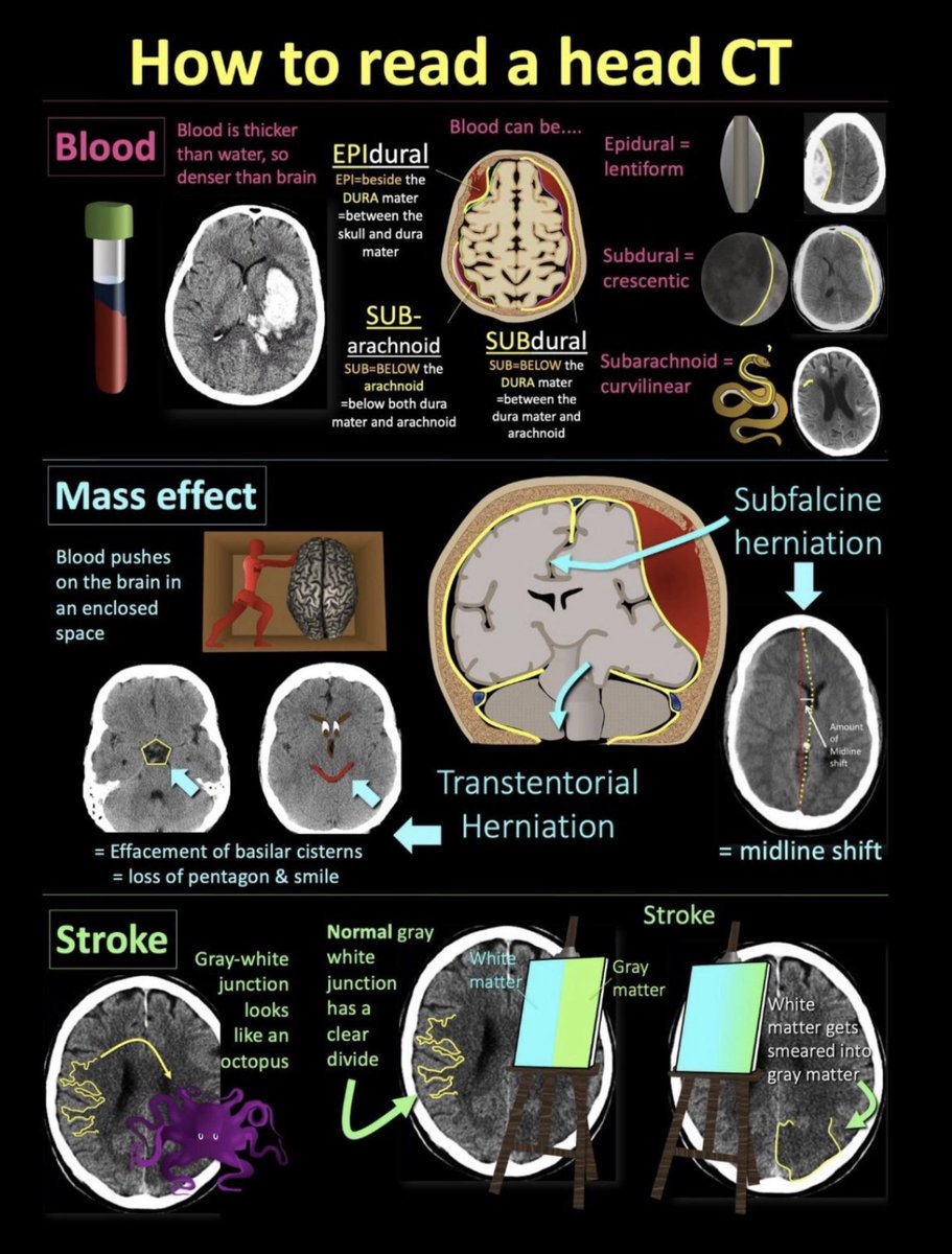
Here's a cheat sheet for a truly critical skill in imaging: How to read a head CT!
If you're looking at head CTs, this is what you NEED to know!
➡️The 3 main things to look for on head CTs are:
🔸Blood
🔸Mass Effect
🔸Stroke
➡️Blood
🔸CT is just a measure of density. Blood is thicker than water, so blood is denser than brain (70% water) on head CT
Blood can be:
🔸EPIdural: looks lentiform
🔸SUBdural: looks crescentic
🔸SUBarachnoid: looks snake-like
➡️Mass effect
🔸You won’t bleed out in your head. Instead, calvarium is a closed space & brain gets compressed
Look for:
🔸Subfalcine herniation = midline shift, ventricles shift to the other side
🔸Transtentorial herniation = Loss of the basilar cisterns--normally look like a pentagon & smiley face
➡️Stroke
🔸Normal gray-white differentiation looks like an octopus & is crispy & well-defined
🔸Stroke looks like someone smeared white matter paint into gray matter paint
You can see the full thread here:
https://t.co/NyrzkWz0Q2
So now you know what to look for on a head CT. Hopefully this short version stays with you for the long haul!