Mi favorito es el Masson. Patóloga de hígado, páncreas y todo lo que se tiña con CDX2. Y un poco pajarera 🐣 #GIPath#Pathology Opinions my own. She/her.
Three best things I learnt from my mentor:
1. Best immunostain is a new Hematoxylin.
2. Be humble.
3. Love what you do, but love your life outside work even more.
#pearlsfrommymentor
Continuing the story of IgG4 disease, here is a study by a group of eminent GI pathologists that explores the role of IgG4 in chronic oesophagitis with ulceration and invites us to recognise a new entity. #openaccess#GIpath https://t.co/b7IYRCKaWZ
1/3 Theres some truth in the dogma that you cant see Helicobacter on digital pathology scans and you need a 40x scan to see them but in my experience its more about a good quality H&E. This was scanned with the 20x objective (0.25 microns per pixel) and Hp can be easily seen.
@drtimbracey@GI_Pearls@PathPro@TobyASchmitt@Pathoutlines We scan everything at 40x and do reflex ihc. It turns out the time spent retrieving blocks to cut them again is not worth it. Anyway I agree, when they are easy to see they pop out in any stain. Digital has nothing to do with it
A flare of Behçet disease, a systemic vasculitis, can result in colorectal bleeding. Small blood vessels can be affected, resulting in patchy ischemic changes.
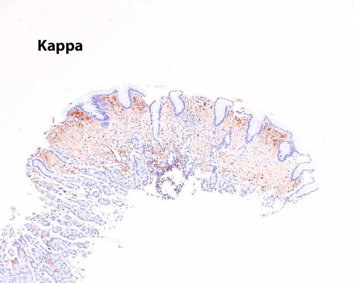
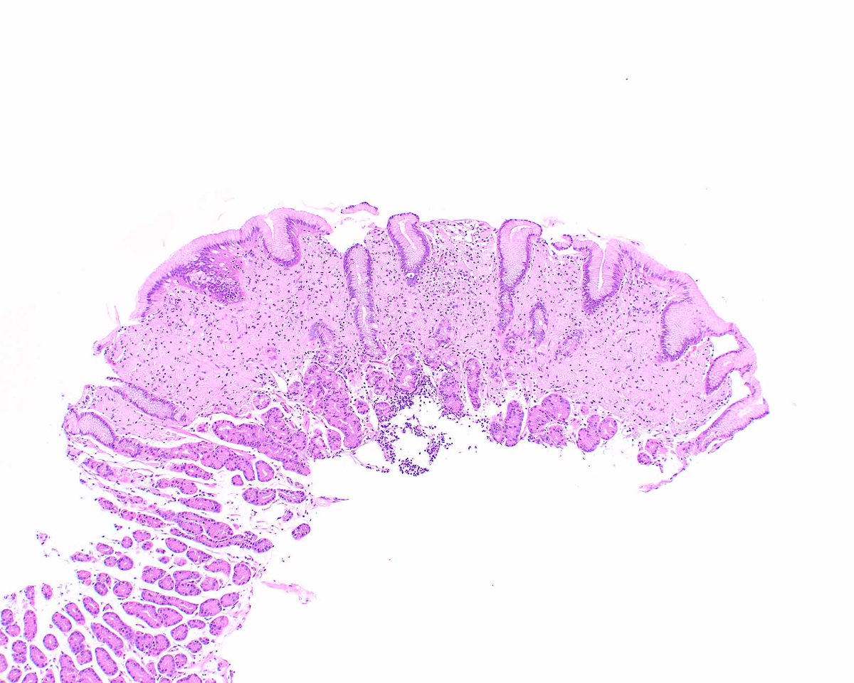
An example of crystal storing histiocytosis; the patient needs to be evaluated for a plasma cell disorder or other lambda-restricted B cell neoplasm.
Arnold CA, et al. Am J Surg Pathol. 2018 Oct;42(10):1317-1324. PMID: 29878935.
University of Miami is interviewing for our GI pathology fellowship for July 2028. We have amazing cases (like this mucosal prolapse polyp) and GI pathologists (M Garcia, R Yantiss, S Al Diffalha, O McDonald, C Milikowski, D Ortiz, and me). Queries: [email protected]
1/3 #GIpath#CritterOnTwitter Look what I found in this inflamed antral hyperplastic gastric polyp 🤓. At first glance, I thought that it is hallucinations or illusions due to end of the week fatigue 😂 then I started to take images of every single one. So all are represented 😅
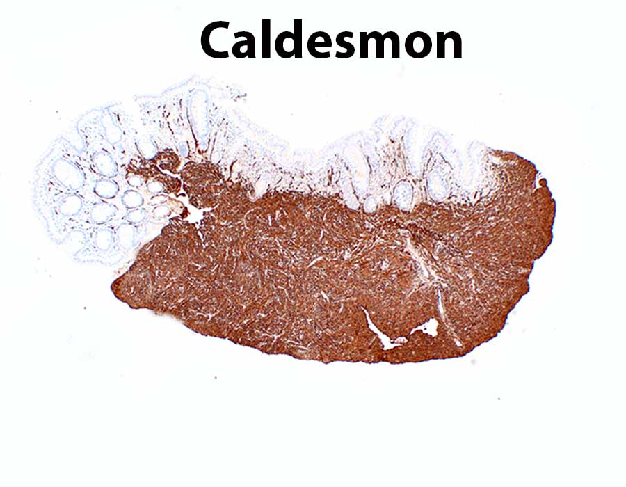
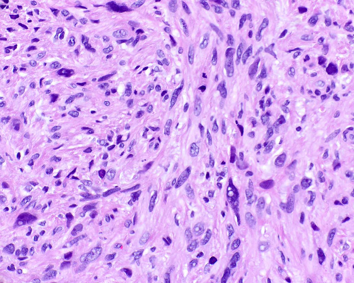
This lesion arose in the small intestine, resulting in obstruction. It probably was related to non-steroidal anti-inflammatory drugs.
Cortina G, et al. PMID: 10555011.
A needle biopsy of this pancreatic solid pseudopapillary neoplasm was labeled "gastric mass". We initially tried to make it into a neuroendocrine tumor.... Sometimes needle placement is misleading.. LEF1 is a nice clean nuclear stain and easier to interpret than beta catenin.
Not everyone is lucky enough to get to work again with her best friend, fifteen years after pathology made us meet.
Bienvenido al equipo @dr_MPrieto! 💜
Ouch! A patient presented with a painful hemorrhagic corpus luteum and hemoperitoneum and "diverticulitis" - except that the diverticulitis was endometriosis that even showed stromal luteinization.
Ayer tuve la suerte de acudir a la reunión anual de la @SEAP_IAP
Algunas ideas fundamentales sobre el #cribado del cáncer de cérvix con el reto de homogeneizar la implantación y reducir desigualdades:
🔹Cribado poblacional más eficaz, equitativo y eficiente que el oportunista
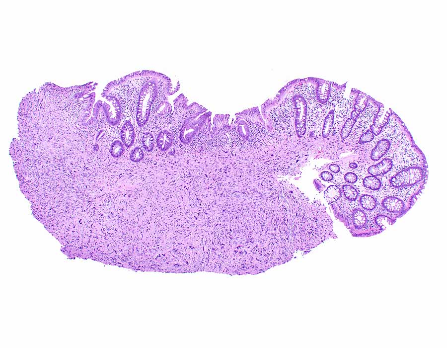
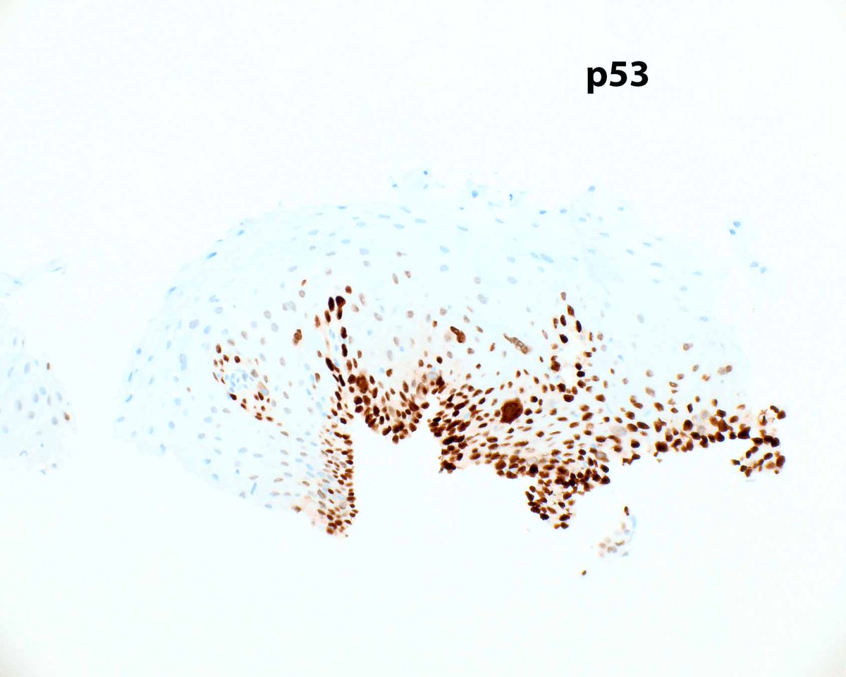
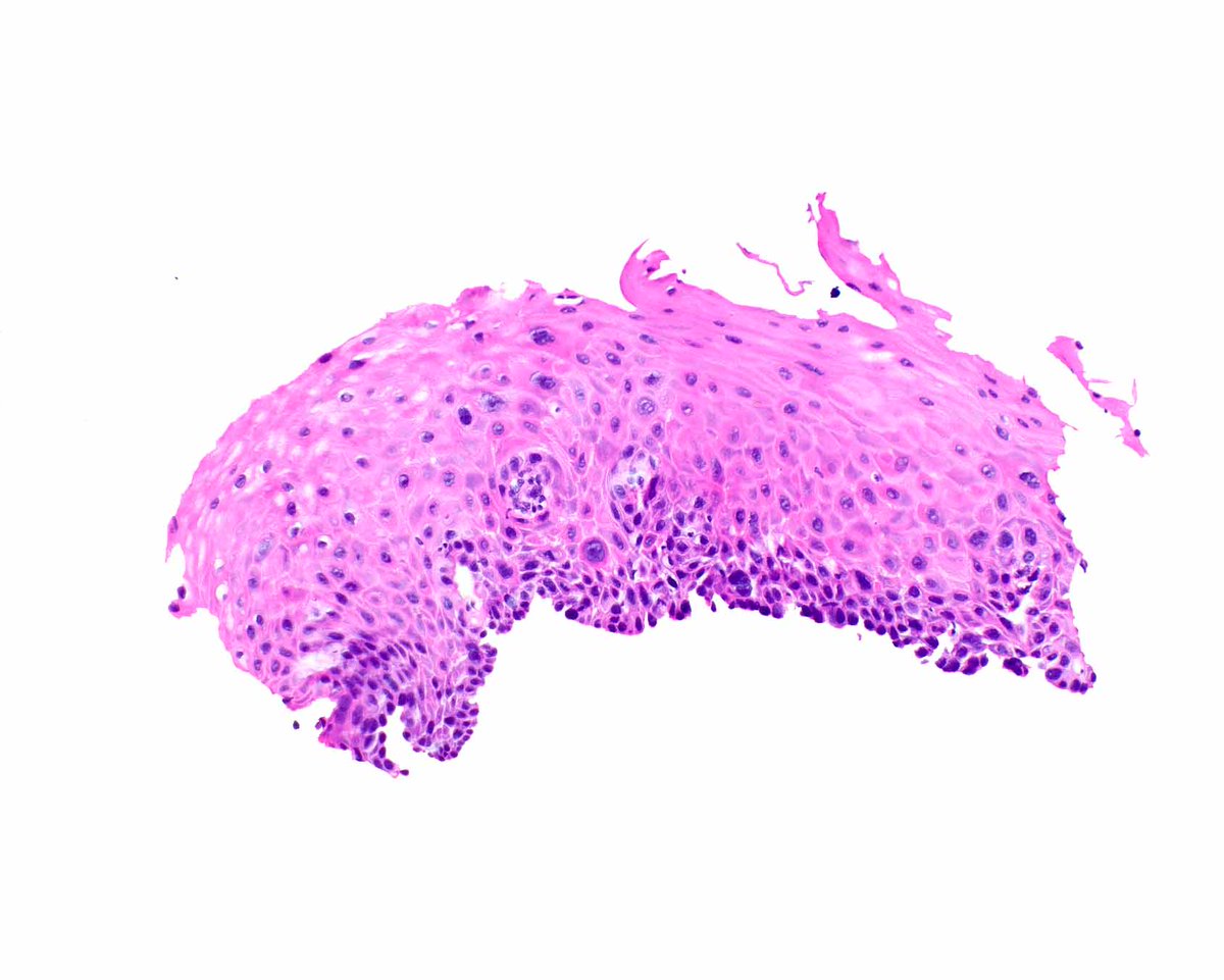
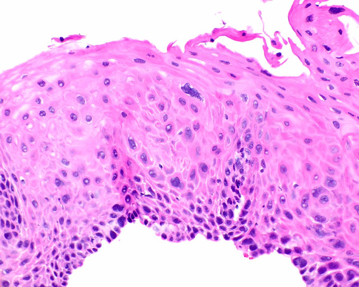
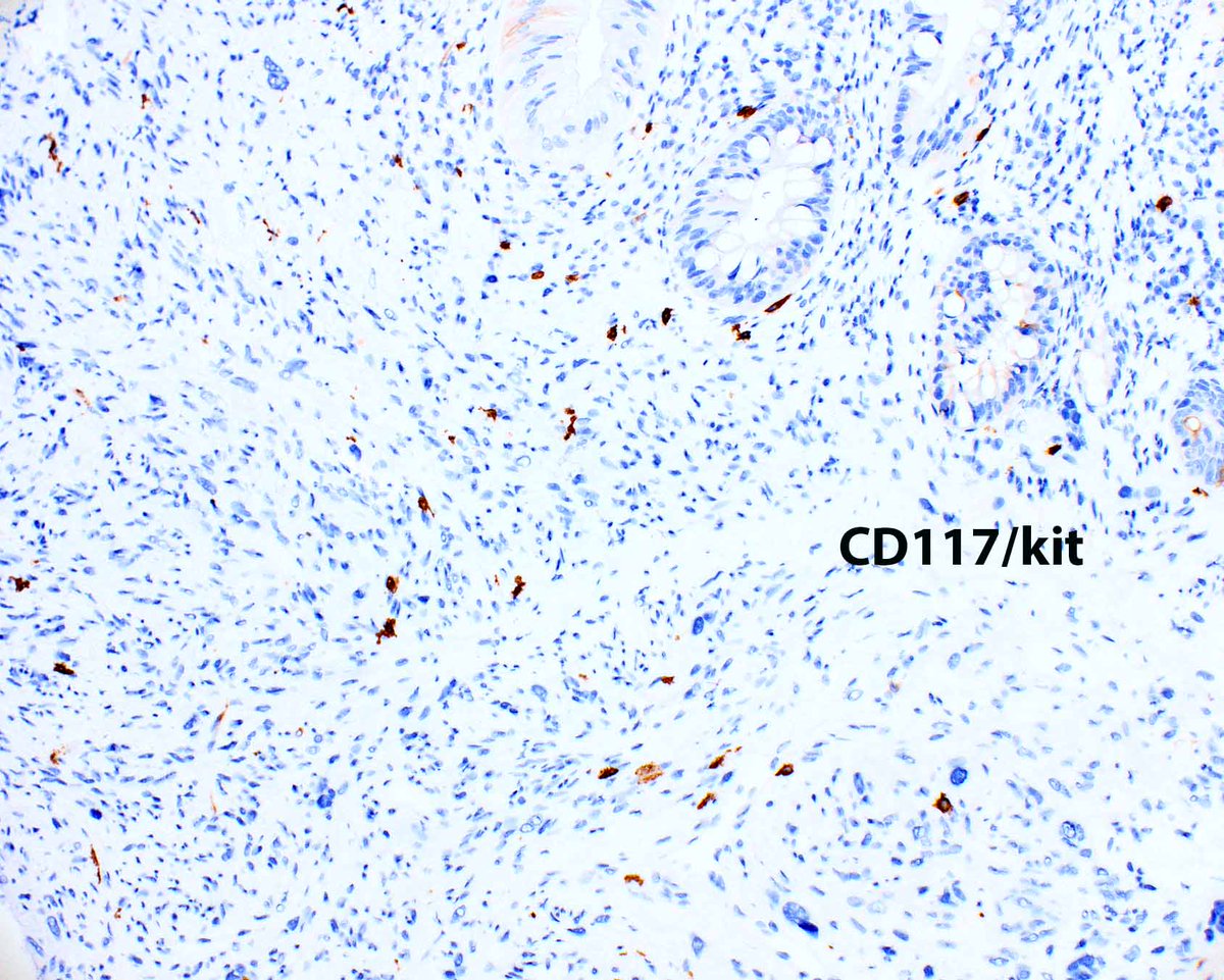
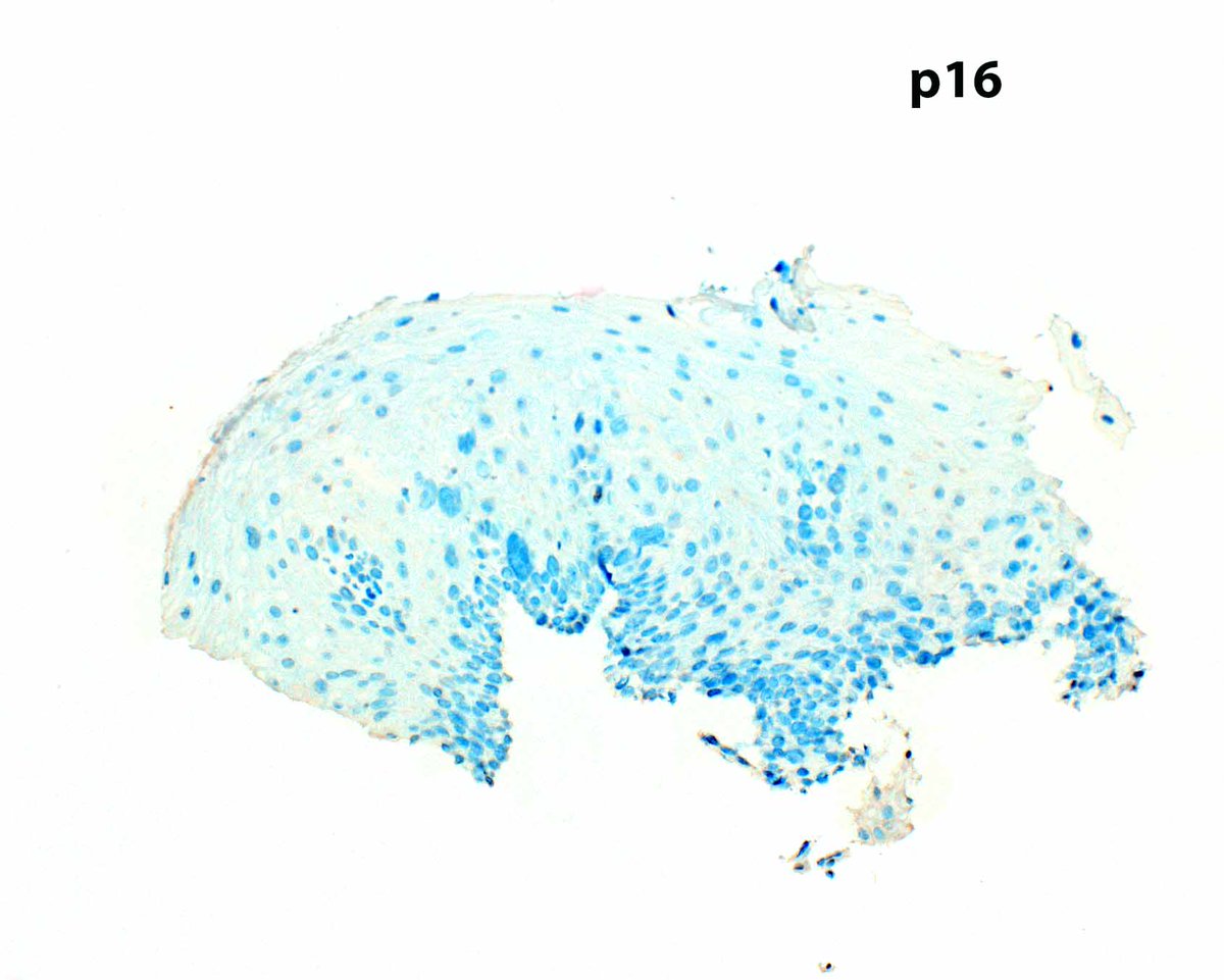
p53 IHC can help confirm a diagnosis of esophageal squamous dysplasia. In occasional HPV-associated cases, p16 staining is strongly reactive. This example is probably driven by a TP53 mutation. Don't expect the full thickness staining typical of glandular dysplasia.
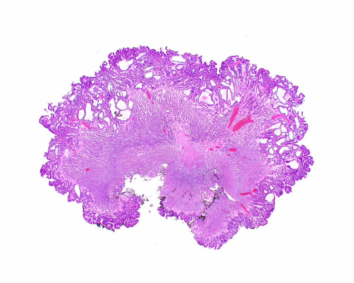
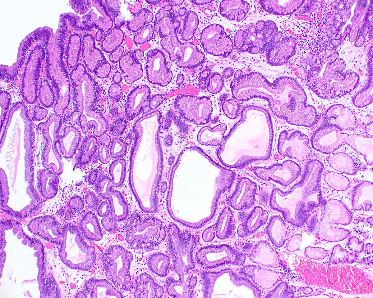
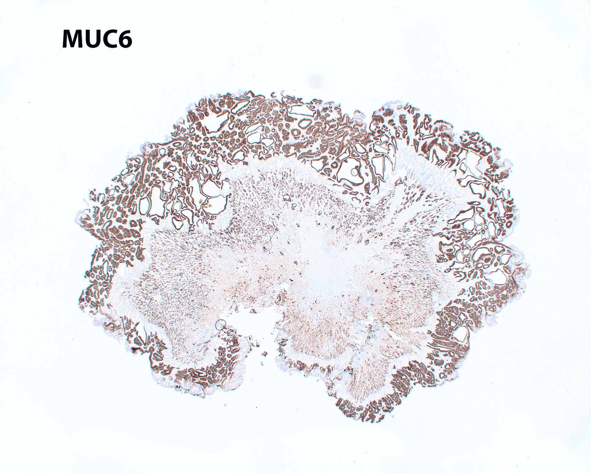
A never-ending obsession with pyloric gland adenomas is a benign problem. This one arose in otherwise normal gastric oxyntic mucosa and coats the surface! They are also associated with autoimmune gastritis and probably arise from pseudo-pyloric metaplasia in that setting.