🚨NUEVO: Artículo de posición de la American Heart Association sobre el consumo de cafeína y su relación con la enfermedad cardiovascular☕️♥️.
👉🏽1) El consumo moderado de cafeína (hasta 400 mg/día o aproximadamente 3-5 tazas de café de 8 oz) está asociado con (sigue abajo👇🏽):
🧵 NEFROPROTECCIÓN EN 2026: lo que TODO internista debe saber.
La ERC ya no se trata solo controlando la presión arterial.
Los nuevos ensayos y KDIGO 2024 han cambiado paradigma. Estas son las claves que más impacto tienen en la práctica clínica. 👇🧵
Objetivos de LDL-C en el paciente renal: De la ERC avanzada a la hemodiálisis 🩸🎯
Las metas de LDL-C se vuelven más estrictas a medida que decae la función renal. El nuevo consenso de la ERA ratifica los pilares terapéuticos en prevención primaria: para ERC estadio 3 (o LDL-C basal elevado), el objetivo es un LDL-C <70 mg/dL usando estatinas moderadas + ezetimiba (COR 1).
Sin embargo, en hemodiálisis, la recomendación pasa a ser individualizada (COR 2b): continuar si ya estaba pautado, pero evaluar con cautela el inicio de novo.
Recuerde el estándar de monitoreo: perfil lipídico a las 4-12 semanas post-ajuste y luego cada 6-12 meses (COR 1).
Diagnóstico genético de la hipercolesterolemia familiar en la práctica clínica actual en 2026.
La genética de la hipercolesterolemia familiar vive un momento de transformación. Durante décadas entendimos este trastorno como una enfermedad monogénica causada por variantes patogénicas en tres genes, pero la práctica diaria nos recuerda que en una proporción muy alta de pacientes con diagnóstico clínico no encontramos ninguna variante causal. Comprender por qué ocurre esto, y qué hacer entonces, es hoy uno de los retos clínicos más interesantes en el manejo del riesgo cardiovascular.
https://t.co/WwOnAmKlit
Residual angina after PCI is common. The explanation depends on the interaction between microvascular dysfunction and the epicardial disease pattern
🎯 Focal CAD: successful PCI unmasks coexisting CMD.
〰️ Diffuse CAD: persistent epicardial resistance remains the dominant cause.
Impact of Coronary Microvascular Dysfunction on Patient-Reported Symptoms After PCI | https://t.co/EYxfNAQNi4
@crfheart@PCRonline@AbbottNews@bouissetfred #PCI #CoronaryPhysiology #CMD #PPG
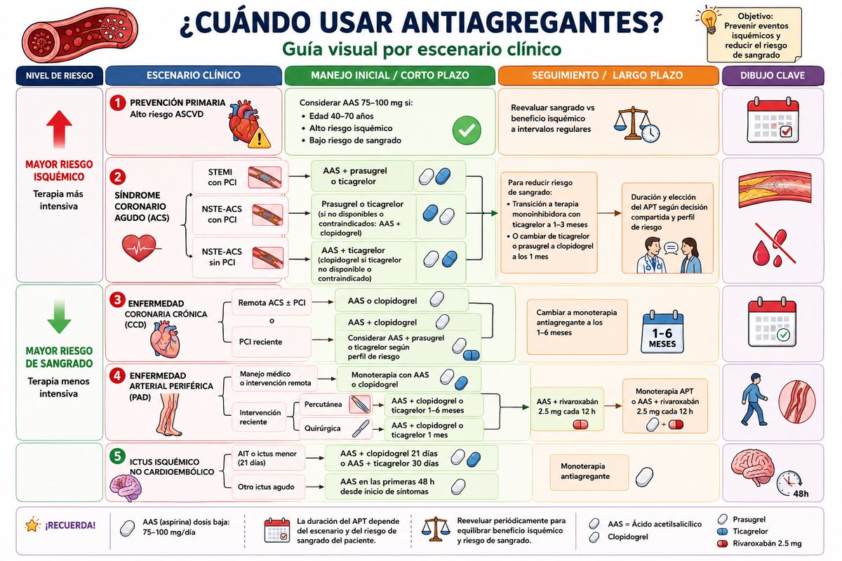
💊¿Prasugrel, ticagrelor o clopidogrel tras una PCI?
Parecía que el asunto de los antiagregantes plaquetarios había quedado claro, pero un nuevo metaanálisis con casi 49.000 pacientes abre de nuevo el debate 🔥🔥🔥
Echemos un vistazo👇
Llevamos años obsesionados con bajar los triglicéridos. Nueva revisión ➡️los TG no son directamente aterogénicos. Son el marcador de las lipoproteínas remanentes ricas en TG, que sí entran en la pared arterial. 🫀
Lo importante no es la cifra, es la carga de partículas apo B
@ESC_Journals
🔗 https://t.co/PfieuYrQSa
Score de Calcio Coronario. 🔎🫀💥
🔹️El Score CAC sigue siendo una de las herramientas más potentes para la estratificación de riesgo cardiovascular en prevención primaria. La evidencia reciente reafirma su utilidad para reclasificar riesgo más allá de los calculadores tradicionales y optimizar decisiones terapéuticas. 📈❤️🔥
🔹️Un sCAC = 0 se asocia con tasas muy bajas de eventos CV a corto y mediano plazo. Permite diferir estatinas en pacientes cuidadosamente seleccionados sin potenciadores importantes de riesgo. ✔️
🔹️Un sCAC >100 generalmente identifica pacientes con riesgo CV suficiente para justificar tratamiento intensivo con estatinas. 💊‼️
🔹️CAC ≥1,000: un nuevo fenotipo de muy alto riesgo. Presentan tasas de eventos comparables a algunas cohortes de prevención secundaria. Requieren control agresivo de LDL-C y seguimiento estrecho. ⚠️🔥
🔹️Otras ventajas: estudios recientes muestran que el sCAC predice mejor eventos coronarios que los polygenic risk scores. Mejora la adherencia, visualizar las calcificaciones coronarias incrementa el apego a cambios en el estilo de vida y al tratamiento farmacológico. ✍️🏻💊
📄🆓️⤵️
DOI 10.3389/fradi.2026.1822303
https://t.co/yZSdiacTI2
Los GLP-1RA reducen las lipoproteínas remanentes postprandiales (apoB48↓, apoCIII↓) de forma independiente del peso.
Y ese remanente postprandial predice MACE mejor que el LDL en ayunas.
Review imprescindible en Circ Res 2026. 📄
https://t.co/CaWEOpNDb0
¿Y si parte del beneficio cardiovascular de los GLP-1 ocurriera dentro de la placa?
➡️Reducen citoquinas (TNFα, IL-6, IL-1β) y moléculas de adhesión, suprimen el inflamasoma NLRP3 y favorecen el flujo de colesterol → placa más estable.
Más allá de la glucosa y el peso
👇 🔗 https://t.co/jVDCknfmi8
Honoring the Giants Who Shaped Modern Cardiac Electrophysiology
Today, I want to pay tribute to two extraordinary pioneers whose groundbreaking work laid the foundation for our understanding of the heart’s electrical system:
Dr. Brian F. Hoffman — A true legend in basic cardiac electrophysiology. Through his pioneering experiments on canine hearts, Dr. Hoffman mapped the anatomical origins of the EKG’s P-R interval and advanced our knowledge of specialized conduction tissues. He is widely regarded as one of the greatest electrophysiological scientists of our time and a key pioneer in invasive heart technologies.
Dr. Anthony N. Damato — Who took foundational basic science and translated it into clinical practice.
Dr. Damato made critical contributions by refining our understanding of the PR interval and developing techniques that enabled the recording of His bundle electrograms in humans — a milestone that essentially launched the field of clinical cardiac electrophysiology.
If the third industrial revolution had happened in the 1960s, Dr. Brian F. Hoffman would most likely have been the inventor of the new ECG.
Their combined legacy continues to influence every ECG we read today. We stand on the shoulders of these remarkable scientists. Their work reminds us that true innovation is built on deep scientific understanding and continuous advancement. Progress in cardiology is only possible because of the visionaries who came before us.
#Cardiology #Electrophysiology #ECG
🫀 CardioNugget
Disproportionate vs Proportionate Secondary MR
➡️ LVEDV (size) vs EROA (severity)
🔴 Disproportionate MR (COAPT)
Smaller LV, high EROA
MR out of proportion to LV dilation
👉 MR is a driver of HF
✅ TEER → benefit
⚪ Proportionate MR (MITRA-FR)
Larger LV, moderate EROA
MR matches LV dilation
👉 MR is a bystander
❌ TEER → limited benefit (focus GDMT)
🟢 Key Takeaway
👉 Same EROA ≠ same disease
👉 Always interpret MR relative to LV size
💡 Memory line:
“Out of proportion → fix the valve
In proportion → fix the ventricle”
#CardioNuggets #MedED #TEER #CardioMed
CAD with Atrial Fibrillation & RBBB
Is ECG Useful? Undoubtedly, yes. No instrument can replace it. However, for more than 120 years, the images recorded by conventional ECG have remained unchanged. 99% of them are high-frequency convoluted waveforms.
Coronary Artery Disease (CAD) was discovered in 1973 and officially named by Harvard in 1976. Yet in clinical practice, doctors face enormous challenges. AMI/CAD/ACS accounts for more than 60% of cardiovascular diseases — even higher in some countries. Doctors frequently encounter cases where the ECG clearly shows a CRBBB pattern, but the final diagnosis is CAD, with coronary angiography (CAG) revealing stenosis or occlusion greater than 70%. The issue is not that the ECG image is poor, but that its resolution has been insufficient for over 120 years.
The new scientific discipline of “signal processing” — a key technology of the Third Industrial Revolution — only developed after the year 2000. Unfortunately, it has not been applied to traditional ECG image processing.
Traditional ECGs do capture all the signals through convolution, but unfortunately, the fine details are not displayed. It is similar to how X-ray and CT images of the same patient contain the same natural anatomical elements, yet reveal different diagnostic information depending on the modality. The same principle applies to traditional ECG versus new-generation ECG — the difference lies in the level of image detail.
Please refer to the attached Figure showing ECG images of 4 patients taken before their CAG results.
PhysioSign USA
La metformina no deja de sorprendernos. 🔬
Nuevo estudio: su diana no es el hígado. Es el intestino.
Lo convierte en una esponja que capta el exceso de glucosa.
70 años después, seguimos aprendiendo. Larga vida a nuestra reina 👸🏻 metformina
@NatMetabolism
🔗 https://t.co/0VMhOGDJYB