Celebrating 100 th Anniversary of Emil Kraepelin 1856-1926 with his Great Great Grandson Dr Christian Schmidt Kraepelin. Emil Kraepelin holds the position of the Father of Biological Psychiatry. #Kraepelin#Psychiatry#MentalHealth https://t.co/c7jVZISLWi
Any condition receives status of a disorder when from persons own account, his family, environment the given behaviour is effecting patients life significantly. This is an alarming issue in western countries , even in Pakistan once in a while such case comes up. #GamingAddiction https://t.co/NxuaPZQLsR
Many people think of the genome as a string of "letters." The human genome, say, has 3.2 billion base pairs of DNA organized across 23 pairs of chromosomes.
But the genome is a 3D object. Genes located on entirely different chromosomes might be clustered together. Mutations in these "distant" genes can lead to disease in surprising ways.
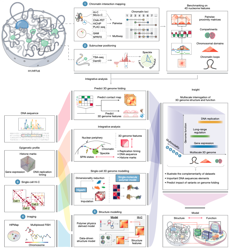
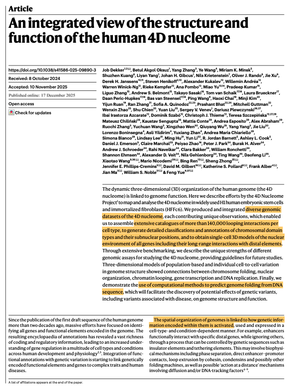
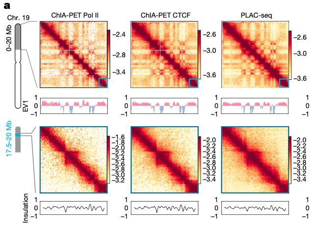
For a new paper in @Nature, researchers released several "maps" of human genomes from two types of cells: embryonic stem cells and fibroblasts. They compared methods to see which ones are least biased, and found many long-range interactions between genes.
The article does a good job explaining how “the genome is organized at different scales”:
> On a single chromosome, histones control which parts of the DNA sequence are accessible and expressed.
> At the scale of hundreds of thousands of bases, “chromatin loops in a dynamic manner,” the authors write, bringing distant genes closer together. > Across chromosomes, sequences "cluster together in space to form subnuclear compartments."
Examples abound. Enhancers, for example, are short DNA sequences that regulate the expression of far away genes. They do this by *physically* touching the genes they control; a protein called cohesin grabs the DNA and tugs it into big loops.
Even promoters, which are thought of as being associated with one gene or operon, can cluster together across many genes! A protein, Ronin, grabs promoters and pulls them together. This is apparently done mostly for genes that tend to be "on," as it helps enzymes find genes faster/not have to diffuse far away to find targets. (This also happens with genes that tend to be "off;" so-called polycomb proteins grab onto promoters, cluster them up, and silence all of them at once. It's a way for the cell to conserve energy.)
One consequence of this spooky "action-at-a-distance" is that diseases might arise from mutations in unexpected locations. Editing these regulatory sequences, in other words, might in turn affect a gene located on an entirely different chromosome that *is* associated with that disease.
Genetic mutations linked to autism, for example, are known to disrupt the 3D organization of the genome. A single deletion at a gene, TAL1, also affects its ability to form long-range chromatin interactions with other genes, leading to leukemia. There are probably many other, as-yet-undiscovered, instances of this.
Many people think of the genome as a string of "letters." The human genome, say, has 3.2 billion base pairs of DNA organized across 23 pairs of chromosomes.
But the genome is a 3D object. Genes located on entirely different chromosomes might be clustered together. Mutations in these "distant" genes can lead to disease in surprising ways.
For a new paper in @Nature, researchers released several "maps" of human genomes from two types of cells: embryonic stem cells and fibroblasts. They compared methods to see which ones are least biased, and found many long-range interactions between genes.
The article does a good job explaining how “the genome is organized at different scales”:
> On a single chromosome, histones control which parts of the DNA sequence are accessible and expressed.
> At the scale of hundreds of thousands of bases, “chromatin loops in a dynamic manner,” the authors write, bringing distant genes closer together. > Across chromosomes, sequences "cluster together in space to form subnuclear compartments."
Examples abound. Enhancers, for example, are short DNA sequences that regulate the expression of far away genes. They do this by *physically* touching the genes they control; a protein called cohesin grabs the DNA and tugs it into big loops.
Even promoters, which are thought of as being associated with one gene or operon, can cluster together across many genes! A protein, Ronin, grabs promoters and pulls them together. This is apparently done mostly for genes that tend to be "on," as it helps enzymes find genes faster/not have to diffuse far away to find targets. (This also happens with genes that tend to be "off;" so-called polycomb proteins grab onto promoters, cluster them up, and silence all of them at once. It's a way for the cell to conserve energy.)
One consequence of this spooky "action-at-a-distance" is that diseases might arise from mutations in unexpected locations. Editing these regulatory sequences, in other words, might in turn affect a gene located on an entirely different chromosome that *is* associated with that disease.
Genetic mutations linked to autism, for example, are known to disrupt the 3D organization of the genome. A single deletion at a gene, TAL1, also affects its ability to form long-range chromatin interactions with other genes, leading to leukemia. There are probably many other, as-yet-undiscovered, instances of this.
THE CELL’S POWERHOUSE
Mitochondria (yellow) make much of the chemical energy that fuels the life of a cell. They also move rapidly as seen in this cancer cell compared to the relatively slow moving nucleus (blue) and cellular adhesions (purple).
#MitoMonday#CellBiology
Chemistry writes the story of life.
Every cell, every second, it tells it again - powered by molecular design.
At the core of this process lies the citric acid cycle, the central hub of metabolism.
Here, acetyl-CoA derived from carbohydrates, fats, or proteins enters a precise series of reactions that convert fuel into energy.
Each step transfers electrons, drives ATP synthesis, and sustains the continuous renewal of life at the cellular level.
What may seem invisible is, in fact, the most constant motion in existence; the quiet rhythm of biochemistry that powers everything we do.
The model of gene expression taught in school is highly misleading!
Transcription factors are proteins that bind to DNA and then help repress, or activate, the expression of genes. Cells have hundreds of different types of transcription factors, each tuned to regulate different genes based on short snippets of DNA located near those genes.
The basic model, taught in school, says that these transcription factor proteins float around the cell and, when they bump into a DNA sequence, either latch onto it strongly (CORRECT SITE!) or fall off quickly (WRONG SITE) and keep searching. All the other DNA in a cell is basically abstracted away as unimportant or irrelevant; mere background noise.
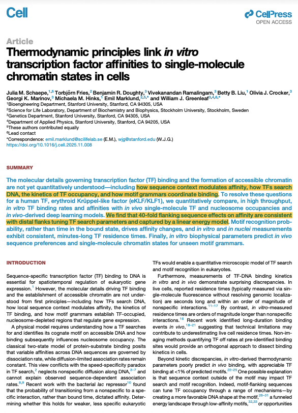
But again, this model is naive! And a new paper, published in Cell, beautifully shows how the sequences SURROUNDING a transcription factor's binding site also matter a great deal.
This won't be surprising to many biologists, as "cracks" in the standard two-state model began emerging decades(?) ago. Biologists have tagged transcription factors with fluorescent tags and then watched them move around living cells. And they have noticed that when transcription factors land in a "wrong" location in the genome, they skip or hop to a nearby location and repeat this until finally connecting with the "correct" sequence. So in other words, there are actually three states that a transcription factor can exist in: free-floating, "searching", or "bound."
(More technically, transcription factors first do a 3D search, then latch onto DNA and do a 1D search to find the correct location.)
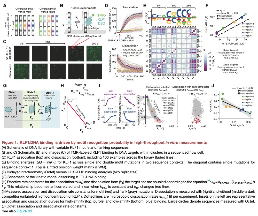
For this new paper, though, scientists exhaustively quantified *how* the sequences flanking a transcription factor binding site influence the search of the protein.
They did a huge in vitro experiment, wherein they placed a specific transcription factor with a known binding site, called KLF1, in a huge library of 11,812 different DNA sequences. These sequences had mutated "core" binding sites and variations in the flanking sequences. They also prepared negative controls. Then, these researchers measured the binding kinetics of KLF1 with each sequence to understand which bases in the flanking sites impact the 1D search.
What they found is that KLF1 has a basically flat disocciation rate from its core sequence, but that the PROBABILITY that it finds this sequence depends a lot on the surrounding context. Even mutations located dozens of bases away from the core site matter a lot, either pushing KLF1 to "hop" faster to find the site, or "trapping" KLF1 and slowing down its search. These flanking sequences can cause up to a 40-fold variation in the affinity of a transcription factor for its target site!
This is just one small part of the paper, though, so I encourage anyone interested to read the whole thing. It is challenging throughout.
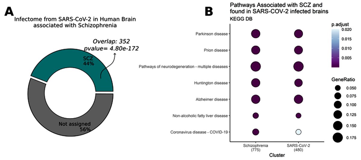
Molecular overlaps of Neurological manifestations of COVID-19 and schizophrenia. 🚨
👉The COVID-19 brain proteome enriches processes that are hallmark features of schizophrenia
👉Shared and distinct molecular pathways were identified in both conditions.
👉Brain ageing processes are likely to present in both COVID-19 and schizophrenia, though possibly driven by different mechanisms
👉Alterations in brain cell metabolism:
-Schizophrenia primarily impacts amino acid metabolism.
-COVID-19 predominantly affects carbohydrate metabolism.
👉Metabolic pathways associated with astrocytic components are enriched in both conditions, suggesting astrocytes' involvement in pathogenesis.
👉Both COVID-19 and schizophrenia influence neurotransmitter systems but with distinct impacts.
Reference: https://t.co/TpJLuW5LFh
#Covid
The cycle of life, every instant, in your mitochondria.
The major electron carrier in mitochondria is NAD+. When it collects an electron from the food you eat, NAD+ is converted into NADH. NADH then feeds the electron to the electron transport chain so the electron can flow to oxygen - the ultimate electron acceptor.
How easily electrons make their way to oxygen determines energy resistance (éR) following the energy resistance principle (ERP) https://t.co/eAL6i73l09
Mitochondrial matrix and cell cytoplasm are two different "sealed" compartments, but there is a special system that connects the NAD+/NADH pools between mito and cytoplasm: the Malate-Aspartate shuttle
The Malate-Aspartate shuttle shown below is how NADH from the cytoplasm (made by glycolysis) is carried into mitochondria. The electron transport chain can then regenerate NAD+ by respiring the flowing electron.
Everywhere in biology: energy transfer. Sometimes without molecular carriers. Here with a number of enzymatic intermediates, creating an integrated electrical circuit.
The circuit is in service of the flowing electron looking for a place to rest. Getting back onto oxygen to become H2O (water) again, as it was initially before getting ripped off by light energy and stuck onto a carbon backbone in a green leaf.
Animation by @janetiwasa lab
https://t.co/V7R07dVJbi
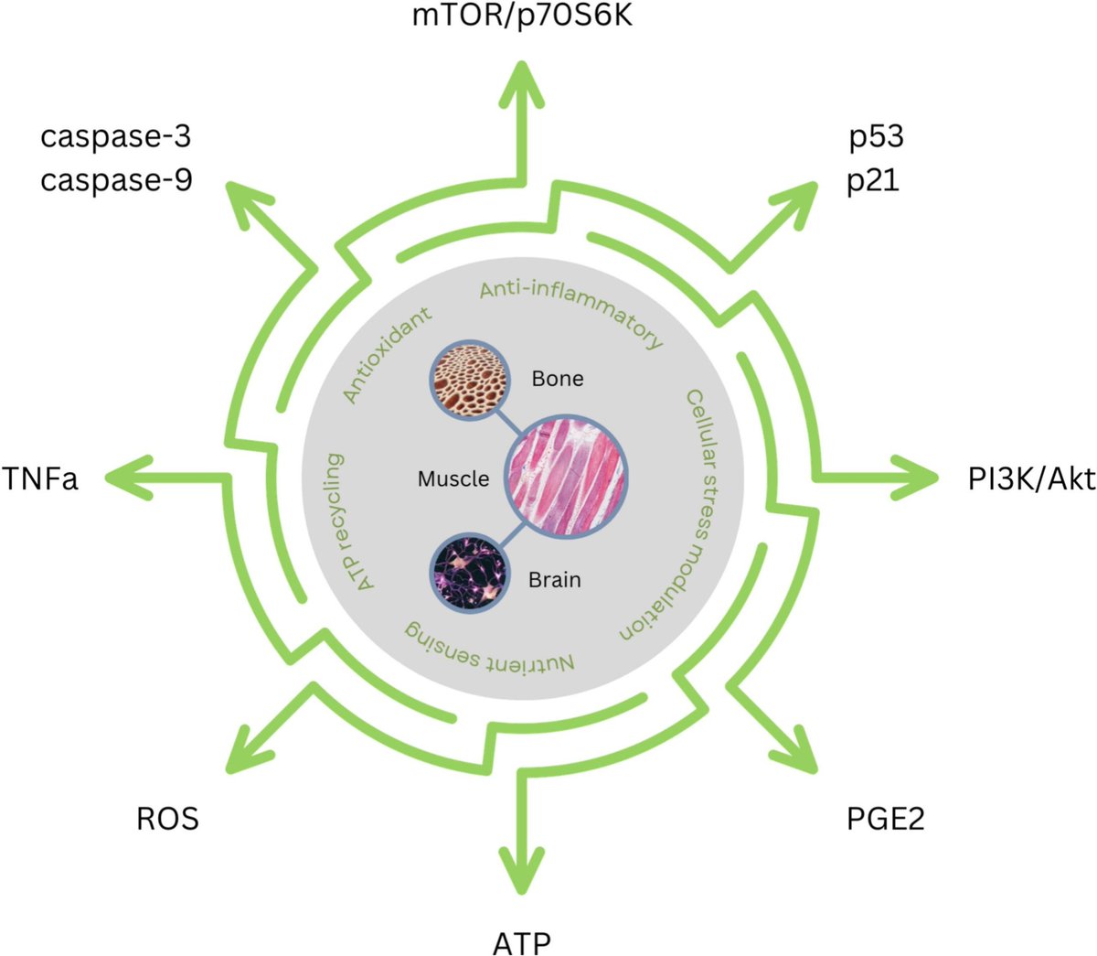
Creatine & cellular senescence: from molecular pathways to populational health
▶️Creatine holds significant promise as a therapeutic agent for mitigating cellular senescence & its associated conditions.
▶️By addressing key drivers of senescence, such as mitochondrial dysfunction, oxidative stress, & inflammation, creatine supplementation may offer a novel approach to promoting healthy aging & managing age-related diseases.
https://t.co/EGzjxHG4Vg @_atanas_@drfherediaz@DrGrimaldesJ@DrRPalmquist@DHPSP
Key tissues & molecular targets for creatine in senescence biology & therapeutic applications:
Class I HDAC inhibitors markedly improve persistence and antitumor efficacy of CAR-T cells by regulation of the HDAC1-H3K27ac axis and activation of the canonical Wnt/β-catenin signaling pathway @CellReports
https://t.co/iF7EWZ1XZx
Wrong. Biomarkers in Melancholic Depression.
So what do the ‘biomarkers’ tell us? (Spoelma et al, 2023)👇
1. Distinctive Symptom Pattern:
👉It presents with a specific set of symptoms and signs ( e.g psychomotor change, ruminative thinking etc )
2. ⬆️Response to Biological Treatments:
👉Preferential response for physical treatments, such as medications and electroconvulsive therapy, over psychological interventions.
3. Biological factors Over Psychosocial:
👉Genetic and biological factors are more significant in melancholic depression than psychosocial influences, marking a critical distinction from non-melancholic depression.
✅Biomarkers and Illness Correlates:
1. Dexamethasone Suppression Test (DST-Related Variables) :
👉Including cortisol non-suppression and elevated cortisol levels
2. HPA dysregulation :
👉further links to inflammation and elevated inflammatory markers
3. Reduced REM Latency :
👉Poor sleep quality
4. Cognitive Domain Involvement:
👉Reduced activity in the prefrontal cortex (PFC) and subgenual anterior cingulate cortex (ACC).
Note how these markers are directly associated with clinical symptomatology 👇
1. HPA Axis Dysfunction:
👉Higher rates of cardiovascular disease, cerebrovascular involvement and other inflammatory conditions.
👉You may remember how elevated CRP acts as a marker to consider Broad Spectrum Antidepressants✅
2. Sleep Dysfunction:
👉REM dysfunction linking to poor sleep quality and early morning awakening
3. Cognitive Impairment:
👉PFC and ACC involvement reflected via executive dysfunction, negative emotional appraisal of events and ruminations / overvalued ideation.
Which makes complete sense why the authors write 👇
“Perhaps the most valid measures are ones incorporating illness correlates as well as clinical symptoms.” ✅
Our brain isn’t just made of neurons. It runs on a whole neighborhood of cells that can either protect your mind or quietly push it toward inflammation.
This diagram reveals how astrocytes, microglia, neurons and oligodendrocytes talk to each other during stress, illness, and injury. Their conversations shape memory, mood, cognition, and long-term brain resilience.
Here is what this graphic shows in plain language:
🧠 Astrocytes act as the central switchboard
They decide whether the brain environment becomes supportive or inflammatory. When they sense danger signals, they activate NF kappa B and release molecules that influence the other cells around them. When conditions are safe, they release factors that help neurons grow and help new oligodendrocytes mature.
🔥 Microglia can protect or damage depending on the signals they receive
They can release IL 1 beta, GM CSF, and other proinflammatory signals, or they can shift to a more supportive state depending on what they detect from astrocytes and neurons.
⚡ Neurons suffer when inflammation rises
Oxidative stress, nitric oxide, and loss of metabolic support weaken them. Glutamate handling becomes impaired, which increases excitotoxic stress.
🧩 Oligodendrocytes and their precursor cells respond to what the environment tells them
Inflammatory signals slow their support for neurons, while regenerative signals encourage new myelin formation and better neuron stability.
The health of your brain depends on how these cells interact, not just on neurons alone.
doi: 10.1126/scitranslmed.adi7828
Reference brain map as a resource for marrying neurobiology to complex biological systems data in a new model organism the clonal raider ant
📹 Dominic D. Frank & Lindsey E. Lopes et al @RockefellerUniv in @CurrentBiology
➡️ https://t.co/LI9fkAbzu8
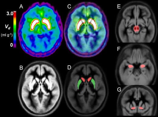
🧵How neurobiology connects to clinical practice in psychosis 🚨1/9
A recent 18F-DOPA PET Study
shows why psychotic depression and schizophreniform psychosis require such different treatment strategies.
The study concluded:
“Dopamine function differed across psychotic disorders when different mood states were present, particularly in the limbic striatum. Transdiagnostically, positive psychotic symptoms were associated with dopamine synthesis capacity in the associative striatum.”
( Jauhar et al,2025)
This matters for how we treat.
Let’s link phenomenology → striatal neurobiology → pharmacology. 👇
Studies suggest that up to 50% of adults with ADHD meet criteria for an anxiety disorder (Fu et al., 2025).
Anxiety is not simply “comorbid”; it is embedded in ADHD’s neurobiology and developmental trajectory.
Let’s explore how anxiety and ADHD intersect, and why recognising this link can improve diagnostic clarity, treatment planning, and patient outcomes. 👇🧵
Note: image is a conceptual illustration (uncertainty ↔ arousal ↔ anxiety), not a validated biomarker/model.
The risk of major depressive disorder (MDD) is determined by the combination of biological susceptibility and the risk or protective factors present in the environment.
Improving our understanding of the neurobiology of MDD is essential in developing new and personalized treatment options for individuals living with MDD.
Download and share our educational slide deck developed by Professor Dr. Elisabeth Binder (Germany): https://t.co/sU3RtoiU1I
#depression #mentalhealth