The L-Wave Sign 📈
🩺 What is the L-Wave Sign?
The L-wave is a mid-diastolic transmitral flow wave seen on pulsed-wave Doppler, occurring after the E wave and before the A wave.
Instead of the normal E���A pattern, you may see:
E → L → A
📍 Where is it seen?
Thread 👇
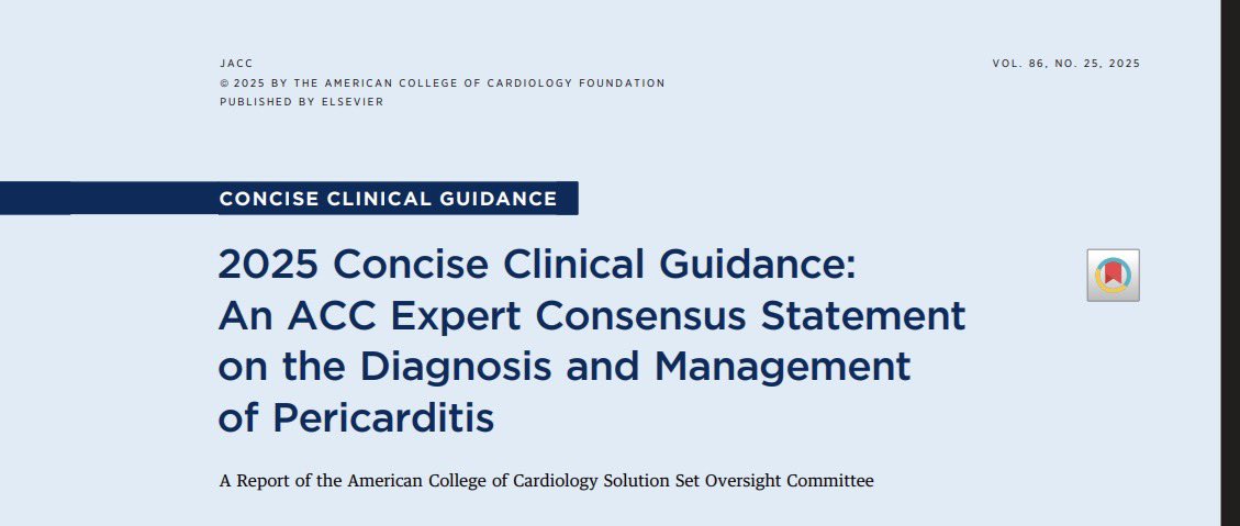
🚨 LA PERICARDITIS HA CAMBIADO.
Ya no se trata igual a todos los pacientes.
El manejo moderno depende del fenotipo biológico:
🔥 Inflamatorio
🧬 No inflamatorio
Identificarlo cambia el tratamiento, evita recaídas y reduce el abuso de corticoides.
⬇️
🫀Congestion is the disease. Diuresis is only the treatment.
For decades, the success of acute heart failure therapy has been measured by urine output, weight loss, or symptom relief. However, these surrogate endpoints do not necessarily indicate that congestion has resolved. Residual congestion at discharge remains one of the strongest predictors of rehospitalization and mortality.
This review proposes a more standardized and physiology-based strategy for decongestion. The objective is not simply to increase urine output, but to achieve effective natriuresis, restore euvolemia, and eliminate congestion before discharge.
The first message is urgency. Intravenous loop diuretics should be administered within 60 minutes of hospital arrival whenever acute heart failure with congestion is diagnosed. Delays may reduce the effectiveness of treatment and worsen outcomes.
The second message is even more important. Early treatment response should be measured objectively. Rather than waiting for body weight to change over several days, clinicians should evaluate:
• Spot urinary sodium after 2 hours, targeting ≥50to70 mmol/L
• Urine output after 6 hours, targeting ≥100to150 mL/hour
Failure to achieve these targets should prompt early intensification of therapy instead of continuing an ineffective regimen.
The review also supports the growing concept of early sequential nephron blockade. Randomized trials such as ADVOR and CLOROTIC demonstrated that adding acetazolamide or thiazide therapy to loop diuretics can improve decongestion in selected patients, particularly when diuretic resistance develops. Acetazolamide appears especially useful in patients with elevated serum bicarbonate, a marker of increased proximal sodium reabsorption and neurohormonal activation.
Another clinically relevant finding is that continuous loop diuretic infusion offers no clear advantage over intermittent bolus administration, consistent with the DOSE trial. Success depends more on achieving an adequate natriuretic response than on the method of administration.
Perhaps the most important lesson is that patients should not leave the hospital while still congested. Successful decongestion must be followed by rapid optimization of guideline directed medical therapy and early outpatient follow up, as demonstrated by the STRONG HF strategy.
Modern heart failure management is evolving from simply prescribing diuretics toward measuring the physiological response to therapy and adapting treatment early. Congestion should no longer be treated empirically. It should be monitored, quantified, and resolved before discharge.
Reference 📚
Bilgeri, V., Spitaler, P., Puelacher, C., et al. (2024). Decongestion in Acute Heart Failure: Time to Rethink and Standardize Current Clinical Practice? Journal of Clinical Medicine, 13(2), 311. https://t.co/fic1FPNt7x
The pulmonary valve isn’t called the “neglected valve” for nothing.
In this sexagenarian with a BAV, it’s easy to focus all your attention on the aortic valve - and miss the pulmonary valve pathology.
Don’t forget to look beyond the obvious. #echofirst
🕵️♂️ intraop TEE
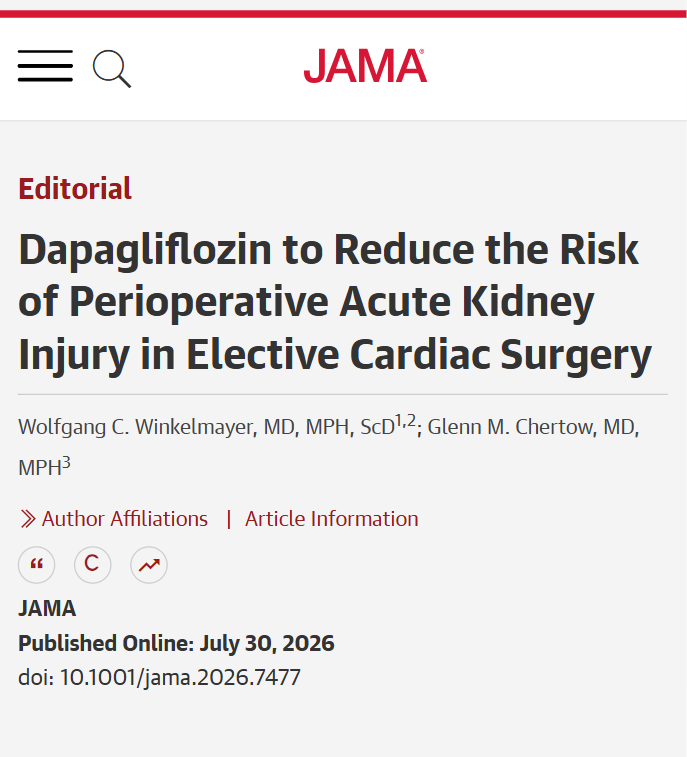
💬 Editorial by Wolfgang Winkelmayer, MD, MPH, ScD, and Glenn Chertow, MD, MPH: Perioperative #dapagliflozin reduced postoperative #AcuteKidneyInjury after elective cardiac surgery, supporting further trials of sodium-glucose cotransporter 2 inhibitors for kidney protection.
https://t.co/eqeqOthmsp
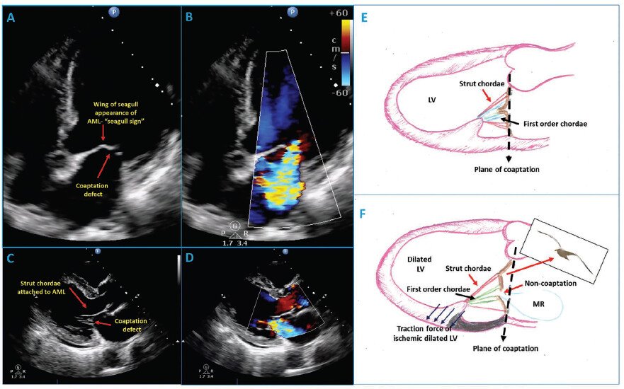
The Seagull Sign of Anterior Mitral Leaflet
🩺 What is the Seagull Sign?
In echo, the mitral valve seagull sign refers to a distinctive wing-like distortion or "kink" in the middle of the anterior mitral valve leaflet during systole, mimicking the wings of a flying seagull.
#JADEL
New in CASE @CASEfromASE:
https://t.co/y09aQHSCms
Emphasizing the value of MMI in assessment of heart failure, check out the latest finding in this low LVEF:
A. ILVNC (noncompaction)
B. HCM (hypertrophic)
C. DCM (dilated)
D. VHD (valves)
E. Other (cors)
@ASE360
El ECO puede AYUDAR a elegir candidatos a Terapia de Resincronizacion Cardiaca
No es solo el retraso eléctrico (QRS)…
Es la dissinconía mecánica la que importa
Qué buscar en el eco:
✅ Flash septal (SF) → contracción septal temprana + rebote
✅ Balanceo apical (ApRock) → movimiento bifásico del ápice
✅ Juntos = SFoAR = dissinconía mecánica
1️⃣ Adquisición multiviendo de alta calidad
2️⃣ Detectar flash septal
3️⃣ Detectar balanceo apical
4️⃣ Integrar → diagnosticar dissinconía
Por qué importa?
❗ 20–45% de los pacientes con CRT son no respondedores
Presencia de SF/ApRock = mayor probabilidad de respuesta
Evaluación visual = herramienta simple, reproducible, al lado de la cama
Dos conceptos: Eléctrico ≠ mecánico
(No es lo mismo)
El eco puede refinar la selección de pacientes más allá de las guías
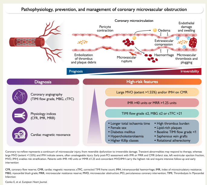
Consenso de la ESC (European Society of Cardiology) 2026, sobre Obstrucción Microvascular Coronaria (o fenómeno de no-reflujo). Lo más sintetizado que puedo:
🟢Fenómeno No-reflow coronario: perfusión tisular miocárdica inadecuada a pesar de la recanalización exitosa de la arteria coronaria epicárdica sin obstrucción mecánica. Frecuente y lo pensamos poco (hasta 60% post infarto con elevación del ST tienen perfusión subóptima). Es un problema microvascular que clínicamente puede evolucionar a falla cardíaca.
🟢 El predictor más importante para no-reflow: tiempo en isquemia (puerta-balón). Ser mujer también es un factor.
🟢 Es un marcador pronóstico importante. Un IMR (index of microvascular resistance) >40 casi triplica el hazard de muerte cardiaca. La hemorragia intramiocárdica triplica muerte.
🟢 Casi ninguna terapia farmacológica cambia el desenlace. Adenosina, nitroprusiato y calcioantagonistas mejoran marcadores angiográficos, no mortalidad. Aspiración de trombo rutinaria no se recomienda (riesgo de EVC).
🟢 Así que el enfoque es: diagnóstico temprano, reducir tiempo en isquemia, Estatina de alta dosis temprana, P2Y₁₂ potente, control de presión, glucemia y factores de riesgo.
Es un tema muy interesante. Pueden ver el artículo completo en el canal (https://t.co/3O93s10lns).
سؤال يحيرني...
إذا كان الكوليسترول لا يفرق بين شريان وآخر أثناء دورانه في الدم.... فلماذا تبدو شرايين القلب والدماغ هي هدفه الأول، بينما لا نرى شرايين العين تتأثر بنفس الكثرة رغم أنها أصغر وأدق وحيوية جداً؟
من يساعدنا في الإجابة
CardioNugget™:
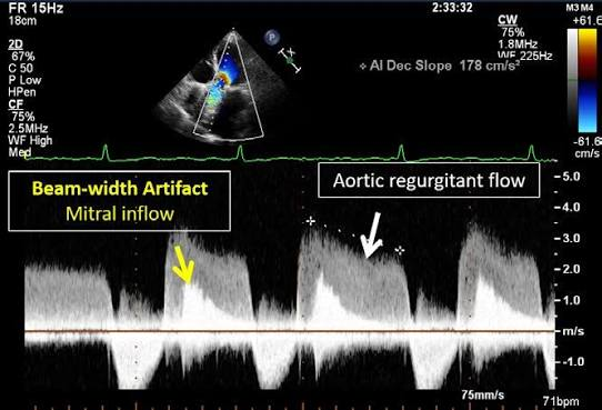
Beam-Width Artifact on CW Doppler
💡 Beam-width artifact = Doppler signal contamination from structures outside the intended ultrasound beam.
In this image:
The CW cursor is aimed at the aortic regurgitation (AR) jet.
Because CW Doppler has a finite beam width (not an infinitely thin line), it also intercepts the mitral inflow (E wave) adjacent to the LVOT.
The machine records both high-velocity AR flow and mitral inflow simultaneously.
Fix: Reposition the transducer or change the imaging window so the adjacent flow is excluded.
#CardioNuggets #MedEd #EchoBoards
Color Doppler demonstrates a left-to-right shunt
What is the most likely diagnosis?
A. Muscular VSD
B. Inlet VSD
C. Perimembranous VSD
D. Supracristal VSD