@rajivxgulati Agreed! If it was as simple as inflammation causing atherosclerosis we would be using steroids and nsaids to treat atherosclerosis! Rise in inflammatory markers is a bystander result of atherosclerosis i.e body trying to repair itself rather than the primary cause.
@Turtlesrhappy1@drjohnm Would you agree that the rise in inflammatory markers was a result of the body trying to repair the atherosclerosis rather than the causative reason of the atherosclerosis. The failure of the anti inflammatory drugs clearly shows this. The antioxidant brigade is a sham.
How long should anticoagulants be withheld before elective surgery?
◻️Apixaban: 1 day (low/moderate bleeding risk), 2 days (high risk)
◻️Rivaroxaban: 1 day (low/moderate), 2 days (high)
◻️Edoxaban: 1 day (low/moderate), 2 days (high)
◻️Dabigatran: 1-4 days depending on bleeding risk & renal function
◻️ Fondaparinux: 36-42 hours
◻️ LMWH: 12 h (prophylactic), 24 h (therapeutic)
◻️ UFH: IV 4-6 h; SC 12–24 h
◻️ Warfarin: Stop 5 days before surgery; confirm INR before the procedure.
Adjust timing for renal impairment, neuraxial anesthesia, and individual patient risk. Bridging is reserved for selected high-thrombotic-risk patients.
Reference: 2024 AHA/ACC Guideline for Perioperative Cardiovascular Management for Noncardiac Surgery, Table 13.
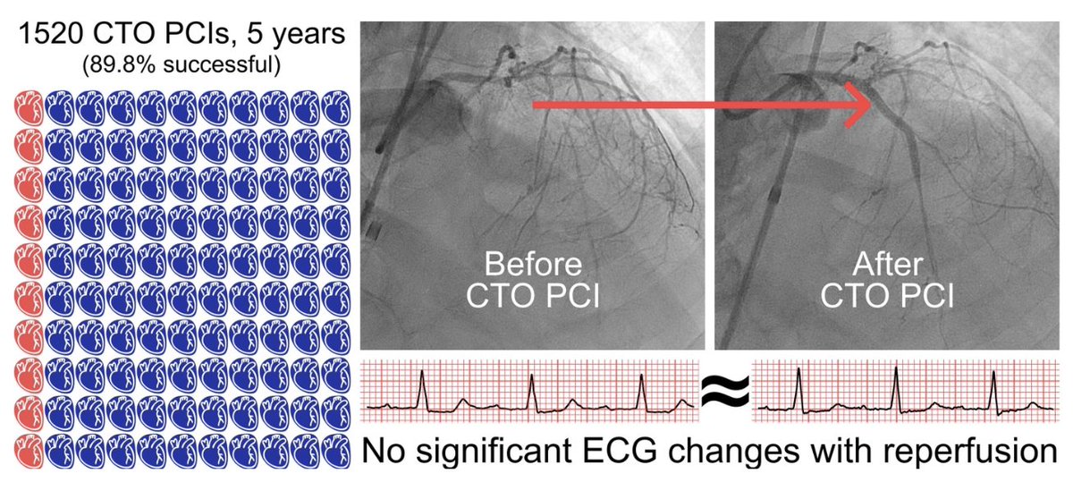
Can reopening a chronic total occlusion (#CTO) make the heart electrically less prone to #ventricular#arrhythmias?
For years, small studies suggested the answer was yes.
We analyzed 7,496 ECGs from 1,232 patients undergoing 1,520 CTO PCIs using automated Python-based signal analysis.
Here's what we found... 🧵
🆕 expert analysis provides a framework for antihypertensive medication selection in pregnancy and the postpartum period. Get the details: https://t.co/olA1uclOsT #Hypertension#CardioObstetrics
CardioNugget™ 🫀 | Noonan Syndrome
🧬 Think Noonan syndrome when you see pulmonary valve stenosis.
High-yield cardiac associations:
🔹 Pulmonary valve stenosis (most common, often with dysplastic leaflets)
🔹 Hypertrophic cardiomyopathy (second most common)
🔹 ASD, VSD, AV canal defects, and coarctation are less common.
💡 Board pearl:
If a child has short stature + webbed neck + hypertelorism + pulmonary stenosis, think Noonan syndrome (a "male Turner-like" phenotype—but with a normal karyotype).
📚 Remember:
Noonan = Pulmonary Stenosis + HCM
make#CardioNugget™ #EchoBoards #Cardiology #CardioTwitter #MedEd #FOAMEd #PedsCardiology #BoardsPrep
Answer: C. Perimembranous VSD
How to identify the different types of VSDs on echocardiography
VSDs are classified by their location within the interventricular septum. Recognizing the surrounding landmarks is the key to identifying each type.
🟧 Parasternal long-axis
- Perimembranous VSD: Located immediately beneath the aortic valve.
- Muscular VSD: Seen within the muscular (trabecular) septum, away from the valves.
🟥 PSAX at the aortic valve level
- Perimembranous VSD: Seen at the 10 o'clock position, adjacent to the septal leaflet of the tricuspid valve.
- Supracristal (outlet) VSD: Seen at the 1 o'clock position, just below the pulmonary valve.
🟦 PSAX at the ventricular level
- Muscular VSD: Located entirely within the muscular septum between the LV and RV.
🟩 Apical 4-chamber view
- Inlet VSD: Adjacent to the tricuspid and mitral valves.
- Muscular VSD: May be seen in the mid-muscular or apical septum.
An important clue is that patients with an inlet VSD caused by an endocardial cushion defect often have the tricuspid and mitral valves inserted at the same level, frequently with an associated primum atrial septal defect.
Reference: Otto CM. Textbook of Clinical Echocardiography
CardioNugget™ 🦀
The Crab View (Suprasternal Coronal View)
📍 How to obtain
Start in the suprasternal notch
From the long-axis aortic arch ("candy cane") view, rotate ~90° into the coronal plane and make slight tilt adjustments until the LA and pulmonary veins come into view.
👀 What you'll see
❤️ Left atrium = Crab's body
🦵 4 pulmonary veins = Crab's legs
🎯 Why it matters
✅ Confirms all pulmonary veins drain into the LA
✅ Detects Total anomalous pulmonary venous return and partial anomalous pulmonary venous return
✅ Evaluates pulmonary venous obstruction/stenosis
#CardioNugget™ #EchoBoards #CongenitalHeartDisease #ASE #CardioTwitter
Actual message received from a patient this morning. Her primary doctor ordered an Lp(a) which came back slightly above normal. We in medicine should consider the psychological harm that we impose on people through overmedicalization and overtesting. A new disease has been created and inflicted on the public (“Lp(a)itis”) for which there is no treatment. But Big Pharma assures us one is coming . . .
“I slept poorly last night. [My PCP] wrote me a note last night saying I had to tell all my first-degree relatives to get checked for Lp(a) and that I should have coronary calcium scoring. It’s weighing heavily on me that I could have passed this to my kids. Because there is nothing to do about it, I feel like it’s my death sentence.”
@DrMShujat 'Widaplik' triple combination is not new, We have been using triple combinations with ACEI/ARB for many years. Apart from aprocitentan and baxdrostat the third new approval in line is Entresto (valsartan +sacubitril) for hypertension (approved in UAE already).
🫀 Pulmonary Vein PW Doppler: A Hidden Gem in Diastology
Pulmonary vein pulsed-wave Doppler provides valuable insights into left ventricular filling pressures, left atrial function, and mitral regurgitation severity.
📍 How to Perform Pulmonary Vein PW Doppler?
Thread 👇
Most echocardiography laboratories have adopted the 2016 diastolic guidelines. With the release of the 2025 update, an important question arises:
What are the key conceptual differences between the 2016 and 2025 approaches to diagnosing diastolic dysfunction?
https://t.co/FPkUqRtmYd
@JGrapsa
📄 Can we finally measure RV volume accurately with simple echocardiography?
🔗 DOI: https://t.co/cHII0FgTLH
🫀 The right ventricle (RV) remains one of the most challenging chambers to assess—especially in congenital heart disease (CHD).
👉 Gold standard? Cardiac MRI
❗ But: expensive, time-consuming, and often requires sedation
✨ This study proposes a simple, fast, and accurate 2D echo-based method for RV volumetry—bringing us closer to true bedside quantification.
✨ The key idea:
👉 Model the RV using a cone-based geometric approach
➡️ Using only 2 standard echo views:
Apical 4-chamber (A4CH)
Parasternal short-axis (SAX)
📐 With just a few parameters:
✔ Cross-sectional areas (A4CH, ASAX)
✔ Tricuspid valve diameter
📊 Performance vs MRI:
🔥 Excellent agreement:
Systolic volume → ICC 0.98
Diastolic volume → ICC 0.96
📉 Minimal bias:
Δ systolic volume ≈ 0.1 mL
Δ diastolic volume ≈ 5.2 mL
➡️ Clearly outperforms traditional 2D models
💡 Why this matters clinically:
👉 Enables:
✔ Rapid bedside RV assessment
✔ Reduced need for repeated MRI
✔ Easier follow-up in paediatric & CHD patients
👉 Particularly valuable in:
Post-operative monitoring
Serial evaluations
Resource-limited settings
🚀 Key innovation:
👉 A mathematically robust yet practical model
➡️ Balancing accuracy + simplicity
➡️ Adaptable to different RV shapes
🚨 Bottom line:
2D echocardiography—when combined with smart modelling—can approach MRI-level accuracy for RV volumetry.
#Cardiology #Echocardiography #RightVentricle #CongenitalHeartDisease #CardiacImaging #CMR #Innovation #MedTech #PediatricCardiology 🫀📊
@PWesslyMD@LucySafi@PriyaPanday27@NadeenFaza@ASE360@JournalASEcho Prolapsing valves pull on chords/papillary muscles, stretching the LV wall inward. This causes "curling" (LV crest rotates posteriorly) & the Pickelhaube sign -- a sharp late-systolic S’ wave >16 cm/s on TDI. Both are markers of increased arrhythmia risk! #ASEchoJC