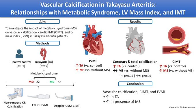
A recent study identified increased vein wall thickness (VWT) as a novel #vascular feature in systemic #lupus erythematosus (SLE), which may indicate subclinical venous involvement, regardless of antiphospholipid antibody or antiphospholipid syndrome status:
◦ VWT was notably increased in SLE patients compared to healthy controls and rheumatoid arthritis patients, emphasizing the unique vascular involvement in SLE.
◦ Portal vein wall thickness was independently associated with #thrombosis, suggesting its potential as a vascular risk marker.
◦⭡P-selectin, growth differentiation factor 15, and citrullinated histone 3 correlated with venous changes, highlighting biochemical markers of thrombo-inflammatory processes.
*Yildirim D, Erden A, Ibrahimkhanli N et al. A new characteristıc of systemic lupus erythematosus: subclinical vein involvement—an in-depth biochemical and imaging study. Rheumatology (Oxford), 2025.
🔗https://t.co/6n8vFV6mhF
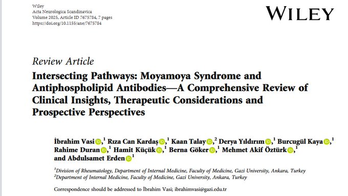
This raises an important question: should #APS be considered in the differential diagnosis for all patients presenting with #moyamoya-like findings? Is this speculation valid and supported by the evidence?
Alternatively, we aimed to explore whether the radiological findings resembling #moyamoya observed in #APS patients could represent ischaemic and haemorrhagic lesions caused by APS rather than true moyamoya syndrome.
Alternatively, we aimed to explore whether the radiological findings resembling #moyamoya observed in #APS patients could represent ischaemic and haemorrhagic lesions caused by APS rather than true moyamoya syndrome.
One of the critical objectives of our study was to determine whether the association between #moyamoya syndrome and conditions like #APS is coincidental or if, as suggested by the diagnostic criteria, these patients should indeed be classified as having moyamoya #syndrome.
One of the critical objectives of our study was to determine whether the association between #moyamoya syndrome and conditions like #APS is coincidental or if, as suggested by the diagnostic criteria, these patients should indeed be classified as having moyamoya #syndrome.
#CXCL-13 correlate with #SLEDAI scores, anti-#dsDNA antibodies, and #C3/C4 levels. Thus, CXCL-13 may serve as a promising diagnostic biomarker for #NPSLE’s challenging diagnosis and management. #SLE#LUPUS
🚨 New pub alert!
#CXCL-13 levels are significantly higher in #NPSLE patients—especially in active cases—than in those without #neuropsychiatric involvement and healthy controls. #SLE#LUPUS
https://t.co/t7gIz3V9Qt
Serum levels of #HMGB1, a nonhistone nuclear protein that has an important role in #psoriasis, are significantly higher in #patients with #psoriatic#arthritis than in patients with #psoriasis and healthy controls. @fsgbmm
https://t.co/mMjT29dR4N
Our study (#Colchicine-intolerant familial Mediterranean fever patients: A comparative study between different colchicine doses and #IL-1 inhibitor monotherapy) is published in the International Immunopharmacology. #FMF#anakinra#canakinumab https://t.co/6uSQqKRI12
#SLE is the most commonly associated disease with #APS, being present in approximately 35% of cases. Since the majority of the patient group in our study had #APS that was secondary to #SLE, non-criteria antibody positivity may be linked to the #immunological activity of #SLE.
Accepted for publication:
Demonstration of diffuse venulitis on MRV might help with diagnosis of Behcet's Disease in this case-control study.
https://t.co/chIGhQ6HfR
Paradoxical Rxns (PR) & biologics in TReasure database: 2867 RA & 5316 SpA pts; 5-yr FUV found 136 pts (1.7%) developed the PRs; 60% w/ 1st biologic, after median 12 mos, 93% psoriais (pustular 60%, palmoplantar 31%), many better w/ another TNFi https://t.co/5tybXZZVKW
Happy to share our editorial:
''The outcome of COVID‐19 in patients with autoimmune rheumatic diseases: Comparable to the general population or worse?'' @EzgiDenizBatu1 https://t.co/aE4Dv3oV4W