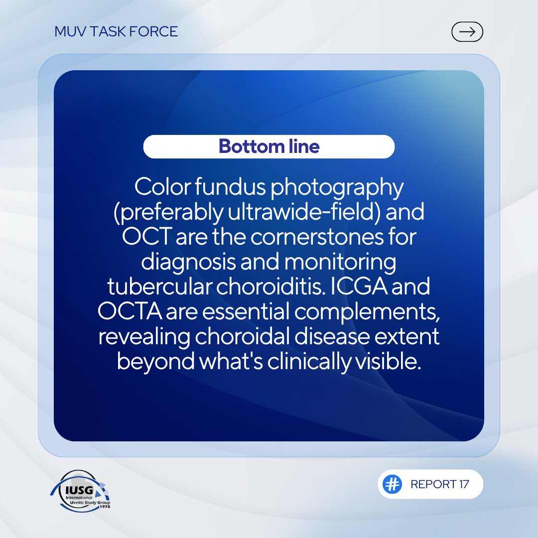
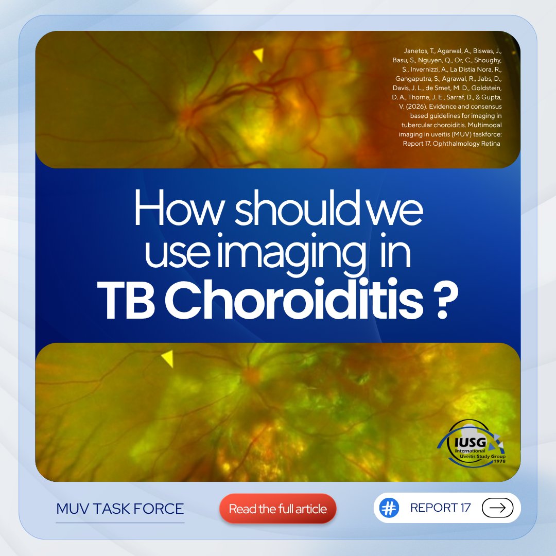
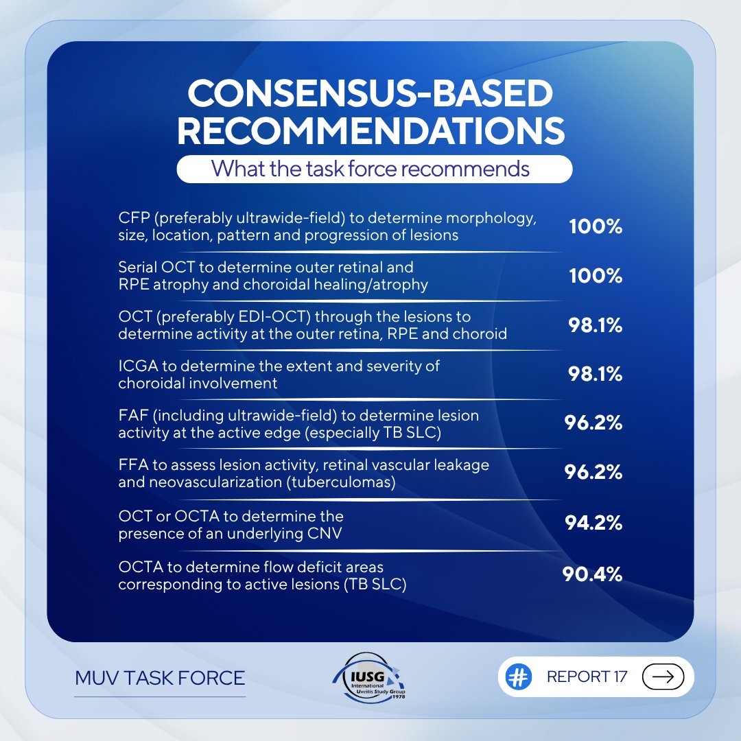
This report brings together a systematic review and structured consensus voting from an international expert panel with recommendations
Read the full report via the link, and follow the IUSG for future reports: https://t.co/moHf769WLh
#Uveitis#Retina#TubercularChoroiditis
The Multimodal Imaging in Uveitis (MUV) Taskforce has published Report 17: Evidence and Consensus-Based Guidelines for Imaging in Tubercular Choroiditis, now available in Ophthalmology Retina.
https://t.co/moHf769WLh
#IUSG#MUVTaskforce#multimodalimaginginuveitis
In 1958, this man’s heart kept stopping—20 times a day.
Doctors had no solution.
Then his wife demanded an experimental surgery that had only been tested on dogs.
What happened next changed medicine forever. 🧵
Be aware of how to identify Yasunari nodules in Neurofibromatosis Type 1 (NF1) using Near-Infrared (NIR) reflectance and Optical Coherence Tomography (OCT). NIR reflectance imaging highlights these nodules as hyperreflective spots, while OCT provides cross-sectional images
Retinal Detachment:
Separation: Involves the separation of the neurosensory retina from the underlying RPE.
Appearance: Often presents with a corrugated, wrinkled appearance of the retina. The detached retina may undulate with eye movements.
Mobility: The detached retina is usually mobile.
Location: Can occur anywhere in the retina, often starting at a tear or break.
Symptoms: Patients may experience sudden onset of floaters, flashes of light, and a shadow or curtain effect.
Hemorrhage: May be associated with retinal or vitreous hemorrhage.
OCT: Shows separation between the neurosensory retina and the RPE, with possible subretinal fluid.
B-scan: Useful in media opacities. It shows a detached, mobile retina with undulating movements.
Laser Uptake: The retina in a detachment typically shows good laser uptake. It creates a chorioretinal adhesion to seal retinal breaks.
Retinoschisis:
Separation: Involves splitting within the layers of the retina, typically between the outer plexiform layer and inner nuclear layer.
Appearance: The split retina appears smooth and taut without the corrugated appearance seen in retinal detachment.
Mobility: The schisis cavity is typically immobile.
Location: Commonly found in the peripheral retina, especially the inferior temporal quadrant. It often has a more stable, predictable configuration.
Symptoms: Often asymptomatic, but may cause gradual vision loss if it affects the macula or if the schisis cavity progresses.
Hemorrhage: Typically not associated with hemorrhage.
OCT: Reveals splitting within the retinal layers, with a schisis cavity and no separation of the entire retina from the RPE.
B-scan: May show a smooth, immobile, dome-shaped elevation of the retinal layers.
Laser Uptake: Retinal tissue in retinoschisis often shows poor laser uptake. This is due to the splitting within the retinal layers, which prevents effective transmission of the laser energy to the deeper layers where adhesion is needed. Photocoagulation is generally less effective and often not indicated for retinoschisis unless there is a coexisting retinal break or a risk of progression to RD. When applied, the laser spots may not achieve the same level of chorioretinal adhesion due to the schisis cavity.
Other imporatant features include:
History and Progression: Retinal detachment often has a sudden onset with a rapid progression, while retinoschisis tends to progress slowly and may remain stable for long periods.
Risk Factors: Retinal detachment is more common in myopia, trauma, or after ocular surgery, while retinoschisis is more common in older adults and may have a genetic predisposition.
#MedTwitter #MedEd #ophthalmology #retina
Images from google
SF6 COMPARED WITH C2F6 FOR INFERIOR RHEGMATOGENOUS RETINAL REPAIR
The Manchester Pseudophakic Retinal Detachment Study
Moussa, George FRCOphth; et. al.
Retina 44(5):p 791-798, May 2024
https://t.co/b0npYdk8Jq
#retina