Arrived in Finland and super excited for the start of #EED2024 and the amphioxus satellite meeting!
In the meantime, recovering from jetlag in a very sunny Helsinki! 😎
@thibaut_brunet So many memories of Claus in Kristineberg. It was amazing to look at the plankton with him, there was an untold story hidden in every single scoop.
Excited to see last Giacomo's @gattoni_giacomo work in the bioRxiv! Fun collaboration with Silvia Mercurio at Roberta Pennati's lab. Thank you all for your hard work, and giving the entire world another beautiful model system to work with 👏😀
Very excited to share this new paper, fruit of a great collab with Silvia Mercurio, out in preprint! 🎉
We set up new methods for in vitro culture and gene/protein detection in the feather star A. mediterranea and used them to investigate its development. ⭐️ Tweetorial below!
This feather star has it all: beautifully colored body, 10 arms, great at regeneration. Plus, it’s a #crinoid, the sister group to other living #echinoderms (starfish, sea urchin..), making it an ideal comparative system to understand how the puzzling echinoderm body plan evolved
We then optimized whole-mount immunofluorescence, which allowed us to study how the different tissues of the crinoid #larva develop. Here for example we labelled the skeleton (yellow) and cilia (green, note the five ciliary bands and apical tuft!) of the doliolaria larva
Next, we focused on our favorite, the nervous system, and looked at the distribution of serotonin, GABA and glutamate 🧠. We characterized the complexity of the larval apical organ (AO), and identified glutamatergic cells in the neural plexus
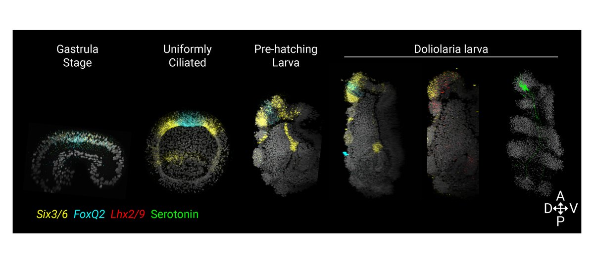
We then optimized in situ HCR (@HCRimaging) to test the molecular patterning of the crinoid AO. 🧬 We found that serotonergic neurons form in a domain co-expressing Six3/6 and FoxQ2 (from early development) and Lhx2/9 as in other echinoderms, supporting AO conservation
At metamorphosis, larvae settle from the anterior pole, neurons degenerate and a new nervous system develops. We showed that cells in the oral/vestibular ectoderm, where the ectoneural system will form, start to express anterior markers in the post-metamorphic cystidean stage!
This work has been an amazing collaboration between my PhD @CamZoology and Master’s @LaStatale labs, and I want to thank Silvia, both supervisors @eBGLab and Roberta Pennati, @MauriceElphick for all his help and support, all the authors, and all members of both labs! 😄
☕️NEW: Ton, Keitley et al. provide a rabbit development atlas and propose that combining rabbit and mouse atlases can help dissect early primate development.
@mailinh_ton@DanKeitley@MarioniLab@BertieGottgens@eBGLab
https://t.co/d4hHQFmjnJ
https://t.co/YX34QEZkni
Proudly presenting our new bioRxiv:
The cell type atlas of the regenerating and asexually reproducing annelid Pristina leidyi
We show that a piwi+ cell population with a pluripotent stem cell signature underlies adult differentiation
https://t.co/t6PwbYv8Et
A tweetorial 🧵👇
I want to thank my wonderful (and patient) labmate @DanKeitley, all the @eBGLab, @CamZoology, @CRUK_CI and the Cambridge #EvoDevo community for their help and support!
If you have any questions/comments, please let us know! 😄
Our work shows that an aGRN patterning the anterior neurectoderm is ancestral to bilaterians. In the chordate lineage the aGRN was incorporated into neurulation to specify and position the forebrain anteriorly, linking the evolution of the AO to that of the chordate brain.
10/10
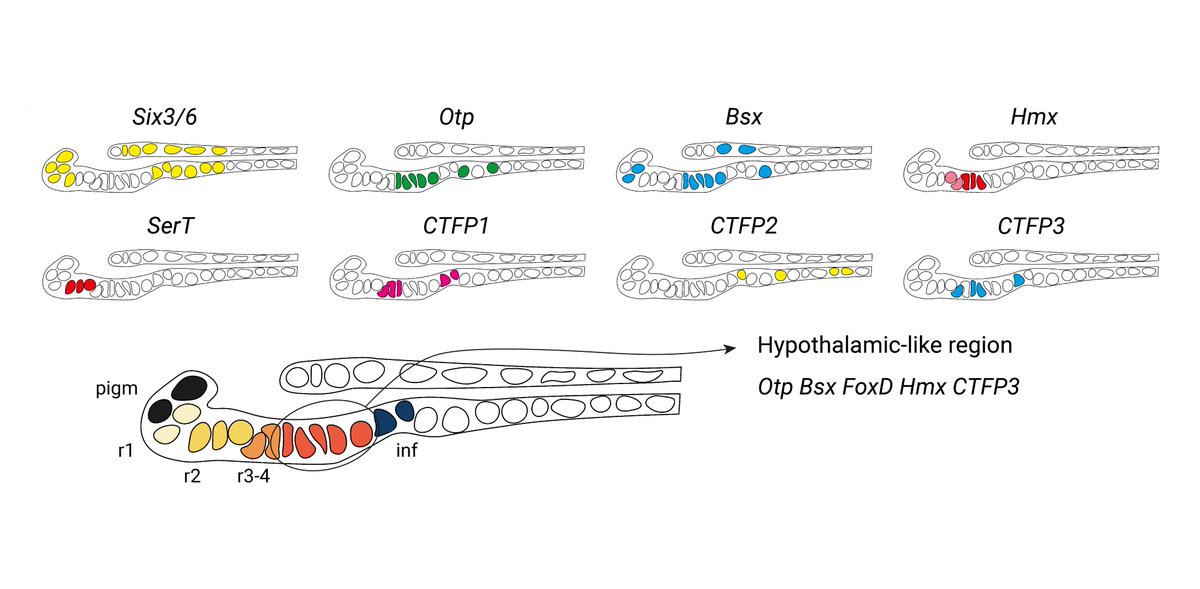
But what exactly is this anterior identity? Here we describe a hypothalamic-like region in the amphioxus larval brain expressing Otp, FoxD, Bsx, Hmx, miR-7 and calcitonin-type neuropeptides. It forms in the area where aGRN is active and is lost following Wnt overactivation.
9/10