It’s not good when your mandibular lymph node aspirate is grossly pigmented: metastatic melanoma in a 12 year old Beagle (arrows both images: neoplastic melanocytes; arrowheads both images: melanophages). The primary tumor was in the oral cavity.
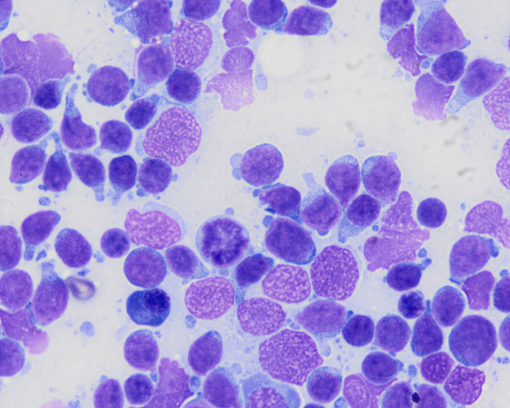
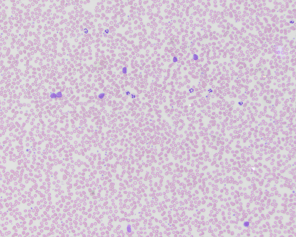
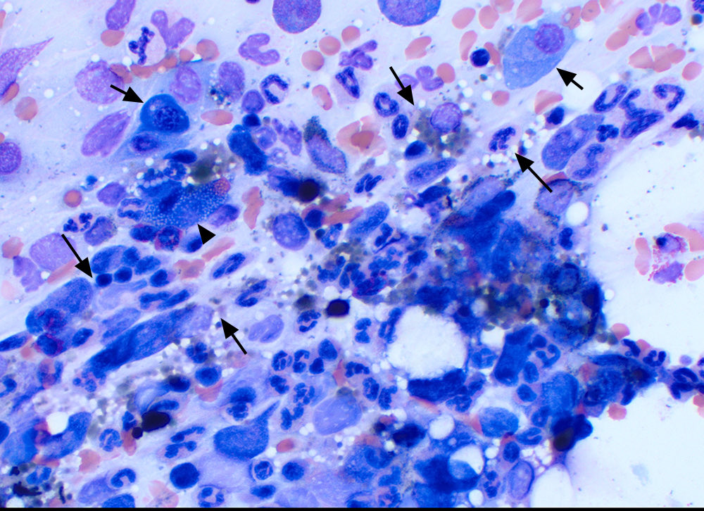
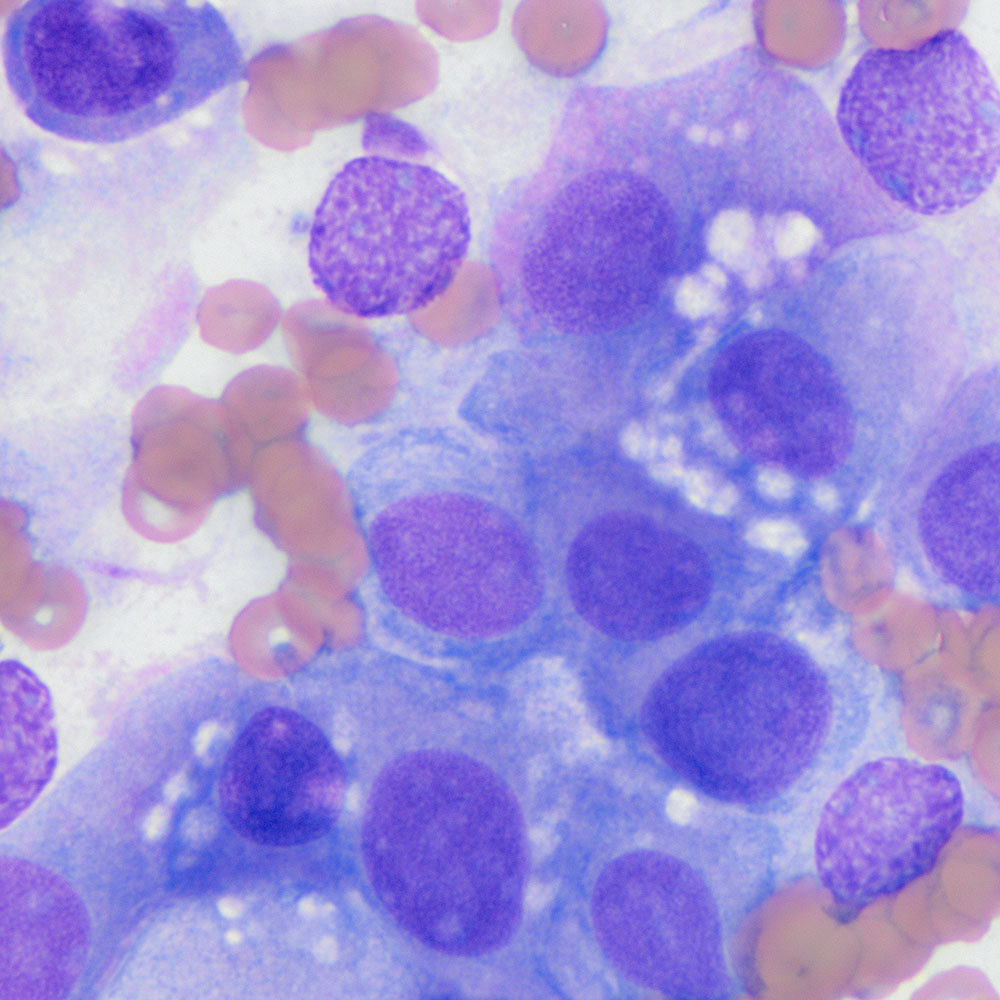
Our latest diagnostic challenge from our first year resident! A blood smear and lymph node aspirate from a 12 year old dog with seizures (first image: blood; second to fourth images: lymph node). Read the case at https://t.co/6UMVUVpLaI.
Pure red cell aplasia in a dog: Fibrosis is supported by streaming of the cells (long arrows). There was a plasmacytosis (short arrows), including Mott cells (arrowhead) and increased marrow iron. All of these findings can be features of this syndrome.
Apocrine gland of the anal sac adenocarcinoma with interspersed normal melanin-containing keratinized squamous epithelial cells (arrows, first image, 20x objective) from the anal sac lining and mixed bacteria from the sac lumen. Second image was taken with the 50x objective.
Ever wanted to know what Aruba Island rattlesnake blood cells look like? Here is your chance with a bonus hemoparasite thrown in. We are missing a basophil though.
A “sneaky” sarcoma in a dog’s spleen: Neoplastic cells stream off splenic stroma (arrow, both images) and are mitotic (arrowhead, image 2). The tumor cells are admixed with normal splenic elements (lymphoid and mixed hematopoietic cells). A hemangiosarcoma was suspected.
Another image from our chicken post from last week (ovarian adenocarcinoma): This time it is an aspirate from the neoplastic ovary, showing the tumor cells.
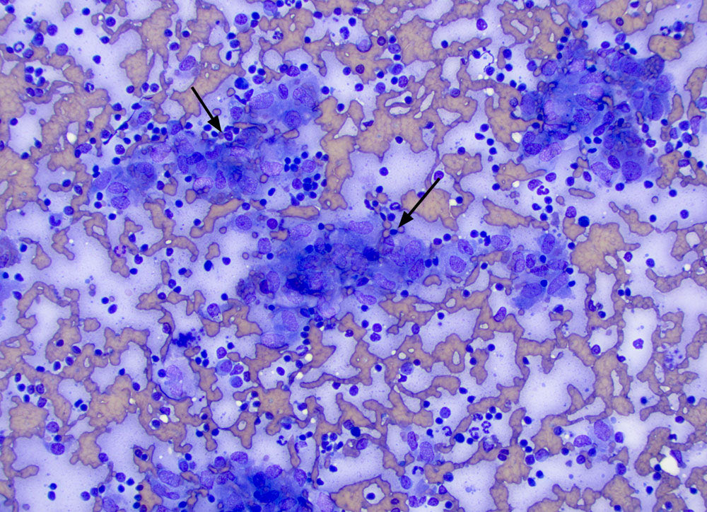
Coelomic fluid from a chicken: Heterophils and macrophages along with groups of cells with blue cytoplasm and large lipid droplets. The latter were suspected to be carcinoma cells. Similar cells were seen in aspirate of an ovarian mass that was identified on ultrasound.
Dysplastic megakaryocyte in the bone marrow of a dog with pure red cell aplasia (the most severe form of a precursor-directed immune-mediated anemia. The cell has multiple separate nuclei (arrow).
Gelatinous transformation of fat in a bone marrow aspirate from a ferret. Normal adipocytes (arrow, first image) and purple smooth mucoid matrix, indicating gelatinous transformation (arrowhead first image, second image). This stromal change occurs with cachexia or anorexia.
Blood from a cat: Morulae are present in the neutrophils (arrow), compatible with an Anaplasma phagocytophilum infection.
Seen just this week as a serendipitous finding on smear examination by our awesome techs.
Aspirate of splenic nodules in a 5 year old cat. There were aggregates of macrophages (arrows, image 1) with intercalated neutrophils (arrow, image 2). Non-staining bacterial rods are in the macrophages (image 3), indicating a mycobacterial infection.
Thymic lymphoma in a 2 year old Heifer. The heifer was presented with neck and ventral edema and jugular distention. Intermediate to large lymphocytes were the main cell type in smears of an aspirate of a thoracic inlet mass. There were also a few macrophages (arrow, 2nd image).
Urine sediment from a young cat with acute kidney injury and failure: “Picket fence”-shaped calcium oxalate monohydrate crystals were present. These crystals are usually seen with ethylene glycol toxicity, which was the suspected clinical diagnosis in this case.
Our belatedly “scary” post of a metastatic carcinoma with concurrent necrosis in a mandibular lymph node aspirate from a cat. The primary tumor was in the cat’s ear and was presumably a ceruminous gland carcinoma. Check out all those nuclei and nucleoli!😳
A blood smear from a cow with a regenerative anemia due to a hemoparasite (arrows).
The organisms have linear to tailing shapes. These features are compatible with Theileria. A PCR for this organism was strongly positive.
Phagocytes eating bad things…
A: Bacterial rods in a neutrophil; B: Bile cast in a macrophage; C: Erythrocyte and platelet in a macrophage (A-C are images of a blood smear from a dog with immune-mediated hemolytic anemia); D: Mycobacteria in histiocytes (cat, spleen).