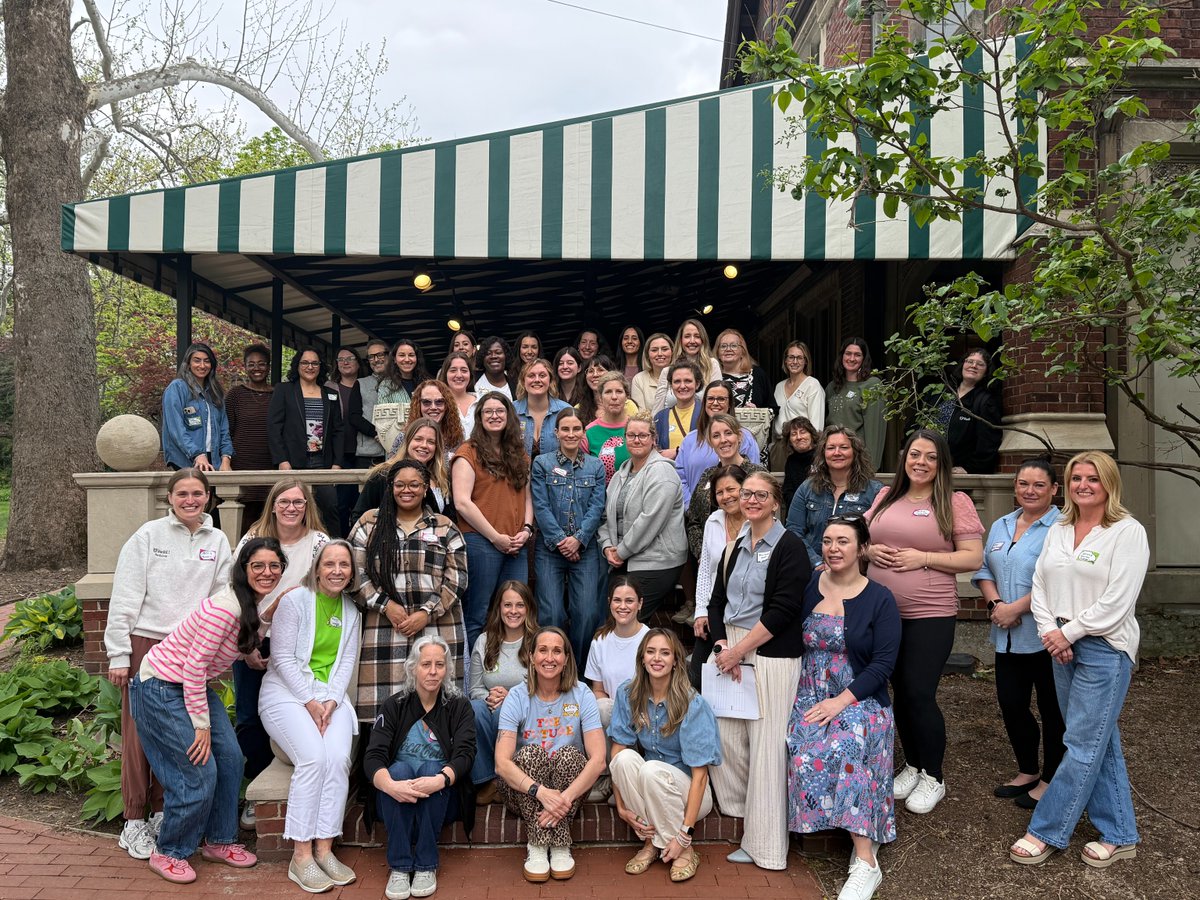
Last weekend, Women from #WashUAnesthesiology gathered for the fourth annual Supporting Women of WUDA Retreat. The day included a keynote from Dr. Sariya Saabye, breakout sessions, and a clothing and book swap. Read the highlights: https://t.co/bygalIfFWz
Each year, National Deaf History Month is celebrated to commemorate the achievements and contributions of people who are deaf and hard of hearing. For more info, local events, and resources visit: https://t.co/YS1ofVDsK9
Join us tomorrow for the H.E.R. Symposium: Honoring Excellence in Research celebrating Women’s History Month and gender equity in STEM.
🗓 March 31 | ⏰ 12–6 p.m.
📍 Connor Auditorium, FLTC
This March, we celebrate Women's History Month with the theme "Leading the Change: Women Shaping a Sustainable Future." Let's honor the incredible women who have transformed our world & continue to inspire us daily. Info, resources, and events: https://t.co/vj9xTRU7j4.
Congratulations to Dr. Anne Drewry on her installation as the inaugural Llorin-Roa Professor of Anesthesiology! A well-deserved honor recognizing outstanding leadership in Critical Care Medicine.
New Free Lecture!
“A Case for POCUS TCCD”
1. How to obtain and optimize the transtemporal acoustic window
2. Identifying MCA flow and waveform patterns
3. Recognizing signs of cerebral circulatory arrest and elevated ICP
4. Integrating TCCD into POCUS protocols for neuro-critical care and brain death evaluation
🎥 Watch now: https://t.co/WV8wJAE7dl or https://t.co/W68bIlBmzM
Thanks to @drmohansai and @YubSedhai for leading the way on this. I hope to collaborate again soon!
@NephroP
Perfect for ICU, Neuro, and POCUS enthusiasts.
#POCUS #TCCD #NeuroICU #CriticalCare #UltrasoundEducation
Congrats to Megan Moseley, PA-C, on being named a 2025–26 AAPA-PAEA Research Fellow! She’s the first PA from @WashUmedicine to earn this honor & will develop a scalable POCUS curriculum for APPs in critical care. https://t.co/fVv6ckG9Qj
1/ Anaesthetic challenge.
Patient turns up at a district general hospital for urgent (cancer) surgery. 2 months earlier TTE showed this pericardial effusion considered to be neoplastic in origin. Colchicine and steroids had been prescribed. A repeat echo the day before surgery was missed. You look at the scan 👀🫀”
(1/x) In fellowship, I managed a peri-arrest patient in the middle of the night who changed my understanding and appreciation for hemodynamics, ultrasound, and TEE.
I've seen similar cases dozens of times now, yet this commonly gets missed, even at top institutions worldwide.
A 🧵
(4/x) Immediately after intubating I inserted the TEE probe.
The anatomy and physiology was immediately obvious --> severe dynamic LVOT obstruction with SAAM.
The epinephrine was killing them, not helping. I bolused phenylephrine, vasopressin, and started fluid boluses... things improved, but not by much.
Hear about Martin's inspirational journey of living without the excruciating pain associated with sickle cell disease and receiving a breakthrough gene therapy thanks to @washumedicine and @STLChildrens@WashUOncology
I have created this simple guide on the use of vasopressors and inotropes in patients with cardiogenic shock, combining both European and American guidelines.
I look forward to your kind opinion and feedback.
If you’re measuring LVOT diameter, be careful, any small error gets squared in the πr² equation. One workaround? Grab the diameter from a previous echo report.
And if you’re just assessing fluid responsiveness, you don’t even need the diameter, just track VTI changes before and after a leg raise or fluid bolus.
But don’t forget to watch the heart rate too, changes in HR can throw off your cardiac output comparison at different time points.
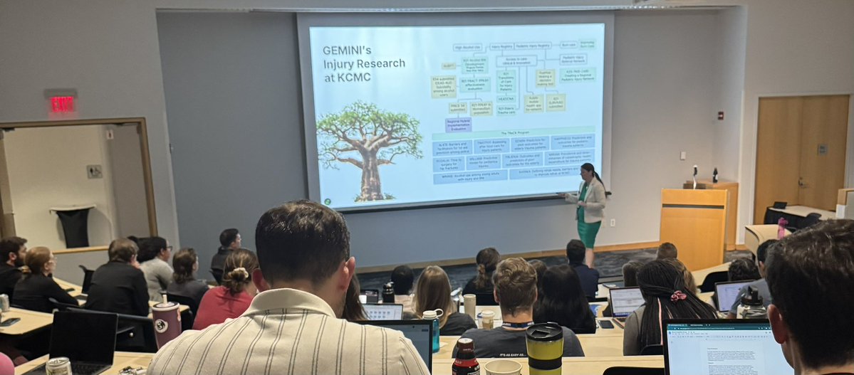
Fantastic Grand Rounds from Dr. Staton and her teams journal in injury prevention as an implementation scientist. Thanks for sharing your many wins & inspiring us during our 22nd Annual Allen P. Klippel Honorary Lecture. @WUSTL_EM@TheTechDoc@TeddyDanielz@dukeemergency
Continuing the formula infographics...
Estimating aortic valve area by continuity equation.
#POCUS#echofirst#Nephpearls#FOAMed
LVOT = left ventricular outflow tract
VTI = velocity time integral
PWD = pulsed wave Doppler - because you want VTI at a specific location (LVOT)
CWD = continuous wave Doppler (because you want peak gradient across the valve)