11/
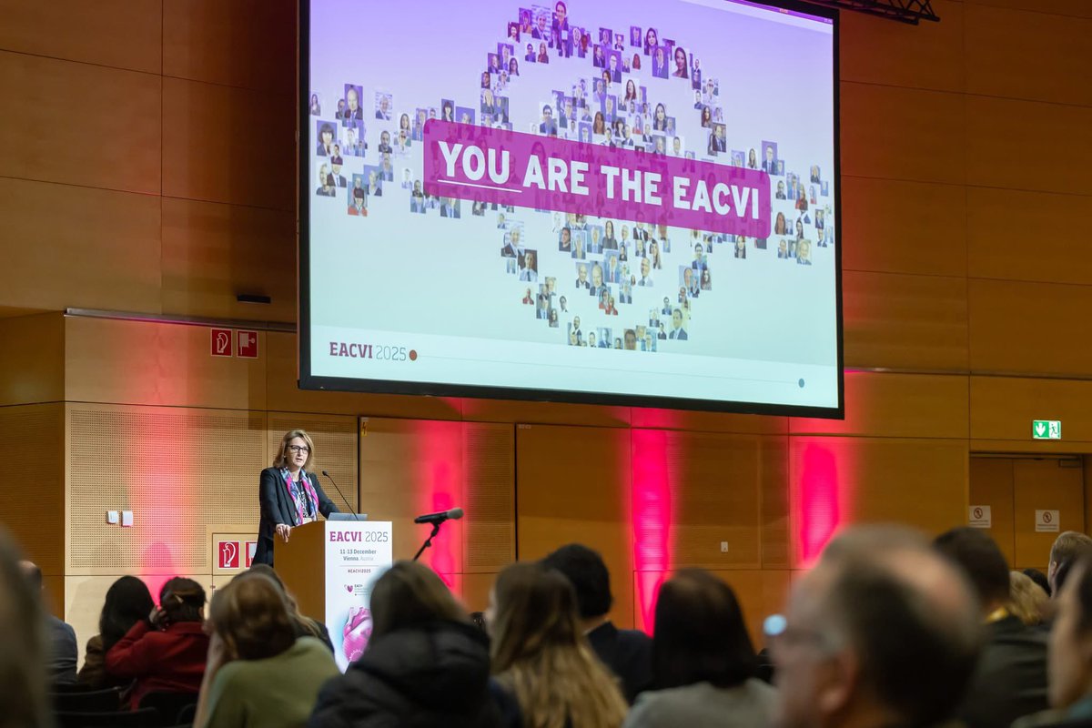
Until then, I wish you all some well-deserved downtime, a joyful festive season, and happy holidays.
Thank you for being part of the #EACVI family.
@VDelgadoGarcia
📢 Cardiac CT is much more than coronary imaging.
Delighted to be a co-author of the new #EACVI / @escardio Council of Cardiology Practice Clinical Consensus Statement on the implementation of #CCT in everyday practice, published in the @ehj_ed@ESC_Journals .
We focus on four practical questions:
1️⃣ What can cardiac CT detect and assess?
2️⃣ How can CT contribute to risk stratification?
3️⃣ What information should be included in the referral?
4️⃣ What should clinicians look for in the CT report?
From coronary and valvular disease to cardiomyopathies, cardiac masses, great vessels and congenital heart disease, the document highlights how #CCT can provide clinically actionable anatomical, functional and prognostic information.
@gpontone1@StefanoNistri@drkharabish@JRodriPalomares@denisamuraru@s_e_petersen@VictoriaDe32503@EACVIPresident
https://t.co/rdMfRBzmxr
🎥🫀 What if cardiac CT could show valves in motion… like real life?
A new study introduces the first use of 4D cinematic rendering (CR) in cardiac imaging 👇
✨ What’s new:
🧠 3D cinematic rendering + time = 4D visualization
🎬 Full cardiac cycle in motion
👁️ Enhanced depth perception & realism
📊 As shown in the videos and images (pages 1–2):
👉 Dynamic visualization of:
🔁 Valve prolapse (tricuspid & mitral)
📍 Annular disjunctions
⚠️ Malcoaptation
🌊 Even regurgitant jets (not visible in standard CT!)
🔥 Game-changing advantage:
👉 Not just anatomy… but dynamic valve behaviour
💡 Additional insight:
📍 Clear relationship between tricuspid annulus & RCA
→ crucial for procedural planning
🚨 Clinical message:
Standard CT = static anatomy
4D CR = real-time functional anatomy
🎯 Big take-home:
From “seeing structures”
➡️ to
“watching the heart move in 3D space and time”
📄 Read the full paper:
👉
🔗 DOI: 10.1093/ehjimp/qyaf165
#Cardiology #CardiacCT #4Dimaging #CinematicRendering #ValvularHeartDisease #ImagingInnovation #CardioTwitter #MedTech #DigitalHealth 🧠💓
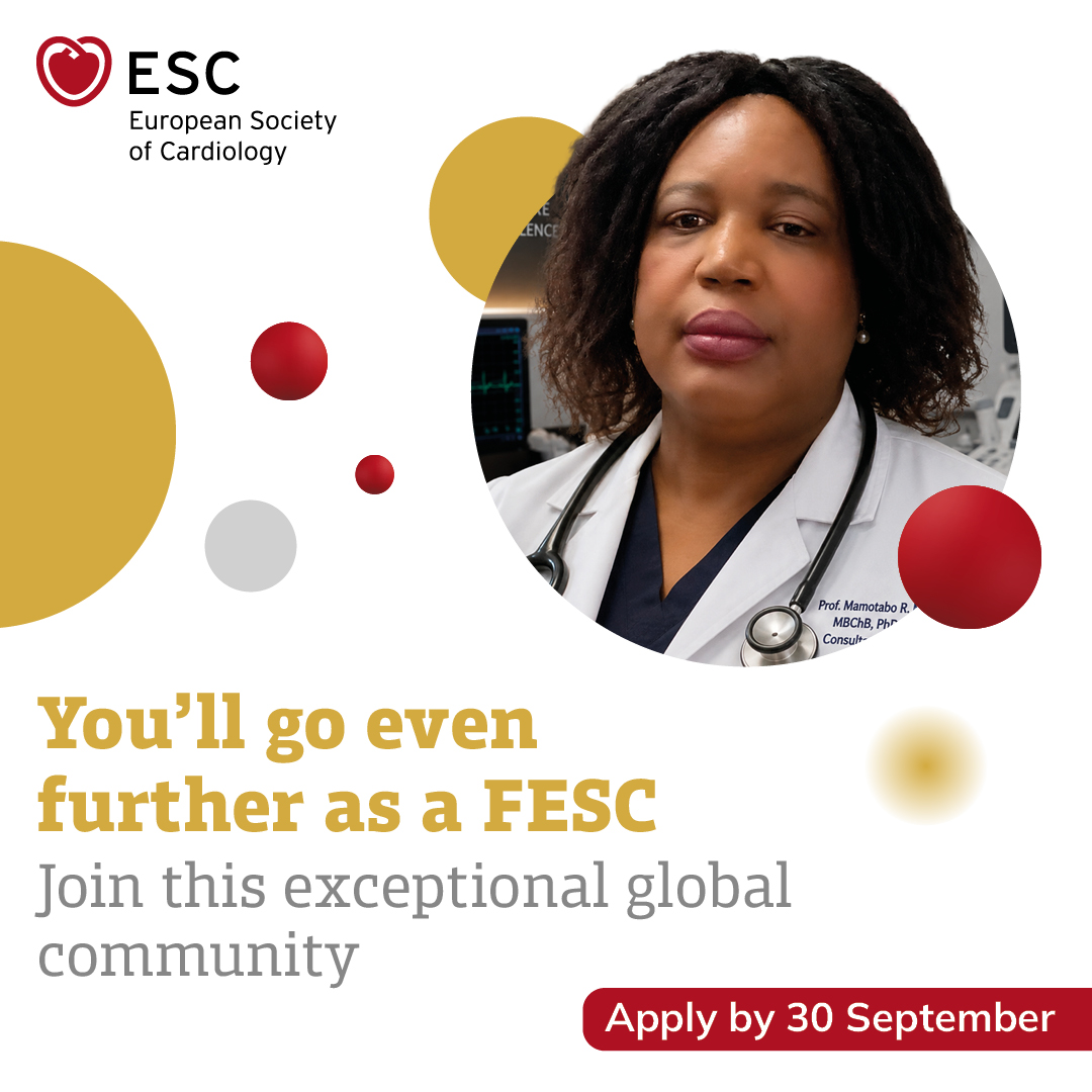
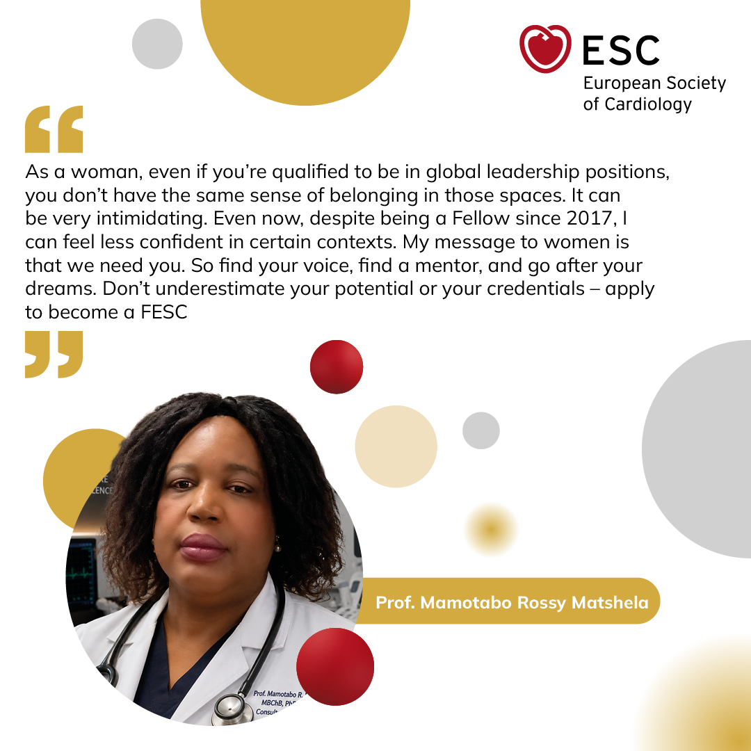
Recognition. Community. Leadership.
As Prof. Mamotabo Rossy Matshela reminds us, "Don't underestimate your potential or your credentials."
Becoming a Fellow of the ESC (FESC) is an opportunity to gain international recognition, connect with a global network of cardiovascular professionals, and contribute to shaping the future of cardiology.
Apply by 30 September https://t.co/DcER2JY5oo
New in @ESC_Journals - Cardiovascular Imaging
Which patients with aortic stenosis (#AS) benefit most from exercise hemodynamics?
🧵1/8
We propose a simple stepwise approach:
📍Cardiac damage staging (@PhilGenereuxMD )
📍Exercise hemodynamics in patients with early cardiac damage
🚨 Registration for #EACVI l2026 is OPEN! 🇮🇹
Join the global cardiovascular imaging community in Milan, Italy, from 3–5 December 2026.
This year’s congress brings together the excellence of #EuroEcho, #EuroCMR, and #ICNCCT under one roof, offering:
🔹 Dedicated single-modality tracks
🔹 An optional multimodality track to broaden your expertise
🔹 Expert-led case discussions and live demonstrations
🔹 Opportunities to connect with leading imaging professionals from around the world
💡 Register by 30 September to secure the best available rates, with additional discounts for #EACVI Members, nurses, allied professionals, and trainees.
Don’t miss this opportunity to advance your knowledge, strengthen your clinical practice, and be part of one of the world’s premier #CVImaging meetings!
@denisamuraru@s_e_petersen@NAjmoneMarsan@gpontone1@EchoSoliman@RFontesCarvalho@rmanka_@Matte_Cameli@DrGEMandoli@anatimoteo46040@AnnaBaritussio@andgiannopmd@PezelT@Ph_Bertrand@Sarah_Moharem@VictoriaDe32503
#EACVI2026 #CardiovascularImaging #Echocardiography #CardiacMRI #CardiacCT #NuclearCardiology #Cardiology #MedicalEducation #ESC
One congress, every perspective – deepen your expertise while embracing the bigger picture at #EACVI26 🩷
Join us in Milan from 3–5 December, where EuroEcho, EuroCMR and ICNC-CT come together to deliver world-class education across cardiovascular imaging.
Register today and benefit from the best rates 👉 https://t.co/At2SxK9wVF
#EACVI @EACVIPresident
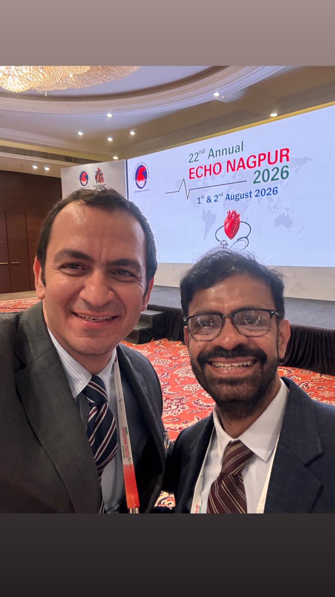
I am glad to share that Prof Delgado, A/P Wong myself are conducting The Hands-on Echocardiography Workshop takes place in Brunei Darussalam as part of AFCC 2026. this workshop covers advanced techniques like AI integration, 3D echocardiography, and valve evaluations. Do join us
🚨 New Research | Acute Decompensated Heart Failure
Can combining venous congestion assessment (VExUS) with lung ultrasound (LUS) improve risk stratification in acute decompensated heart failure?
A newly published study in the @EHJIMPEiC evaluated 104 patients with acute decompensated heart failure and found that early serial changes in VExUS and lung ultrasound, integrated into the novel ΔVExPLUs score, were strongly associated with clinical outcomes. Patients with less improvement in congestion had a significantly higher risk of adverse events.
💡 Key takeaway: Combining systemic venous congestion and pulmonary congestion into a single bedside ultrasound assessment may provide a more comprehensive and clinically meaningful way to monitor decongestion and identify high-risk patients early during hospitalization.
This study highlights the growing role of multiorgan point-of-care ultrasound in guiding heart failure management and improving prognostic assessment.
Read the article here:
https://t.co/re3TSKKJ8N
@rafavidalperez@YBououdina@MSBBrandao@ydaryani@Elizabeth_antos@pfelissamburu@CharlesFauvel@alexsfelixecho@galzeranod@andgiannopmd@hrt01a@WilliamKokFaiK1@M_Marwan_@drahmedmohsen85@aniela_petrescu@benayozbay@slumberbell@senguptasp@ElizabetaK10533@samsrivastava77@MihaiTrofenciuc@C_VanDeHeyning@drozgeozden@Giulia_Vinco@VazyurVasquez
#HeartFailure #POCUS #Ultrasound #Cardiology #VExUS #LungUltrasound #Echocardiography #MedicalEducation #FOAMed #CardioTwitter
🧬🫀 We image the scar… but do we really understand it?
A new review explores how molecular contrast agents are redefining imaging after myocardial infarction 👇
✨ Key insights:
❤️ Post-MI remodelling drives heart failure & mortality
📊 Conventional imaging (CMR, PET) shows:
👉 infarct size
👉 function
❌ but NOT the underlying biology
🔬 Molecular imaging targets the real drivers:
👉 inflammation
👉 fibroblast activation
👉 scar formation
💡 As shown in the graphical abstract (page 2):
👉 Conventional imaging = what we see
👉 Molecular imaging = what is happening
🔥 Game changer:
Detect disease earlier (before structural changes)
Identify stage of remodelling (inflammatory → proliferative → fibrotic)
Guide targeted therapies (e.g. anti-inflammatory, anti-fibrotic)
🚀 Examples of next-gen tracers:
🧲 SPIONs → macrophage activity (MRI)
🔥 FDG / DOTATATE → inflammation (PET)
🧵 FAPI → fibroblast activation
🧱 Collagen/elastin tracers → scar maturation
🎯 Big take-home:
From “how big is the infarct?”
➡️ to
“what biology is driving the remodelling?”
📄 Read the full paper:
👉
🔗 DOI: 10.1093/ehjimp/qyag019
#Cardiology #CMR #CardiacPET #MolecularImaging #HeartFailure #PrecisionMedicine #CardioTwitter #Research #MedTech #PostMI 🧠💓