Please subscribe my YouTube channel: https://t.co/G6ZVTzN7H2
I would like to present my project (echocardiography step by step®). With my project, I want to take you into the world of echocardiography through the prism of my experience.
@echo_batman🏆📝
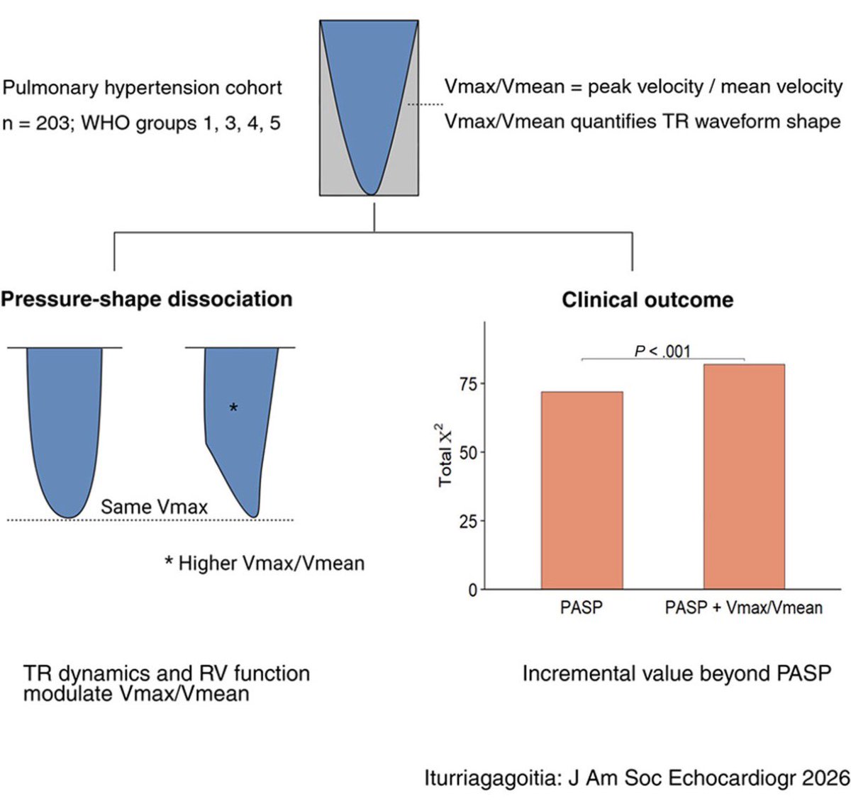
Vmax/Vmean reflects TR CW Doppler & RV adaptation to pulmonary vascular load
Even when Vmax is similar,
Vmean can differ, w distinct TR CWD shapes
Persistent ⬆️ after PH-targeted Rx ids pts w ⬆️ risk for ☠️ or HFH https://t.co/x0FjCQsFSo
🫀📡 The stethoscope is evolving… and it’s now an ultrasound probe.
A new review highlights how Point-of-Care Ultrasound (POCUS) is transforming bedside cardiopulmonary diagnosis 👇
✨ Key insights:
⚡ POCUS = real-time, bedside, physiology-driven imaging
🫁 Lung ultrasound (LUS) → detects congestion better than CXR & exam
🩸 VExUS score → quantifies systemic venous congestion
❤️ FoCUS + LVOT-VTI → assess structure, function & cardiac output
🔄 Enables dynamic monitoring and therapy guidance
💡 As shown in the graphical abstract (page 2):
👉 HF, STEMI, shock → one tool, multimodal assessment
👉 From congestion ➝ to perfusion ➝ to haemodynamics
🚨 Clinical message:
Traditional exam = limited sensitivity
POCUS = immediate + actionable information
Not a replacement… but a powerful extension of clinical reasoning
🎯 Big take-home:
From “suspecting haemodynamics”
➡️ to
“seeing haemodynamics at the bedside”
📄 Read the full paper:
👉
🔗 DOI: 10.1093/ehjimp/qyaf147
#Cardiology #POCUS #Echocardiography #HeartFailure #CriticalCare #Ultrasound #CardioTwitter #MedTech #BedsideMedicine #DigitalHealth 🩺📊
🫀📡 PET myocardial perfusion is not just imaging… it’s precision physiology.
A new EACVI consensus statement redefines how we perform and interpret PET/CT myocardial perfusion imaging (MPI) 👇
✨ Key insights:
🥇 PET = most advanced non-invasive tool for CAD assessment
🔍 Detects:
👉 ischaemia
👉 viability
👉 scar
⚡ Goes beyond anatomy → provides quantitative myocardial blood flow (MBF)
📊 MBF reserve <2.0 = abnormal → powerful for risk stratification
💡 From the figures (pages 8–9):
👉 Integration is key: perfusion + function + MBF + CT calcium
👉 17-segment model + polar maps = standardized interpretation
🔥 Game changers:
🧠 Quantitative MBF improves detection even when perfusion looks “normal”
⚖️ PET allows true physiological assessment of coronary disease
🧑⚕️ Strong focus on patient-centric protocols & preparation
⚠️ Important practical points:
☕ Avoid caffeine before test
💊 Medications may need adjustment
🧪 Choice of tracer = crucial (half-life, availability, accuracy)
🎯 Big take-home:
From “is there a stenosis?”
➡️ to
“is this myocardium truly underperfused?”
📄 Read the full paper:
👉
🔗 DOI: 10.1093/ehjimp/qyaf157
#Cardiology #CardiacPET #NuclearCardiology #MyocardialPerfusion #PrecisionMedicine #CardioTwitter #Imaging #CAD #MedTech #EACVI 🩺📊
A middle-aged woman with long history of joint pains and recent transient vision loss undergoes TEE. Modified long axis view shown. What tests would you order next?
@DHZB_Akademie "3D Advanced Echocardiography Workshop" (happening January 22- 24, 2027) makes it clear why this is way "more than just 3D."
The program bridges advanced imaging with intensive clinical realities”specifically targeting ICU management, complex valvular heart disease, mechanical circulatory support (MCS), and heart failure.
Perfect for clinicians who want practical utility, not just pretty 3D pictures.
Yes, it’s a 3D workshop but we are taking it straight to the ICU
At the #echofirst Workshop , we aren't just looking at static valves. We’re tackling the tough cases:
Echo in ICU: Deep dives into ECMO hemodynamics and using echo to guide acute fluid management.
Critical Decision Making: Using acute heart failure echo to trigger therapy (from diuretics to MCS).
Live Knobology: Real-time mastering of GE & Philips systems.
Register & view the full program: https://t.co/tqmJDqUuqQ
Just take a look at this GOAT line up 🔝
@alex1708ander@alexsfelixecho@dr_benoy_n_shah@iceman_ex@echo_batman@FGraziani_Grace@VictoriaDe32503@Ph_Bertrand@StellEkaterina@echo_stepbystep@fiore_corrado@LunaGargani@ArgaizR@papadocardio@eromerodorta@mauripieroni72@KemalogluOz@JanKnierim@echotainment@DonalErwan@FH_Verbrugge
🧬🫀 We image the scar… but do we really understand it?
A new review explores how molecular contrast agents are redefining imaging after myocardial infarction 👇
✨ Key insights:
❤️ Post-MI remodelling drives heart failure & mortality
📊 Conventional imaging (CMR, PET) shows:
👉 infarct size
👉 function
❌ but NOT the underlying biology
🔬 Molecular imaging targets the real drivers:
👉 inflammation
👉 fibroblast activation
👉 scar formation
💡 As shown in the graphical abstract (page 2):
👉 Conventional imaging = what we see
👉 Molecular imaging = what is happening
🔥 Game changer:
Detect disease earlier (before structural changes)
Identify stage of remodelling (inflammatory → proliferative → fibrotic)
Guide targeted therapies (e.g. anti-inflammatory, anti-fibrotic)
🚀 Examples of next-gen tracers:
🧲 SPIONs → macrophage activity (MRI)
🔥 FDG / DOTATATE → inflammation (PET)
🧵 FAPI → fibroblast activation
🧱 Collagen/elastin tracers → scar maturation
🎯 Big take-home:
From “how big is the infarct?”
➡️ to
“what biology is driving the remodelling?”
📄 Read the full paper:
👉
🔗 DOI: 10.1093/ehjimp/qyag019
#Cardiology #CMR #CardiacPET #MolecularImaging #HeartFailure #PrecisionMedicine #CardioTwitter #Research #MedTech #PostMI 🧠💓
Want to publish in medical journals but unsure where to start?
Read my editorial in @BrJCardiol on publishing: why, what, where, when, how and more...
My thanks to Henry Purcell & the excellent BJC team!
#GetPublished
@RitikaTuliMD The color Doppler vena contracta should be interpreted with caution. It reflects more than regurgitant volume, including radial flow entrainment, and represents only a single 2D slice of a complex 3D regurgitant orifice. #EchoFirst@JaeKOh2@NMerke@dr_benoy_n_shah@iamritu
🫀🔥 Is atrial disease… already metabolic before it becomes electrical?
A new study using CMR + FDG-PET reveals a hidden layer of atrial disease 👇
✨ Key findings:
📉 Higher atrial FDG uptake → lower LA strain
📈 Higher FDG uptake → larger LA volume
🔗 Strong correlations (see scatter plots on pages 6–7) between:
👉 metabolism (SUV, aTBR)
👉 function (strain)
👉 structure (LAVi)
⚠️ These associations persist independent of AF or sarcoidosis
💡 Big message:
👉 Atrial myopathy is not just structural or electrical
👉 It has a metabolic substrate
🚨 Even more interesting:
Patients with AF = higher FDG uptake + worse strain
Suggesting metabolism may precede clinical arrhythmia
🎯 Clinical insight:
We might be looking at a new early marker of atrial disease
→ before AF develops
→ before irreversible remodeling
🎶 “The atrium doesn’t just remodel… it rewires its metabolism.”
📄 Read the full paper:
👉
🔗 DOI: 10.1093/ehjimp/qyag037
#Cardiology #AtrialFibrillation #CMR #FDGPET #CardiacImaging #AtrialMyopathy #PrecisionMedicine #CardioTwitter #HeartResearch #Metabolism 🧠💓
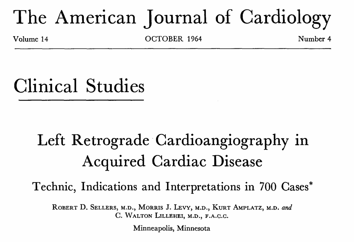
Dear community, I'm looking for a photograph of Dr. Robert D. Sellers, who worked at the @UMNews with C. Walton Lillehei in the 1950s–1960s and published a landmark paper in October 1964. I haven't been able to find one anywhere. Any help would be greatly appreciated! 🙏#MedHist.
🫀🤖 Heart failure is not just about the heart anymore.
A new study shows how AI + multi-organ ultrasound can redefine congestion across the HF spectrum 👇
✨ Key insights:
🌊 Congestion = multi-organ phenomenon (lungs, kidney, liver, IVC)
🧠 AI integrates clinical + echo + lab data → better phenotyping
📊 A simple 3-item model (IVC + LUS + RVF) predicts multi-organ congestion
🎯 Key predictors:
👉 LV function (S’)
👉 Pulmonary pressures
👉 LA volume
👉 Metabolic + clinical factors
💡 As highlighted in the graphical abstract (page 2):
AI doesn’t just detect congestion… it explains it
🚨 Clinical message:
👉 Congestion is often missed clinically
👉 Ultrasound detects it earlier
👉 AI turns it into a precision tool
🎯 Big take-home:
From “is the patient congested?”
➡️ to
“Where, how, and why is the patient congested?”
📄 Read the full paper:
👉
🔗 DOI: 10.1093/ehjimp/qyag036
#Cardiology #HeartFailure #AIinMedicine #Echocardiography #Ultrasound #PrecisionMedicine #CardioTwitter #DigitalHealth #MedTech 🩺📊
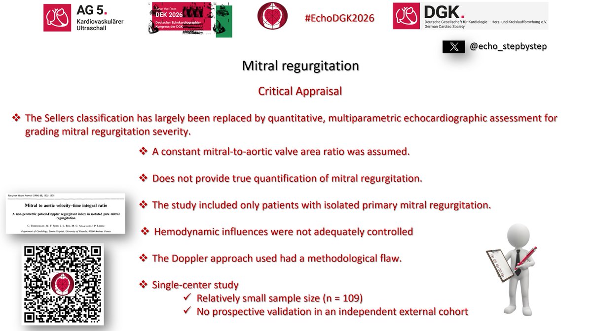
@WilliamZoghbi In my opinion, the methodological limitations of this study should be acknowledged whenever it is incorporated into contemporary clinical guidelines. Prospective multicenter validation using modern reference standards (volumetric) is warranted. Until then… #EchoFirst#WhyCMR
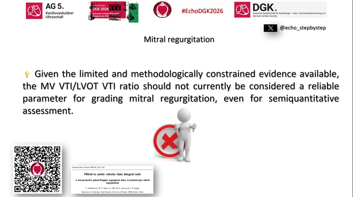
Current #guidelines recommend the mitral inflow/LVOT VTI ratio as an easy-to-measure semiquantitative index for isolated primary MR (>1.4 severe, <1 mild). But how robust is the evidence behind it? Let's take a closer look at this widely cited parameter. #EchoFirst Part 1
@WilliamZoghbi Let me summarize the critical appraisal of the landmark study by Christophe Tribouilloy et al. (1994), which laid the foundation for the MV/LVOT VTI ratio and has been cited in international echocardiography guidelines for more than 30 years. https://t.co/lqsvgG3Bsc Part 12