Having started my career in basic neuroscience research, I have always understood Alzheimer disease to mean the pathophysiology associated with amyloid plaques and tau tangles. As a clinician, I appreciate that the relationship between Alzheimer disease and cognitive impairment is complex.
1. Clinically diagnosing the cause of cognitive impairment is different than diagnosing that the patient has Alzheimer disease (pathology). Many of my patients with cognitive impairment have multiple diagnoses (e.g., sleep apnea, medication-related cognitive dysfunction, depression, previous cerebral infarction), and sometimes have Alzheimer disease (based on biomarker testing). I do what I can to mitigate the effects of all of these conditions, and when Alzheimer disease is present I do not assume that it is the sole or even the primary cause of cognitive impairment. This is true across multiple potential etiologies: for example, I may diagnose someone with cerebrovascular disease and a prior infarct based on MRI findings, but determine that this condition is asymptomatic and not contributing to their cognitive impairment.
2. Given the simplicity of some biomarker tests and the difficulty of clinical assessment, there is a risk that clinicians may diagnose the etiology of cognitive impairment as Alzheimer disease without doing a full work-up. This is why appropriate use recommendations for biomarkers always mandate that biomarker results must be integrated with a clinical evaluation and not used as a “stand-alone.”
3. Alzheimer disease pathology accumulates silently for ~10-20 years before the onset of cognitive impairment. However, during this pre-clinical phase the accumulation of amyloid pathology is associated with many other biomarker changes that appear pathological (e.g., abnormal CSF synaptic biomarkers, brain atrophy, brain hypometabolism). While the brain is remarkably resilient to damage and individuals may or may not develop symptoms, the brain is sick and the pre-clinical phase is a disease state.
4. Suggesting that a disease only exists when organs are severely damaged and failing (dementia) seems counter to what we have learned in other areas of medicine. For example, patients diagnosed with hypertension or asymptomatic coronary artery disease may change their diet and medications to avoid a heart attack. Individuals with asymptomatic chronic kidney disease may or may not go on to require dialysis, but they can be monitored and sometimes treated.
5. There are valid concerns about the stigma and risks of asymptomatic individuals being labeled as having Alzheimer disease given that they may or may not develop cognitive impairment. The solution is simple—we don’t perform biomarker testing in asymptomatic individuals outside of research studies or clinical trials. Again, this has been mandated by appropriate use recommendations for biomarkers.
We can help patients by promoting accurate understanding and appropriate use of biomarkers. #EndAlz
The morphological descriptions and openly available spatial probability maps provided here are important for the accurate identification of this sulcus in structural and functional neuroimaging studies examining this region.
Paper alert‼️Check out our recent publication on the examination of the morphology of the inferior frontal sulcus (ifs) in the human brain.
👇
https://t.co/Fhwv7DbQqu
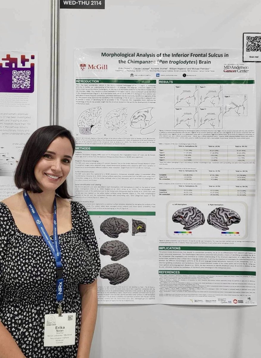
Last poster session today at #OHBM2024. Come visit my poster #2114 on the sulcal morphology of the inferior frontal sulcus in the chimpanzee (pan troglodytes) brain and lets discuss cross-species similarities and potential relevance for language! 🧠
New paper alert! @ExpPhysiol
A @char_lab collab, synthesizing the evidence of elevated sympathetic activity (muscle sympathetic nerve activity) in #PCOS. Check it out 😁.
https://t.co/TOTwhhoCEY
Such an enriching experience at #OHBM2023! Met some truly remarkable individuals, including the esteemed Michael Petrides and Kevin Weiner, leading lights in neuroscience 🌟🧠 Grateful for this opportunity to connect and learn from these inspiring minds. #Networking#Inspiration"
Exciting job opportunity! @TheNeuro_MNI@mcgillu is seeking an Assistant Professor (Tenure-Track) in #Cogneuro Please apply & RT. For more information and to submit your application please visit: https://t.co/oXSgnoTQmo
Authors from @BorrellLab examine the processes that lead to folding of the #cerebralcortex, a phenomenon that is fundamental to the cognitive function of the human #brain and that is altered in pathological dysfunction:
https://t.co/U90RvnmpNs
#evolution#neurogenesis
Check out Jennifer's article on optimal blocking of the cerebral cortex for cytoarchitectonic examination. We use a neuronavigation method to determine the most optimal sectioning angles for cyto studies based on the orientation of sulci @erikanolan16 🧠👇 https://t.co/TJpEN1DEri
@BrainImagingLiv Absolutely. My poster and 3-minute presentation are on the conference’s virtual platform Fourwaves. Please let me know if you would like me to send it to you another way. Do not hesitate to email me if you have any comments or questions! Au plaisir.
Last day at #OHBM2022! Had very stimulating exchanges yesterday and look forward to hearing more of your insightful thoughts today. Come visit me and lets talk science! 🧠
Excited to be going to #SfN22 to present the first results from my postdoc research @TheNeuro_MNI on sulcal-cytoarchitectonic relations in the human vs chimp medial frontal region!
🔥🧠🏄
Interested in the morphology of the inferior frontal sulcus? Go check out the poster of Petrides lab PhD candidate @erikanolan16 at #OHBM2022 tomorrow and Thursday from 1:45-2:45pm, poster WTh714. 🤓🧠