Esto de lo más importante que podes llegar a ver hoy. 4 consejos de una persona que a sus 53 años de un día para el otro le diagnosticaron una enfermedad degenerativa mortal.
Escúchenlos con atención
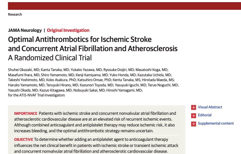
Anticoagulantes y Antiagregacion Plaquetaria en #ACV
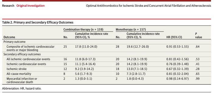
🧠En pacientes con ACVi, FANV y ASCVD
💊Añadir un antiplaquetario a la terapia anticoagulante no ofreció ningún beneficio clínico neto comparado con monoterapia anticoagulante
👹Peor, terapia combinada 📈 riesgo de sangrado.
🔗: https://t.co/yXUdaJS7R4
How to treat orthostatic hypotension in Parkinson and Parkinsonism? Dizziness on standing? Melissa Armstrong nails it in one slide during her plenary lecture this morning at MDS Hawaii. @movedisorder @FixelInstitute
Just because you don’t see it, doesn’t mean it’s not there!!!
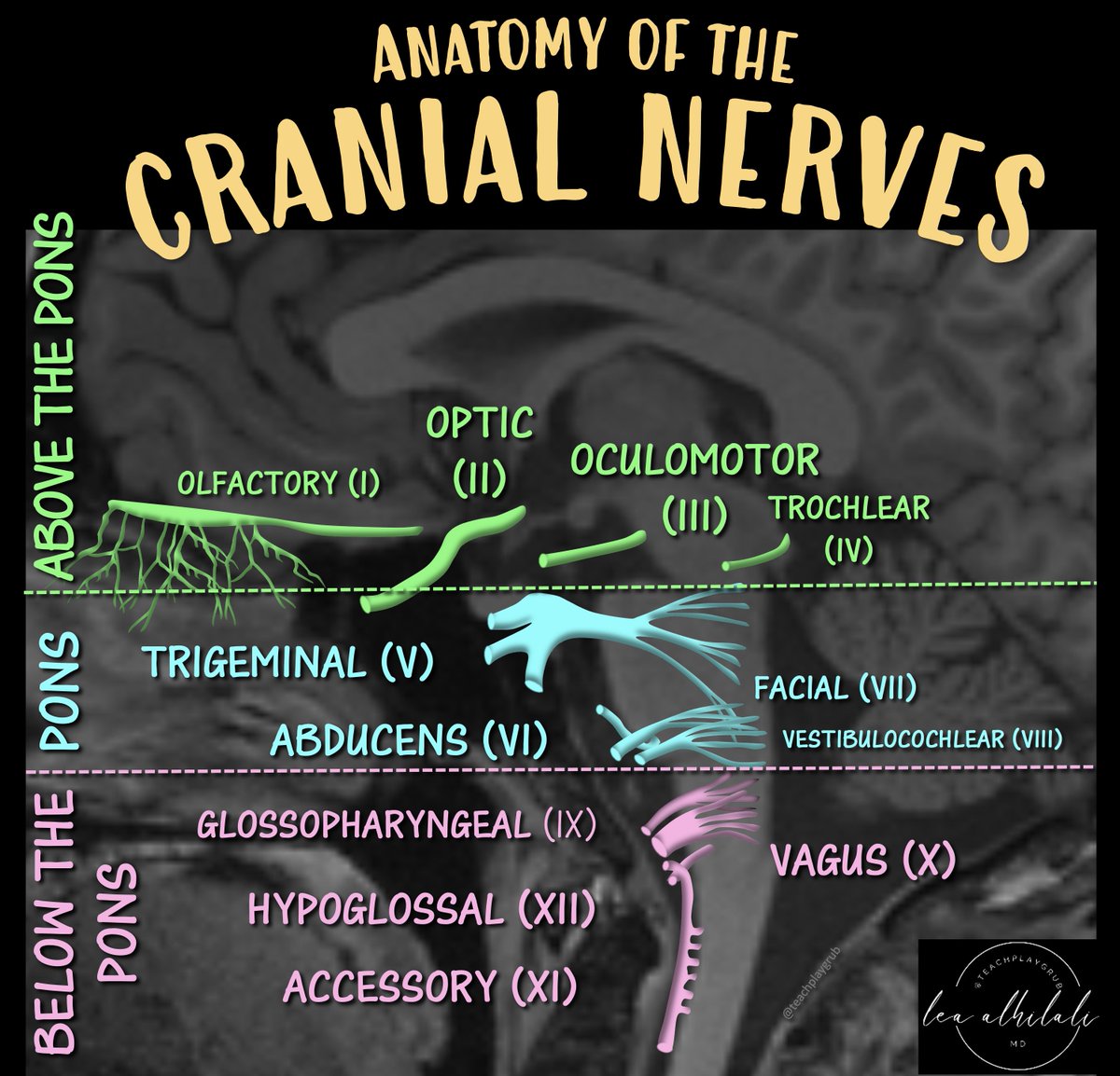
Just because you don’t see all cranial nerves on MRI doesn’t mean they aren’t there!
You must know their anatomy so you know when pathology might affect them.
Here is a figure w/the anatomy you NEED to know!
➡️Just remember this easy rule of four from Peter Gates:
There are:
🔸 4 cranial nerves from above the pons (including 2 from the midbrain)
🔸4 from the pons
🔸And 4 from the medulla oblongata!
Now you won't have be nervous when it comes to this nerve anatomy!
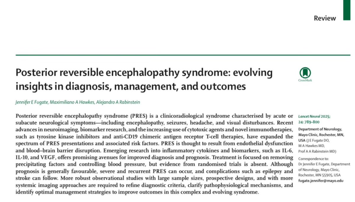
Check out our latest article exploring the advances in posterior reversible encephalopathy syndrome: pathophysiology, diagnosis, and treatment.
https://t.co/ljp992u2Bn
Treatment Effect of Early Intravenous Milrinone for Cerebral Vasospasm or Delayed Cerebral Ischemia After Aneurysmal Subarachnoid Hemorrhage
https://t.co/PmF6QZXPqY
@juneuromd@NeurocritCareJ
MRI Neuroanatomy 101 🧠
The red nuclei (red), mammillary bodies (pink), optic tracts (yellow), & cerebral peduncles (dark blue) are marked here.
Fibers of the cerebral peduncle are organized from anteromedial to posterolateral as:
- frontopontine (FP)
- corticobulbar (CB)
- corticospinal (CS)
- temporo-parieto-occipito-pontine (TPP)
Notice the ICA branching into the first segments of the anterior cerebral artery (A1) and middle cerebral artery (M1).
Try to trace these structures on the opposite side!
#radiology #neurotwitter #Neuroscience #Anatomy #Neurosurgery #education #MedTwitter #AI #MRI #brain #vascular #KnowledgeIsPower #AI #ArtificialIntelligence #algorithm
Can you spot the diagnosis on this head CT? 🚨 Clinical context: sudden left hemiparesis following jugular catheter removal. What’s your take? 🤔 @Radiopaedia@SVINJournal@neuroangio1
#Mondaytip
☝️Stroke recurrence in ICAD isn't just about stenosis severity👉the affected artery matters!
⭕️ICAD trials fail to account for differences in the anatomy of each of the major intracranial vessels
Look at the interesting findings of this review
https://t.co/qEq4BrqmFb
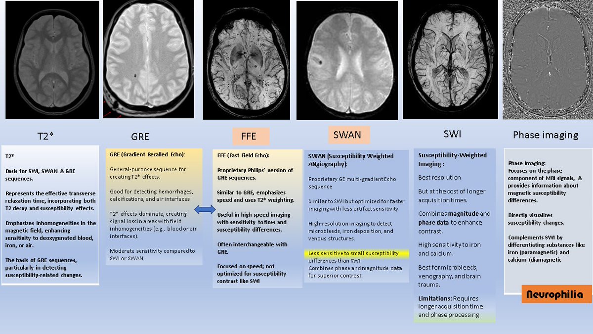
MRI for the non-radiologists. Do the terms FFE, GRE, T2*, SWI, SWAN & Phase imaging confuse you? Here is a handy reference sheet for that. Without going into any of the physics of MRI, the 1st point to understand is;
1. T2* (pronounced "T2-star") is the basis of other sequences, such as SWAN, GRE, and FFE