Advanced Practice Physio, MSK Sonographer, Shockwave Specialist, Works in NHS & Private.
Specialist Interest in Hip & Groin and Running Injuries. Views my own
𝐓𝐡𝐞 𝐡𝐚𝐫𝐝𝐞𝐬𝐭 𝐜𝐥𝐢𝐧𝐢𝐜𝐚𝐥 𝐬𝐤𝐢𝐥𝐥 𝐢𝐧 𝐢𝐧𝐭𝐞𝐫𝐯𝐞𝐧𝐭𝐢𝐨𝐧𝐚𝐥 𝐌𝐒𝐊 𝐢𝐬 𝐧𝐨𝐭 𝐩𝐥𝐚𝐜𝐢𝐧𝐠 𝐭𝐡𝐞 𝐧𝐞𝐞𝐝𝐥𝐞. 𝐈𝐭 𝐢𝐬 𝐭𝐮𝐫𝐧𝐢𝐧𝐠 𝐝𝐨𝐰𝐧 𝐭𝐡𝐞 𝐩𝐫𝐨𝐜𝐞𝐝𝐮𝐫𝐞 𝐭𝐡𝐞 𝐩𝐚𝐭𝐢𝐞𝐧𝐭 𝐜𝐚𝐦𝐞 𝐛𝐨𝐨𝐤𝐞𝐝 𝐟𝐨𝐫.
A young, active patient attended my NHS review clinic after two years of unresolving hip pain following a traumatic fall. Failed conservative rehab, persistent sharp mechanical symptoms, booked in for a guided injection. We had a transparent shared decision-making discussion and agreed not to inject. Here is why:
1. Steroid is an anti-inflammatory, not a structural repair. In a traumatic labral tear driven by acute mechanical disruption, dampening baseline inflammation rarely shifts the underlying mechanical irritability.
2. True mechanical symptoms do not respond to intra-articular steroid. Sharp catching, clicking, and locking sensations are physical impingement or displacement signs, not chemical synovitis.
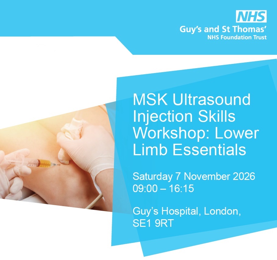
We will be covering complex hip case selection, differing injection approaches, and the critical decision matrix of when to withhold an injection at our upcoming guided injection workshop at Guys Hospital on 7th November, 2026:
https://t.co/iA0ofpcqa9
𝐖𝐞 𝐤𝐧𝐨𝐰 𝐡𝐨𝐰 𝐭𝐨 𝐝𝐢𝐚𝐠𝐧𝐨𝐬𝐞 𝐅𝐀𝐈 𝐚𝐧𝐝 𝐠𝐥𝐮𝐭𝐞𝐚𝐥 𝐭𝐞𝐧𝐝𝐢𝐧𝐨𝐩𝐚𝐭𝐡𝐲. 𝐒𝐨 𝐰𝐡𝐲 𝐝𝐨 𝐬𝐭𝐚𝐧𝐝𝐚𝐫𝐝 𝐫𝐞𝐡𝐚𝐛 𝐩𝐫𝐨𝐭𝐨𝐜𝐨𝐥𝐬 𝐬𝐨 𝐨𝐟𝐭𝐞𝐧 𝐟𝐚𝐢𝐥 𝐢𝐧 𝐟𝐞𝐦𝐚𝐥𝐞 𝐫𝐮𝐧𝐧𝐞𝐫𝐬?
Treating a female runner with hip pain as simply a smaller male runner with FAI or gluteal tendinopathy misses the clinical picture. Standard MSK assessment templates focus almost entirely on local tissue and joint mechanics: labral integrity, femoroacetabular impingement, tendon load.
For female runners, that framing is often incomplete.
1. Systemic drivers dictate tissue capacity. Fuelling status, endocrine fluctuations, and pelvic floor function directly govern tissue load tolerance, not just joint mechanics.
2. The dangerous diagnoses look different. What presents as low-grade, persistent groin or anterior hip discomfort can be a femoral neck or pubic ramus stress fracture, where a missed diagnosis has high clinical consequences.
3. Standard templates create blind spots. Because systemic and female-specific risk factors rarely appear on routine hip assessment forms, early warning signs get overlooked until load tolerance drops entirely.
4. The research has an extrapolation problem. Much of the female-specific literature is derived from military recruit cohorts or elite athletes. Recreational female runners make up the bulk of our caseload, yet remain noticeably under-represented in the data.
If we keep assessing female runners purely through a local mechanical lens, we will continue to miss what is actually driving their presentation.
I will be exploring this in detail during my session, ‘Hip Pain in Female Runners’, at the Lower Limb Masterclass this Thursday (22 October 2026).
Details and registration: https://t.co/AjjvlasZdV
Focus Shockwave Therapy - Pre and Post SWT on Shoulder Calcific Tendinopathy. Patient with persistent shoulder pain - improved pain and movement.
A great outcome following FSWT combined with a progressive loading programme.
Shock It -Load It
#shockwavetherapy
It usually is a horse. But knowing when it's a zebra is part of the job.
@JackAChew Jack Chew on why hip dysplasia in young adults gets missed....
I will be presenting on hip pain in female runners at the upcoming Lower Limb Masterclass next week on 22 October.
When a female distance runner presents to your clinic complaining of a deep groin ache or lateral hip pain, what is your immediate instinct? Is it to isolate the structure—asking whether it’s a labral tear, gluteal tendinopathy, or FAI?
I want to challenge that localised mechanical mindset. Hip and Groin pain in female runners is not just a male presentation in a different body.
Energy availability, hormonal life stage, presence of hyper-mobility and the pelvic floor all change what we look for and how we load.
Thursday 22nd October
6 - 9pm Lower Limb Masterclass
Online + Recorded
@TPMPodcast
------------------
𝐖𝐡𝐚𝐭 𝐢𝐬 𝐲𝐨𝐮𝐫 𝐧𝐞𝐱𝐭 𝐦𝐨𝐯𝐞 𝐰𝐡𝐞𝐧 𝐚 𝐩𝐚𝐭𝐢𝐞𝐧𝐭 𝐜𝐨𝐧𝐭𝐢𝐧𝐮𝐞𝐬 𝐭𝐨 𝐫𝐞𝐩𝐨𝐫𝐭 𝐚𝐧𝐭𝐞𝐫𝐢𝐨𝐫 𝐡𝐢𝐩 𝐩𝐚𝐢𝐧 𝐚𝐟𝐭𝐞𝐫 𝐚 𝐧𝐞𝐠𝐚𝐭𝐢𝐯𝐞 𝐔𝐒 𝐠𝐮𝐢𝐝𝐞𝐝 𝐢𝐧𝐭𝐫𝐚-𝐚𝐫𝐭𝐢𝐜𝐮𝐥𝐚𝐫 𝐢𝐧𝐣𝐞𝐜𝐭𝐢𝐨𝐧?
An intra-articular hip injection should not be the automatic answer for anterior hip pain.
When an intra-articular injection fails, clinicians often assume surgery is the next step.
But anterior hip symptoms frequently sit outside the joint entirely.
Peritendinous pathology around the hip is routinely overlooked. Specifically, the iliopsoas tendon.
Here is how I approach it in clinic before ever considering a needle:
1. Look for the clinical subgroups that actually respond. In my experience, iliopsoas peritendinous injections work best in a very specific cohort: post-operative hip arthroscopy patients with persistent anterior irritation, athletes managing heavy hip-flexor load (dancers, sprinters, martial artists), and patients who have already failed an intra-articular injection.
2. Clinician first, sonographer second. An ultrasound probe is a tool to guide treatment, not a substitute for a thorough clinical examination. If the physical assessment does not clearly implicate the tendon, seeing echogenic changes on a screen is meaningless.
3. Provoke the tendon before you image it. Use targeted clinical testing—such as the HEC test—to confirm pain reproduction under specific tension before attributing symptoms to the iliopsoas.
4. Target the space deep to the tendon. On ultrasound, identify the anterior inferior iliac spine and the echogenic iliopsoas tendon. Using an in-plane approach from lateral to medial, place the injectate beneath the tendon to achieve lift-off.
Sticking a needle near a tendon is straightforward.
𝐓𝐡𝐞 𝐬𝐤𝐢𝐥𝐥 𝐥𝐢𝐞𝐬 𝐢𝐧 𝐤𝐧𝐨𝐰𝐢𝐧𝐠 𝐰𝐡𝐢𝐜𝐡 𝐩𝐚𝐭𝐢𝐞𝐧𝐭 𝐚𝐜𝐭𝐮𝐚𝐥𝐥𝐲 𝐧𝐞𝐞𝐝𝐬 𝐢𝐭, 𝐚𝐧𝐝 𝐰𝐡𝐢𝐜𝐡 𝐡𝐢𝐩 𝐩𝐚𝐢𝐧 𝐬𝐢𝐭𝐬 𝐨𝐮𝐭𝐬𝐢𝐝𝐞 𝐭𝐡𝐞 𝐣𝐨𝐢𝐧𝐭.
If you want to move beyond standard intra-articular injections and develop practical ultrasound injection skills for peritendinous hip targets (iliopsoas, GTPS, proximal ITB), join us on 7th November at Guy's Hospital.
Most hip injection training focuses on the joint capsule. The patients who struggle most often need something else entirely.
I will be leading the hip section, with Michael and Gareth covering ultrasound-guided interventions for the knee, foot, and ankle.
Full details and registration: https://t.co/iA0ofpcXZH
LAST FEW SEATS!
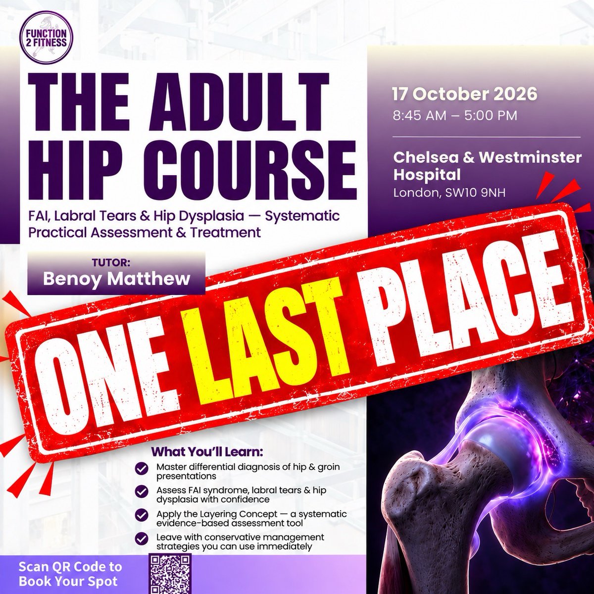
𝐀 𝐬𝐢𝐧𝐠𝐥𝐞 𝐩𝐥𝐚𝐜𝐞 𝐡𝐚𝐬 𝐣𝐮𝐬𝐭 𝐨𝐩𝐞𝐧𝐞𝐝 𝐮𝐩 𝐟𝐨𝐫 𝐭𝐡𝐢𝐬 𝐰𝐞𝐞𝐤𝐞𝐧𝐝'𝐬 𝐡𝐢𝐩 𝐜𝐨𝐮𝐫𝐬𝐞 𝐚𝐭 𝐂𝐡𝐞𝐥𝐬𝐞𝐚 𝐚𝐧𝐝 𝐖𝐞𝐬𝐭𝐦𝐢𝐧𝐬𝐭𝐞𝐫 𝐇𝐨𝐬𝐩𝐢𝐭𝐚𝐥, 𝐋𝐨𝐧𝐝𝐨𝐧
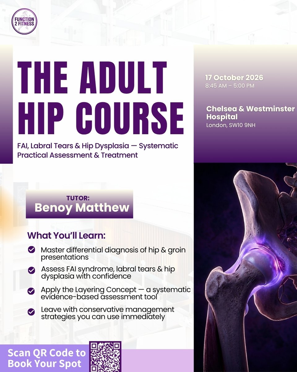
We are wrapping up our UK teaching calendar for Hip courses for 2026, this weekend at Chelsea and Westminster Hospital.
Following a last-minute cancellation, a single place has just opened up for the London hip course this Saturday, 17th October.
Once this weekend concludes, our next face-to-face teaching cohort will be out in Dubai.
If you have been looking to sharpen your hip and groin assessment before the year closes, here is what we are covering:
1. Intra-articular pathology differentiation — moving beyond vague anterior hip pain to isolate labral tears, FAI, and chondral wear with reliable clinical testing.
2. Navigating the clinical grey zones — distinguishing deep intra-articular pain drivers from overlying athletic pubalgia and adductor involvement.
3. The management framework — Access to the exact clinical protocols and rehabilitation progressions I use in clinic with my own patients.
4. Objective milestone planning — structured decision-making around when to progress loading, when to consider image-guided injection therapy, and when to refer onward.
Who this is designed for:
Band 6/7 MSK therapists, FCP clinicians, and private practitioners managing complex or recalcitrant hip and groin presentations who want clear, reproducible clinical pathways.
Because it is a single place, booking will be strictly first come, first served.
Secure the place here: https://t.co/S3Y0eFrjIJ
𝐓𝐫𝐞𝐚𝐭𝐢𝐧𝐠 𝐚𝐥𝐥 𝐬𝐡𝐢𝐧 𝐩𝐚𝐢𝐧 𝐥𝐢𝐤𝐞 𝐦𝐞𝐝𝐢𝐚𝐥 𝐭𝐢𝐛𝐢𝐚𝐥 𝐬𝐭𝐫𝐞𝐬𝐬 𝐬𝐲𝐧𝐝𝐫𝐨𝐦𝐞 𝐰𝐨𝐫𝐤𝐬—𝐮𝐧𝐭𝐢𝐥 𝐲𝐨𝐮 𝐦𝐢𝐬𝐬 𝐚𝐧 𝐚𝐧𝐭𝐞𝐫𝐢𝐨𝐫 𝐭𝐢𝐛𝐢𝐚𝐥 𝐜𝐨𝐫𝐭𝐞𝐱 𝐛𝐨𝐧𝐞 𝐬𝐭𝐫𝐞𝐬𝐬 𝐢𝐧𝐣𝐮𝐫𝐲
We are seeing a noticeable rise in bone stress injuries across runners.
The clinical challenge is that the highest-risk sites masquerade as everyday, lower-risk presentations.
If you treat running injuries, here are three high-risk bone stress injuries you cannot afford to miss:
Femoral neck bone Stree: More prevalent in female runners and strongly associated with low energy availability. It routinely presents as anterior groin or hip pain, closely mimicking intra-articular hip pathology, labral tears, and femoroacetabular impingement.
Anterior tibial cortex bone stress: Frequently misdiagnosed as standard medial tibial stress syndrome. While posteromedial shin pain is common and lower risk, anterior cortex stress carries a high risk of non-union or complete fracture if managed as simple shin splints.
Missing the distinction between a low-risk presentation and a high-risk cortical site turns a manageable deload into a catastrophic fracture.
On Sunday 18th October, we are running an online event, focusing on the two most critical sites: femoral neck and tibial bone stress injuries.
We will cover:
• Early differential triage to stop you missing them on clinic day
• Staged rehabilitation protocols from early offloading to return-to-run criteria
Full details and registration:
https://t.co/0D4U3Kq5QE
(The full recording will be sent to all registrants if you cannot attend live.)
#Physiotherapy #SportsPhysio #BoneStressInjury #RunningInjuries #MSKPhysio #SportsMedicine #Tibia #FemoralNeck
One month to go!
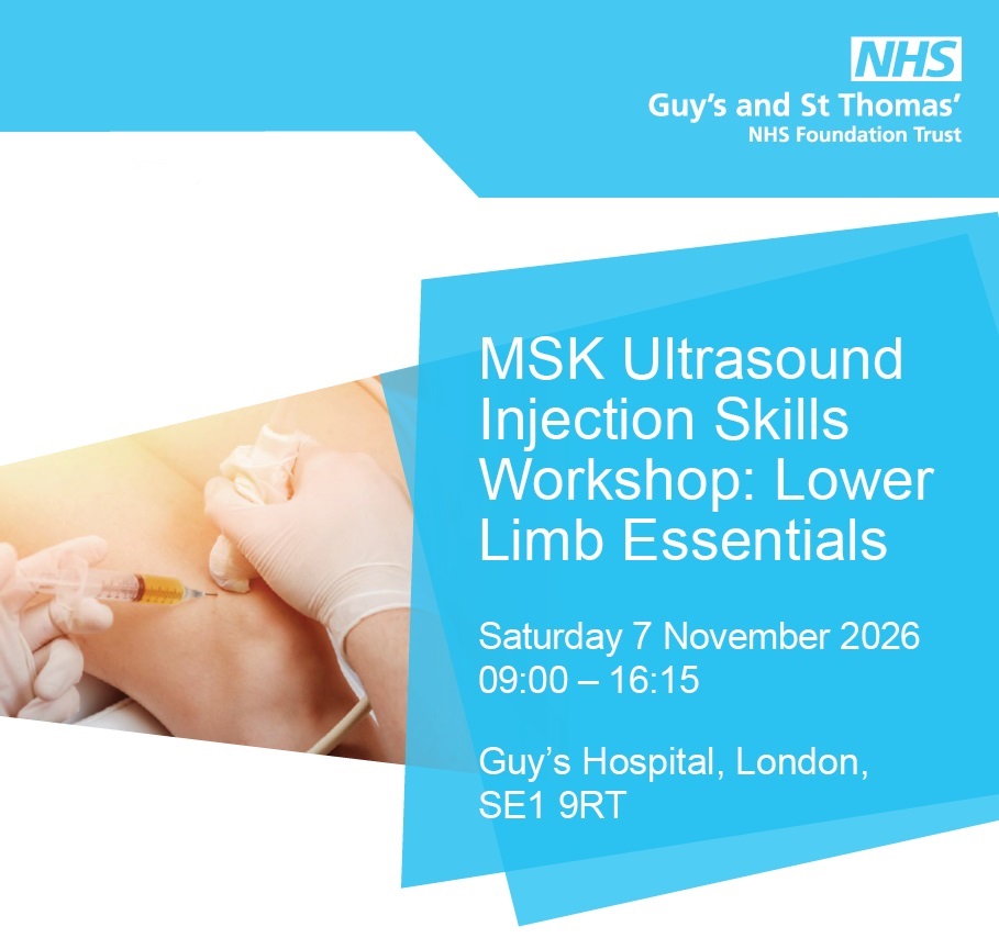
Our MSK Ultrasound Injection Skills Workshop: Lower Limb Essentials takes place at Guy’s Hospital on 7 Nov
Benefit from small-group teaching, tailored expert guidance and hands-on practice across hip, knee + foot
https://t.co/32uBdL1tos
#ultrasound#msk
𝐇𝐢𝐠𝐡-𝐑𝐢𝐬𝐤 𝐁𝐨𝐧𝐞 𝐒𝐭𝐫𝐞𝐬𝐬 𝐈𝐧𝐣𝐮𝐫𝐢𝐞𝐬 (𝐁𝐒𝐈) 𝐢𝐧 𝐑𝐮𝐧𝐧𝐞𝐫𝐬 🦴🏃♀️
Some bone stress injuries in runners are not simply “more severe versions” of common running injuries.
-The location matters.
-The biomechanics matter.
-Specific Rehab and Return to Running Matters
-Addressing Low Energy Availability and Metabolic factors matters
And getting the diagnosis wrong can have serious consequences.
Join Yasmin Palfrey and Benoy Mathew for a focused online session exploring two high-stakes sites for bone stress injury:
🔹 Tibial Bone Stress Injury
Are medial tibial stress syndrome and tibial bone stress injury separate conditions — or points on one continuum?
We’ll explore what imaging and histology actually tell us, and why anterior cortex lesions can behave very differently from posteromedial lesions.
🔹 Femoral neck BSI
Why do tension-side and compression-side injuries require very different management?
And what happens when they’re missed — including the risks of displacement, avascular necrosis and a prolonged return to running?
🎙️ Speakers
Yasmin Palfrey
Specialist Running Physio | MSK Sonographer
Benoy Mathew
Advanced Practice Physio | MSK Sonographer
📅 Sunday 18 October 2026
⏰ 9:00–11:00 AM UK time
💻 Online Event — Recording Available
If you assess runners with lower-leg, hip or groin pain, this is a session worth adding to your CPD calendar.
👉 Book your place:
https://t.co/0D4U3Kq5QE
Tag and share with a MSK colleague who sees runners regularly
𝐓𝐡𝐞 𝐡𝐚𝐫𝐝𝐞𝐬𝐭 𝐩𝐚𝐫𝐭 𝐨𝐟 𝐚𝐬𝐬𝐞𝐬𝐬𝐢𝐧𝐠 𝐭𝐡𝐞 𝐚𝐝𝐮𝐥𝐭 𝐡𝐢𝐩 𝐢𝐬 𝐫𝐚𝐫𝐞𝐥𝐲 𝐟𝐢𝐧𝐝𝐢𝐧𝐠 𝐩𝐚𝐢𝐧; 𝐢𝐭 𝐢𝐬 𝐤𝐧𝐨𝐰𝐢𝐧𝐠 𝐰𝐡𝐢𝐜𝐡 𝐩𝐨𝐬𝐢𝐭��𝐯𝐞 𝐟𝐢𝐧𝐝𝐢𝐧𝐠 𝐢𝐬 𝐝𝐫𝐢𝐯𝐢𝐧𝐠 𝐭𝐡𝐞 𝐩𝐫𝐞𝐬𝐞𝐧𝐭𝐚𝐭𝐢𝐨𝐧.
When symptoms overlap across the joint, adductors, and anterior pelvic structures, running through isolated special tests often produces conflicting findings rather than clarity.
A systematic approach changes how you evaluate these cases:
1. Map the presentation against mechanical versus non-mechanical patterns before testing individual tissues.
2. Group findings by clinical entities rather than relying on single, isolated provocation manoeuvres.
3. Differentiate intra-articular hip pathology from co-existing extra-articular or groin overload early in the exam.
4. Treat the clinical exam as a triage system that dictates whether your next step is load management, imaging, or secondary referral.
When your assessment follows a clear sequence, complex hip and groin pain becomes manageable rather than ambiguous.
This systematic framework forms the core of our workshop at Chelsea and Westminster Hospital, which has been updated annually across deliveries in 16 countries.
This will be our final London date for 2026 on 17 October, with just two seats remaining.
Booking link: https://t.co/8wmp9nylm0
If you have a UK or London-based colleague looking to refine their hip and groin assessment before the year ends, feel free to tag them below.
𝐄𝐩𝐢𝐬𝐨𝐝𝐞 𝟏𝟐: 𝐑𝐢𝐠𝐡𝐭 𝐈𝐧𝐣𝐞𝐜𝐭𝐢𝐨𝐧, 𝐖𝐫𝐨𝐧𝐠 𝐒𝐭𝐫𝐮𝐜𝐭𝐮𝐫𝐞: 𝐌𝐚𝐩𝐩𝐢𝐧𝐠 𝐂𝐨𝐦𝐦𝐨𝐧 𝐇𝐢𝐩 & 𝐆𝐫𝐨𝐢𝐧 𝐈𝐧𝐣𝐞𝐜𝐭𝐢𝐨𝐧𝐬
"𝐈 𝐡𝐚𝐝 𝐚𝐧 𝐢𝐧𝐣𝐞𝐜𝐭𝐢𝐨𝐧 𝐚𝐧𝐝 𝐢𝐭 𝐝𝐢𝐝𝐧'𝐭 𝐰𝐨𝐫𝐤." 𝐁𝐮𝐭 𝐰𝐡𝐢𝐜𝐡 𝐢𝐧𝐣𝐞𝐜𝐭𝐢𝐨𝐧, 𝐈𝐧𝐭𝐨 𝐰𝐡𝐚𝐭, 𝐚𝐧𝐝 𝐰𝐡𝐲?
Many patients can't tell you what they were injected with, whether it was guided, or what it targeted. In this episode, Callum and Benoy go through the common hip and pelvis injections and show why the clinical reasoning around the needle matters more than the needle itself.
In this episode:
-Why a "failed" injection is often the wrong injection for the wrong structure, such as a hip joint injection for adductor-related groin pain, or GTPS injections for ischio-femoral impingement
-The three jobs of an injection: calming pain, opening a window for rehab, and diagnosing what's actually driving symptoms
-Anterior hip and groin: intra-articular hip, pubic and osteitis pubis blocks, peritendinous adductor injections, and nerve blocks for inguinal-related pain
Takeaways:
-Imaging tells you what the hip looks like. The injection can help to identify the pain driver
-The injection isn't the intervention. Your clinical reasoning is.
Essential listening for physiotherapists, osteopaths and S&C coaches managing active patients with hip and groin pain.
🔗 Listen now: https://t.co/5NMOQVG09L
🎓 Want to go deeper on managing complex hip and groin presentations? Join Benoy on the Adult Hip Course, Details and booking links in the show notes.
UK HIP COURSES
https://t.co/8wmp9nylm0
𝐖𝐡𝐞𝐧 𝐚 𝐜𝐨𝐦𝐩𝐞𝐭𝐢𝐭𝐢𝐯𝐞 𝐇𝐲𝐫𝐨𝐱 𝐚𝐭𝐡𝐥𝐞𝐭𝐞 𝐟𝐚𝐢𝐥𝐬 𝐦𝐨𝐧𝐭𝐡𝐬 𝐨𝐟 𝐬𝐭𝐚𝐧𝐝𝐚𝐫𝐝 𝐠𝐫𝐨𝐢𝐧 𝐫𝐞𝐡𝐚𝐛, 𝐭𝐡𝐞 𝐢𝐬𝐬𝐮𝐞 𝐢𝐬 𝐚𝐥𝐦𝐨𝐬𝐭 𝐧𝐞𝐯𝐞𝐫 𝐭𝐡𝐞𝐢𝐫 𝐰𝐨𝐫𝐤 𝐞𝐭𝐡𝐢��.
This morning, Matti (Full Consent to Share) flew in from Malta for a second opinion.
He is a competitive Hyrox athlete who has been sidelined by persistent, pubic-related groin pain that had completely stalled under standard local physiotherapy.
𝐀 𝐩𝐚𝐭𝐢𝐞𝐧𝐭 𝐬𝐡𝐨𝐮𝐥𝐝𝐧’𝐭 𝐡𝐚𝐯𝐞 𝐭𝐨 𝐛𝐨𝐚𝐫𝐝 𝐚𝐧 𝐢𝐧𝐭𝐞𝐫𝐧𝐚𝐭𝐢𝐨𝐧𝐚𝐥 𝐟𝐥𝐢𝐠𝐡𝐭 𝐣𝐮𝐬𝐭 𝐭𝐨 𝐠𝐞𝐭 𝐚𝐧 𝐚𝐜𝐜𝐮𝐫𝐚𝐭𝐞 𝐝𝐢𝐚𝐠𝐧𝐨𝐬𝐢𝐬 𝐟𝐨𝐫 𝐜𝐡𝐫𝐨𝐧𝐢𝐜 𝐠𝐫𝐨𝐢𝐧 𝐩𝐚𝐢𝐧.
We blocked out a two-hour slot to unpick what had been missed.
Here is what that time allows when conservative management fails:
1. Standard protocols often fail chronic groin pain because the clinical label is too vague. Without distinguishing between adductor-related, pubic-related, or inguinal drivers, rehabilitation is just guesswork.
2. A standard 30 or 45-minute appointment rarely leaves enough time to map complex load tolerance against high-demand sporting movement.
3. Point-of-care ultrasound (POCUS) at the initial consult bridges the gap between subjective symptoms and structural load tolerance, letting us evaluate the adductor-aponeurotic plate and pubic symphysis dynamically.
4. Objective capacity testing paired with diagnostic imaging allows us to rule out secondary intra-articular hip pathology before building a progressive loading strategy.
5. Once you establish a precise working diagnosis and an agreed mechanical baseline, high-level rehabilitation does not require weekly clinic visits—it can be managed effectively through remote follow-up.
When rehabilitation fails, the answer is rarely more of the same generic exercises. It requires stepping back, giving the assessment adequate time, and finding diagnostic clarity.
For clinicians treating stubborn groin pain: What specific objective test or imaging threshold triggers you to step back and re-evaluate your initial working diagnosis?
Just over 5 weeks to go!
Join us at Guy’s Hospital London on 7 Nov for our MSK Ultrasound Injection Skills Workshop: Lower Limb Essentials
Hands-on training, live model scanning, expert guidance + advanced simulation
Spaces are filling fast! https://t.co/32uBdL1tos
#physio
𝐀𝐫𝐞 𝐰𝐞 𝐭𝐫𝐚𝐜𝐤𝐢𝐧𝐠 𝐫𝐮𝐧𝐧𝐢𝐧𝐠 𝐥𝐨𝐚𝐝 𝐚𝐥𝐥 𝐰𝐫𝐨𝐧𝐠 𝐛𝐲 𝐨𝐛𝐬𝐞𝐬𝐬𝐢𝐧𝐠 𝐨𝐯𝐞𝐫 𝐰𝐞𝐞𝐤𝐥𝐲 𝐦𝐢𝐥𝐞𝐚𝐠𝐞?
When assessing an injured runner, our first question is almost always: "How much did you run this week?"
We track weekly volume, calculate the acute-to-chronic workload ratio, and monitor week-to-week percentage increases.
Neither week-to-week changes nor the acute-to-chronic workload ratio showed a clear relationship with overuse running injuries.
Tissue does not fail under neat weekly averages. It fails under acute mechanical overload.
If we want to protect runners returning from injury, our subjective assessment needs to shift:
1. Stop relying solely on total weekly mileage as your primary indicator of tissue tolerance. Aggregated data hides the acute spikes that drive structural overload.
2. Ask what their single longest run was over the preceding four weeks. This establishes the true baseline capacity the runner has actually demonstrated recently.
3. Scrutinise the single-session jump. A jump from 10 km to 15 km in an individual run represents a massive surge in continuous load, even if the total weekly volume fits neatly within a 10% guideline.
4. Audit the next planned long run before they lace up. The clinical risk is rarely the baseline training load itself; it is the sudden, unprepared jump in a single session.
5. Treat single-session progression as its own distinct loading paradigm during rehabilitation, keeping session volume strictly proportional to recent tissue exposure.
The question is not just how much work a runner accumulates across seven days, but what specific mechanical demand their tissue had to survive in one single session.
We cover practical load prescription and objective return-to-running progressions in detail in our running rehabilitation modules on PhysioTutors.
https://t.co/ezKvBtHW0o
𝐖𝐡𝐞𝐧 𝐰𝐚𝐬 𝐭𝐡𝐞 𝐥𝐚𝐬𝐭 𝐭𝐢𝐦𝐞 𝐲���𝐮 𝐜𝐡𝐞𝐜𝐤𝐞𝐝 𝐭𝐡𝐞 𝐢𝐥𝐢𝐚𝐜 𝐭𝐮𝐛𝐞𝐫𝐜𝐥𝐞 𝐨�� 𝐚 𝐩𝐚𝐭𝐢𝐞𝐧𝐭 𝐫𝐞𝐟𝐞𝐫𝐫𝐞𝐝 𝐰𝐢𝐭𝐡 𝐧𝐨𝐧-𝐫𝐞𝐬𝐨𝐥𝐯𝐢𝐧𝐠 𝐮𝐧𝐢-𝐥𝐚𝐭𝐞𝐫𝐚𝐥 𝐥𝐨𝐰 𝐛𝐚𝐜𝐤 𝐩𝐚𝐢𝐧?
Not every persistent ache above the lateral pelvis is coming from the lumbar spine.
Over recent clinical courses, a consistent pattern keeps emerging: patients presenting with recalcitrant lateral pain around the iliac crest, carrying an entrenched diagnosis of a "chronic QL strain".
On closer assessment, the lumbar spine and quadratus lumborum are completely clear. The actual culprit is proximal iliotibial band (ITB) syndrome.
Here is why this diagnostic blind spot persists:
1. The anatomy sits right on the borderland between the lower back and the lateral pelvis, meaning clinicians reflexively treat the lumbar spine when the pathology is actually peripheral.
2. True quadratus lumborum strains rarely present as chronic, isolated pain at the lateral iliac crest without acute onset, significant spinal loading reproduction, or back involvement.
3. The proximal ITB origin at the iliac tubercle is often completely omitted during palpation, leaving an inflamed enthesis undiscovered.
4. Proximal ITB pathology is substantially more prevalent than many recognise, easily masquerading as either persistent back dysfunction or refractory lateral hip pain.
5. Labeling this as a muscular strain leads patients into repetitive stretching and soft-tissue work that often continually irritates the tendon insertion.
If a patient presents with long-standing "QL pain" that fails to shift with spinal rehab, take the lumbar spine off the hook and palpate the iliac crest tubercle.
How often are you assessing the proximal ITB insertion in patients presenting with persistent lateral crest pain?
(I will also be breaking down the clinical assessment and differential diagnosis of these lateral hip presentations in depth at Therapy Expo in Birmingham this November, alongside upcoming courses in London and Dubai.)
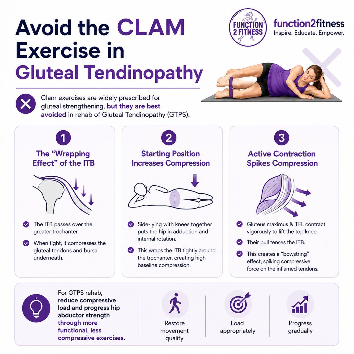
𝐀𝐯𝐨𝐢𝐝 𝐭𝐡𝐞 𝐂𝐋𝐀𝐌 𝐄𝐱𝐞𝐫𝐜𝐢𝐬𝐞 𝐢𝐧 𝐆𝐥𝐮𝐭𝐞𝐚𝐥 𝐓𝐞𝐧𝐝𝐢𝐧𝐨𝐩𝐚𝐭𝐡𝐲
Clam exercises are widely prescribed for gluteal strengthening, but they are best avoided in rehab of Gluteal Tendinopathy (GTPS). The primary reason is that the pathology of GTPS is driven largely by compressive overload rather than purely tensile failure, and the clamshell movement fundamentally amplifies this compressive force.
Here is the exact biomechanical breakdown of why this occurs:
1. The "Wrapping Effect" of the ITB
GTPS primarily involves insertional tendinopathy of the gluteus medius and minimus. These tendons insert onto the greater trochanter directly beneath the iliotibial band (ITB). When tension within the ITB increases, the band acts like a taut strap pulling down over the bony prominence of the greater trochanter, compressing the tendons and bursa underneath.
2. The Starting Position Increases Baseline Compression
The clam exercise is performed in a side-lying position. As the patient rests with their knees together, gravity pulls the top hip into a position of relative adduction and internal rotation. Adduction is the primary movement that causes the ITB to wrap tightly around the greater trochanter, establishing a high baseline of compressive force against the tendons before the active movement even begins.
3. Active Contraction Spikes Compressive Forces
During the active concentric phase of the clam, the hip is abducted and externally rotated against gravity (or a resistance band).
To execute this movement, both the gluteus maximus and the tensor fasciae latae (TFL) must contract vigorously.
Because the superficial fibres of the gluteus maximus and the TFL insert directly into the ITB, their contraction pulls the fascial band completely taut.
This tension creates a compressive "bowstring" effect. The active force translates into immense, localised pressure from the ITB directly against the inflamed gluteal insertions resting on the greater trochanter.
In tendinopathy rehabilitation, the goal is to apply controlled, progressive tensile load to the tendon to stimulate mechano-transduction and structural adaptation, whilst strictly minimising compressive irritation.
Clamshells do the exact opposite. By starting in adduction and then actively tensioning the overlying fascial structures, the exercise reproduces the precise pathomechanical driver that caused the tendinopathy in the first place.
📍 We'll be covering all things hip-related in far more depth on our upcoming Hip & Groin courses.
📍 UK: London (Oct) and Manchester (Sept) courses at:
https://t.co/8wmp9nylm0
Missed diagnoses are one of the trickiest parts of Hip and Running injuries assessments.
I sat down with Charlie Goodchild on his podcast to talk through the ones that slip through most often: Adult Hip dysplasia, Femoral neck bone-stress injuries, proximal ITB syndrome, ischiofemoral impingement, Osteitis pubis and Pudendal Neuralgia.
If Hips or running injuries are part of your caseload, this one's for you. Listen to the full episode at spotify and youtube with link below:
If interested in learning more on how to approach complex hip and groin, I will be covering all key pathologies in detail on my upcoming workshops - Sept (Manchester) and October (London).
Course Info and booking links at
https://t.co/8wmp9nylm0
Podcast Link:
https://t.co/blB8sJzg1q