

Top Tweets for #echoboards

Point related to attenuation.
⭕️ What is half-intensity depth?
🟢 It's the depth at which the ultrasound beam intensity drops to half of its original value.
The simplified formula is: 6/frequency in MHz
🟤 Example #echoboards question - a 3 MHz transducer loses half its intensity by what depth?
- 6/3 = 2 cm
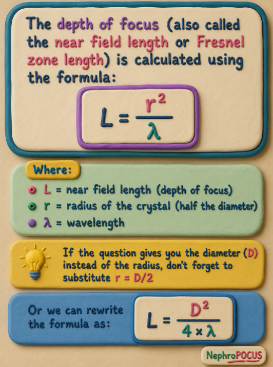
In the above 🧵, we learned that a bigger crystal keeps the beam focused deeper into tissue.
A #echoboards relevant question -
⭕️How do you calculate depth of focus when they give you crystal diameter and wavelength?
🟢L = r2/λ
L = near field length (depth of focus)
r = radius of the crystal
λ = wavelength
🟤For example, if given diameter = 10 mm and wavelength = 0.5 mm,
r = 5 mm; L = 5 square/0.5 = 25/0.5 = 50 mm
Another #echoboards favorite
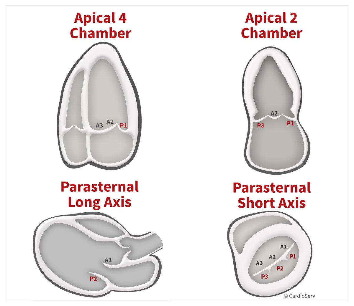
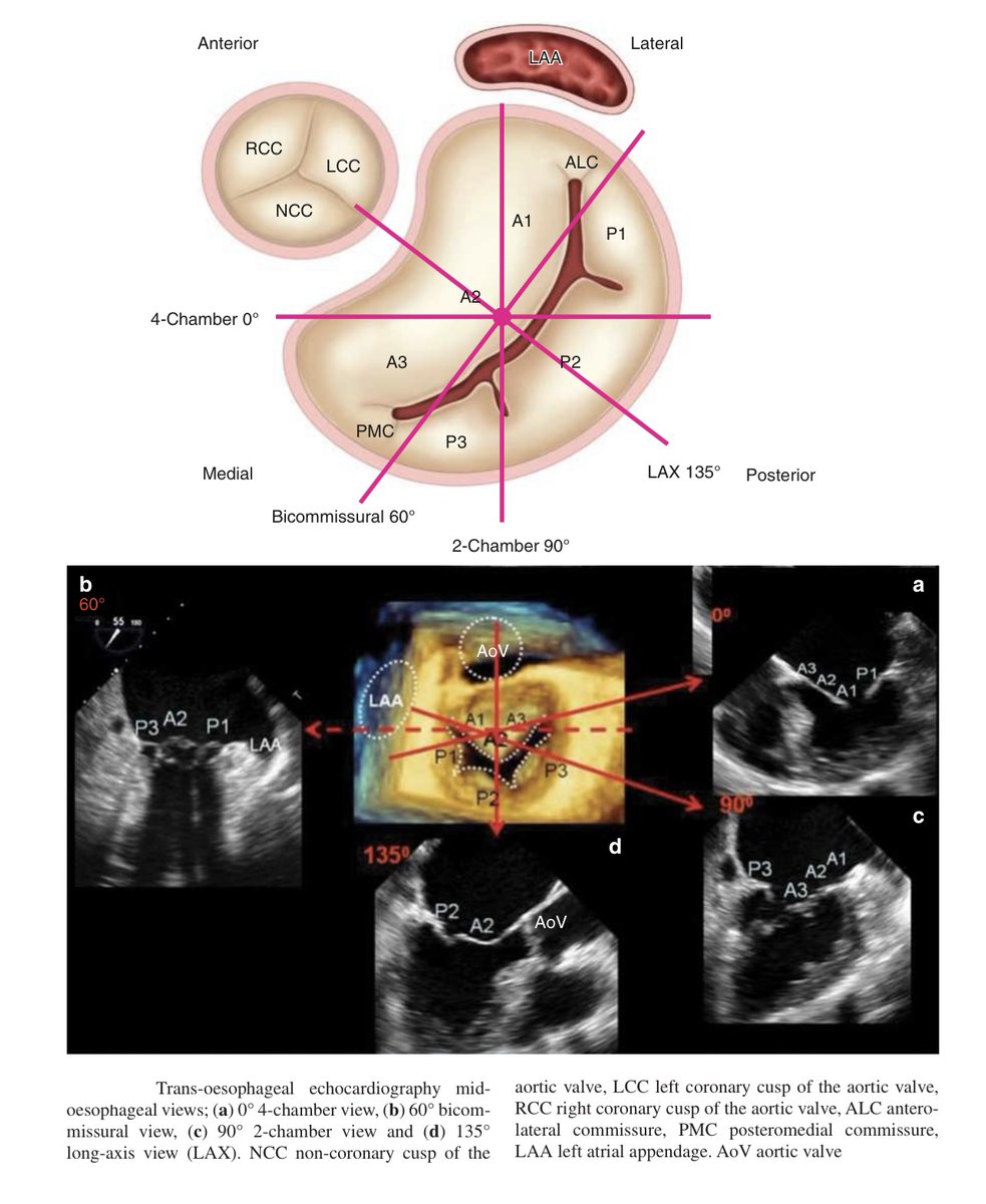
⭕️ Mitral valve segments and scallops seen on various #TEE (& TTE) views.
I’d mainly focus on -
✅ ME 4-chamber, commisural view and 3D short-axis (en face view)
✅ en face view tips:
⭕️12 o’clock: Aortic valve → adjacent to A2 (“Aortic = Anterior”).
⭕️9–10 o’clock: Left atrial appendage (LAA) → adjacent to the A1/P1 (anterolateral) commissure.
⭕️3–4 o’clock: Interatrial septum → adjacent to the A3/P3 (posteromedial) commissure.
Image courtesy: N. Moorjani et al., Operative Mitral and Tricuspid Valve Surgery (2018); Cardioserv.


I'll keep adding some tricky #EchoBoards trivia to this thread.
If you've made it this far, you're probably genuinely preparing for the exam rather than just randomly scrolling through your feed 👍
First, the questions about conservation:
⭕️PISA → Conservation of mass (flow continuity)
⭕️Continuity equation for aortic valve area → Conservation of mass
⭕️Bernoulli equation (ΔP = 4V²) → Conservation of energy
*Both PISA and the continuity equation are applications of the same law
#echofirst #POCUS
Fellows preparing for the #echoboards often say #physics is the toughest part of the exam. I tried to put together a thread 🧵covering the high-yield topics from wave mechanics and resolution to duty factor, tissue harmonic imaging, and more.
Hope you find it useful. Good luck!👍
#Physics notes for those taking #echoboards. Will add more points to this thread 🧵
#POCUS #Nephpearls
⭕️Sound is a mechanical wave that results from the back and forth vibration of the particles of the medium through which the sound wave is moving. These compressions and rarefactions are typically depicted as a sine wave.
⭕️ Wavelength (λ): The "distance" between two consecutive identical points on a wave (e.g., peak to peak or compression to compression), measured in millimeters.
⭕️Frequency: The number of complete wave cycles occurring per second, measured in Hertz (Hz).
⭕️ Amplitude: The maximum pressure variation (height) of the wave above or below the baseline.
⭕️Period: The "time" it takes for one complete wave cycle to occur, measured in seconds. It is the reciprocal of frequency: T = 1/f.
⭕️Spatial Pulse Length: The physical "length" of a pulse in space - the distance the pulse occupies in tissue. Equals the number of cycles in the pulse multiplied by the wavelength: SPL = number of cycles × λ (4 cycles in the example below).
⭕️Pulse Duration (PD): The total "time" the transducer is transmitting a single pulse. PD = number of cycles × period.
⭕️Pulse Repetition Period (PRP): The time from the "start" of one pulse to the "start" of the next pulse, including both the transmit time and the listening (receive) time. Determined by imaging depth.
⭕️Pulse Repetition Frequency (PRF): The number of pulses emitted per second, measured in Hz. It is the reciprocal of PRP: PRF = 1/PRP. Determined by imaging depth - deeper imaging requires lower PRF.

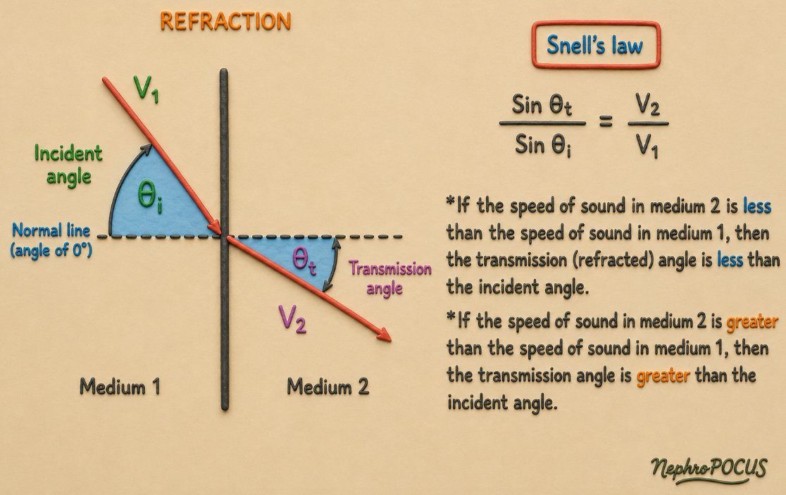
Another potentially testable concept on #echoboards - Snell's law
⭕️Snell's law describes how an ultrasound beam changes direction (refracts) when it crosses a boundary between two tissues with different propagation velocities.
sinθt/sinθi = V2/V1
θi - angle of the incoming beam (angle of incidence)
θt - angle of the transmitted beam (angle of refraction)
V2 - speed of sound in the second tissue
V1 - speed of sound in the first tissue (incident)
🌟Both angles are measured from the perpendicular (normal) to the tissue boundary, not from the boundary itself. *Very important to understand‼️*
🎯 Board pearl - If the speed of sound in medium 2 is less than the speed of sound in medium 1, then the transmission (refracted) angle is less than the incident angle and vice versa.
*Since it violates the machine's assumption that ultrasound travels in a straight line, refraction may result in artifacts (e.g., second copy of a true reflector).
Now that we touched upon contrast, one high-yield #echoboards question is about mechanical index.
⭕️The mechanical index (MI) is essentially a measure of how hard the ultrasound beam "pushes" on tissue - and more importantly for contrast imaging, how aggressively it interacts (mechanically disturb or rupture) with microbubbles.
⭕️ Think of microbubbles as tiny, gas-filled balloons (1-4 μm diameter) 🎈🎈floating in the bloodstream. The MI determines whether those balloons gently wobble, violently oscillate, or pop.
🌟 Recommended MI for real-time contrast echocardiography: 0.1 to 0.3 (basically, low MI is good) 🌟
⭕️ High MI destroys microbubbles. Do we ever use high MI? - Clinically used as brief "flash" impulses (MI 0.8–1.2, lasting 5–15 frames) to deliberately clear contrast from the myocardium, then the rate of replenishment is observed at low MI to assess myocardial perfusion.
Duty factor is another testable concept for #echoboards
⭕️ In diagnostic ultrasound, the transducer does not send out sound continuously. Instead, it works in a "pulse-listen" pattern - sends a short burst (pulse) of sound, then stays quiet and "listens" for returning echoes before sending the next pulse. The duty factor describes how much of each cycle is spent transmitting versus listening.
⭕️ other way of saying it - fraction of time the transducer is actually "on" and transmitting sound, compared to the total time of one pulse cycle (transmitting + listening).
⭕️ Formula: Duty factor = Pulse duration (PD) ÷ Pulse repetition period (PRP)
🟤 In diagnostic imaging, the duty factor is very small - typically around 0.1% to 1% because the transducer spends the vast majority of its time listening for echoes rather than transmitting.
⭐️ In continuous wave (CW) Doppler, the duty factor is 100% (or 1.0), because the transducer transmits continuously (one element sends while another receives).

Tweet no. 13 #echoboards #Physics thread.
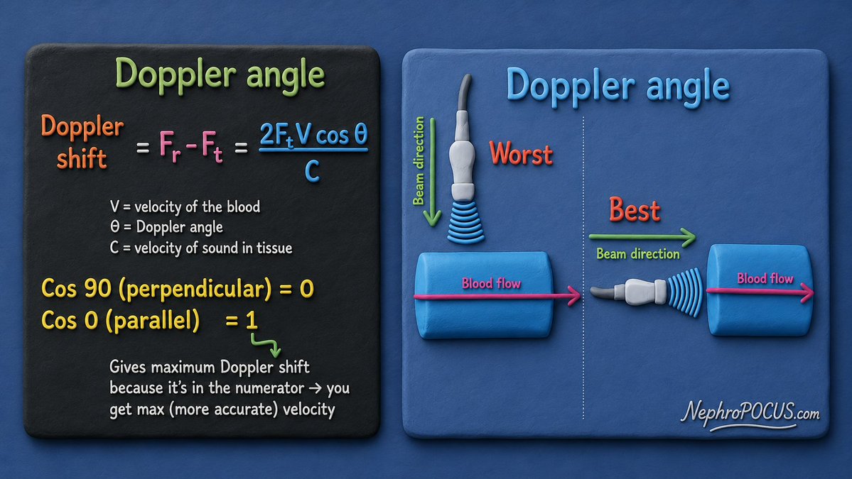
Doppler equation - memorize this ‼️
⭕️The Doppler shift is the difference between the frequency transmitted by the transducer and the frequency returning from moving blood cells.
Simple concept:
🩸 Faster blood flow → Bigger Doppler shift → Higher measured velocity.
🩸 Slower blood flow → Smaller Doppler shift → Lower measured velocity.
Positive vs. Negative Doppler shift
➡️ Blood moving toward the transducer → Returning frequency is higher than transmitted → Positive Doppler shift (flow toward the probe).
⬅️ Blood moving away from the transducer → Returning frequency is lower than transmitted → Negative Doppler shift (flow away from the probe).
✅ 0° (beam parallel to flow) → cos 0 = 1 (= max numerator) → Maximum Doppler shift → Most accurate velocity.
✅ 90° (beam perpendicular to flow) → cos 90 = 0 → No Doppler shift → Velocity cannot be measured.
✅ As the Doppler angle increases, cos θ decreases, so the Doppler shift becomes smaller and the measured velocity is underestimated.
⭐️ Bottomline: Parallel angle is the best for Doppler.
In #echofirst, it is not recommended to use 'angle-correct' feature on the machine.
Instead, obtain a view that makes the beam as parallel to blood flow as possible. #Anatomy is the 🔑
#Physics notes for those taking #echoboards. Will add more points to this thread 🧵
#POCUS #Nephpearls
⭕️Sound is a mechanical wave that results from the back and forth vibration of the particles of the medium through which the sound wave is moving. These compressions and rarefactions are typically depicted as a sine wave.
⭕️ Wavelength (λ): The "distance" between two consecutive identical points on a wave (e.g., peak to peak or compression to compression), measured in millimeters.
⭕️Frequency: The number of complete wave cycles occurring per second, measured in Hertz (Hz).
⭕️ Amplitude: The maximum pressure variation (height) of the wave above or below the baseline.
⭕️Period: The "time" it takes for one complete wave cycle to occur, measured in seconds. It is the reciprocal of frequency: T = 1/f.
⭕️Spatial Pulse Length: The physical "length" of a pulse in space - the distance the pulse occupies in tissue. Equals the number of cycles in the pulse multiplied by the wavelength: SPL = number of cycles × λ (4 cycles in the example below).
⭕️Pulse Duration (PD): The total "time" the transducer is transmitting a single pulse. PD = number of cycles × period.
⭕️Pulse Repetition Period (PRP): The time from the "start" of one pulse to the "start" of the next pulse, including both the transmit time and the listening (receive) time. Determined by imaging depth.
⭕️Pulse Repetition Frequency (PRF): The number of pulses emitted per second, measured in Hz. It is the reciprocal of PRP: PRF = 1/PRP. Determined by imaging depth - deeper imaging requires lower PRF.
#echoboards prep question
What abnormality is shown here?
Clue: h/o Kawasaki disease

#echoboards prep question
What type of prosthetic valve is shown here?
POLL below.
#echoboards prep question
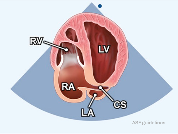
Which tricuspid valve leaflets are typically seen in this view?
POLL in thread.
#POCUS
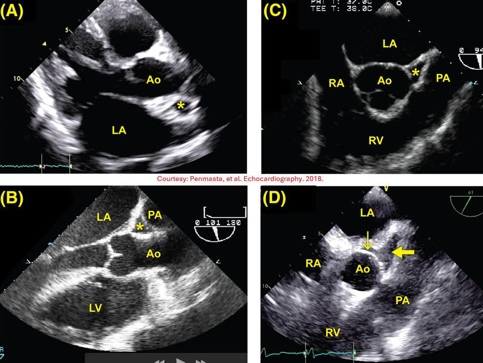
🫀#echoboards prep question
The asterisk in panels A-C and the arrow in panel D all indicate the same anatomic structure. What is it?
📌 Panel A is a transthoracic echocardiogram
📌 Panels B–D are transesophageal echocardiograms
Recommendation of the day: If you're preparing for advanced peri-op #TEE #echoBoards, follow @alex1708ander.
His cases are excellent for training your 👁️ to recognize a wide range of pathology. I wish I had found his handle before my exam, but I'm sure it'll be a valuable resource for the next generation of #POCUS enthusiasts.


🫀 CardioNugget™
😴 Hibernating myocardium
Chronic reduction in blood flow at rest
❤️ Myocardium "downregulates" contraction to survive
Improves only after revascularization
🧠 Think: "The heart is conserving energy."
#EchoBoards #Cardiology #CardioNugget™ #BoardPrep #CardioTwitter

🫀 CardioNugget™
⚡ Stunned myocardium
Brief severe ischemia → reperfusion restores blood flow
💥 Temporary contractile dysfunction despite normal perfusion
Recovery: hours to days (sometimes weeks)
🧠 Think: "The heart is shocked."
#EchoBoards #Cardiology #CardioNugget™ #BoardPrep #CardioTwitter

#echoboards #POCUS
❓PEDOF (probe) stands for: (POLL in thread)
A. Pulsed Echo Doppler Oscillation Frequency
B. Pulsed Echo Doppler Flowmeter
C. Phased Echo Doppler Output Filter
D. Pulsed Emission Doppler Oscillation Flow

Important artifacts for #echoboards
Mirror image
Beam takes a longer path → structure placed too "deep".
Refraction
Beam bends → structure placed in wrong "lateral" position.
#POCUS #FOAMed
(*Animations from ASE artifact guidelines 2026)
Trends for you
Most Popular Users

Elon Musk 
@elonmusk
240.8M followers

Barack Obama
@barackobama
119.2M followers

Donald J. Trump
@realdonaldtrump
111.8M followers

Cristiano Ronaldo
@cristiano
111.4M followers

Narendra Modi
@narendramodi
107M followers

Rihanna
@rihanna
97.8M followers

NASA
@nasa
92.2M followers

Justin Bieber
@justinbieber
91.1M followers

KATY PERRY
@katyperry
88.1M followers

Taylor Swift
@taylorswift13
81.9M followers

Lady Gaga
@ladygaga
73.5M followers

Virat Kohli
@imvkohli
70.5M followers

Kim Kardashian
@kimkardashian
70M followers

YouTube
@youtube
68.7M followers

Bill Gates
@billgates
64.1M followers

Neymar Jr
@neymarjr
63.4M followers

The Ellen Show
@theellenshow
62.4M followers

CNN
@cnn
61.9M followers

Selena Gomez
@selenagomez
61.1M followers

X
@x
60.8M followers