Top Tweets for #molPath

Paratesticular biphasic mesothelioma harboring BRAF V600E mutation 🧬🧬
🚨Check out our case report: The First Report of BRAF V600E Mutation in Paratesticular Mesothelioma @IJSPjournal @MGBpathology
➡️https://t.co/trjcKtB1LC
#GUpath #MolPath


Great fun presenting at the @IAP_AUS molecular pathology session today!
Managed to paraphrase @gradecricketer while drawing parallels between methylation profiling 🧠🧬and DRS 🏏
#molpath #neuropath


A great reminder 💭 that recognizing subtle cytologic clues can directly impact diagnosis, prognosis, and even targeted therapy options.
#CytoJ #SalivaryGland #FNAC #SecretoryCarcinoma #MolPath https://t.co/cUcPzQZLYE"

Now there are 3.
Thyroid neoplasms harboring a novel OCLN::PRKCI gene fusion
https://t.co/N0ZmiTlV1n
https://t.co/p3uzbkgExG
#EndocrinePath #moldx #molpath


Happy to see our @Pathoutlines article on PATZ1-rearranged sarcoma published 👇🏼
https://t.co/uziryeRBEN
3 things to remember about PATZ1-rearranged sarcoma:
💎Pseudoalveolar/(micro)cystic change->Swiss 🧀-like appearance
💎Hyalinized blood vessels
💎Rhabdomyoblastic differentiation often seen -- skeletal muscle marker expression tends to be accentuated around the pseudoalveolar spaces
Check out our article online (link above) 😊
#PathTwitter #BSTPath


#MolPath #PathTweetAward2026
Apologies for the delayed repost — I’ve been away from #PathX for a bit and will do my best to catch up over the coming days.
Huge congratulations @avash22 on winning the March #PathTweetAward ! Your mTOR tweetorial is outstanding: clear, thoughtful, and highly educational. A very well-deserved recognition for such a valuable contribution to the #PathX community.
Many thanks for the time and effort you put into this educational thread. It is truly appreciated 🙏💫💫💫

Monthly #PathTweetAward trainee standout (March 2026) 🏆
https://t.co/KN818hwEpP
👏 Congrats on your impact on #PathTwitter!
@adi_agnihotri @ariella8 @archibhat3 @PoojaSMD @cebulka26 @LakshmiKoulmane @pembeoltulu @VijayPatho @VHNguyenMD @smlungpathguy @ahsanuitis @jsamsoondar


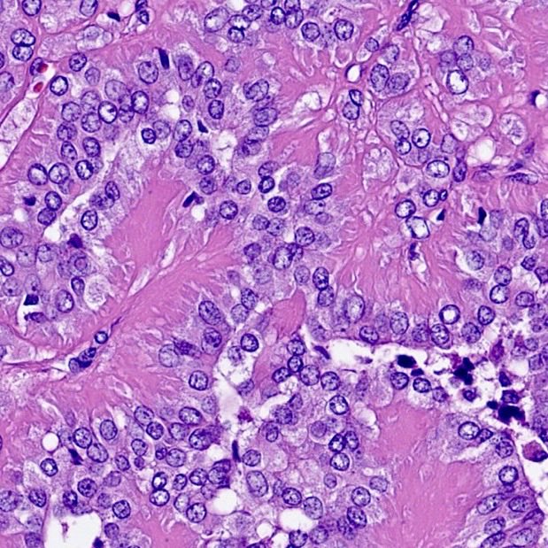
One gene, many faces 🧬 🎭
#gupath🤝#molpath #pathreads
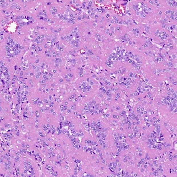
DICER1-associated renal tumors can mimic a wide spectrum of renal neoplasms — another reminder that molecular testing is becoming indispensable in modern renal pathology
🔗 https://t.co/ihz4hmuQyh


Lets raise awareness regarding the spectrum of Dicer1-mutated kidney tumors.
Free access link: https://t.co/uyxOo1lEDR


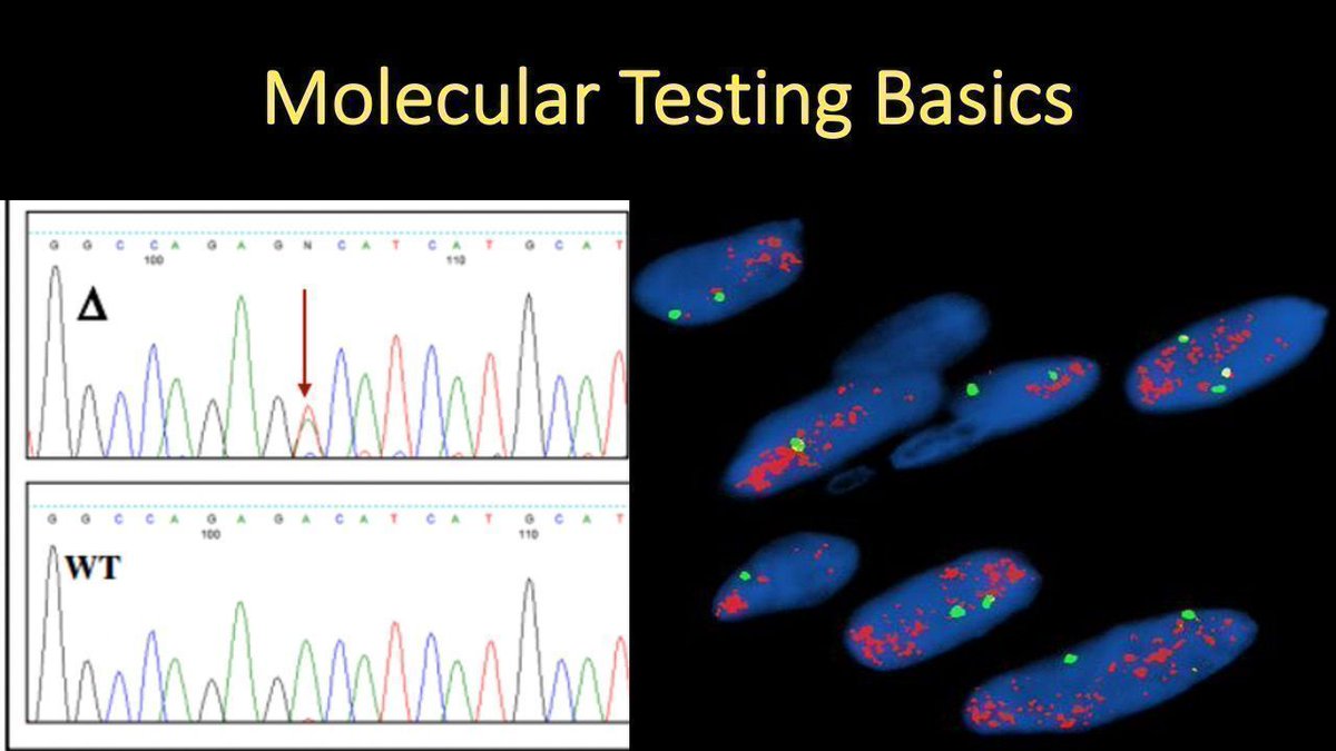
Molecular Testing Basics explained in 15 minutes (video): https://t.co/wcwsYeKYIl . A simple explanation of the main types of molecular tests #pathologists use, when we use them, & why. #pathology #pathTwitter #MedTwitter #MedEd #MolPath #dermtwitter #dermpath

We have an opening for the Molecular Genetic Fellowship Program at MD Anderson for 2027-2028 academic year. Contact info 👇 for anyone who is interested in applying
#molpath #pathology
https://t.co/OIUP9WbYzE

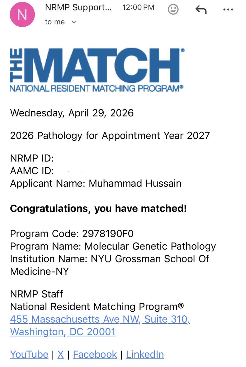
NYU remains home for the next two years. 🧬 ✨
Grateful and excited to share that I’ve matched in Molecular Genetic Pathology at @NYUGSOM_Path ! 🧬
Looking forward to continuing my journey here after my Hematopathology fellowship. #pathx #molpath

Very pleased to share an online piece brought to you by the CAP Personalized Health Care Committee. By Drs. Pearl Audon and Gagan Mathur. Cell-free DNA in transfusion medicine! 🤯 🧬 🩸 #PathTwitter #molpath #clinpath
@Pathologists
https://t.co/B19MRNQt8s
#breastpath #Gynpath #Molpath #Diagnexia
Excelente oportunidad para ver a la Dra. Janira Navarro en un tema actual :
Patología molecular práctica para neoplasias endometriales y mamarias
30 de abril de 2026 | 11:00 a. m. EST | 4:00 p. m. GMT | En español
Una sesión enfocada en consideraciones diagnósticas prácticas en patología molecular para neoplasias endometriales y mamarias.¡Webinario en español!
A registrarse ↘️

Join Dr Janira Navarro for:
Practical Molecular Pathology for Endometrial and Breast Malignancies
April 30, 2026 | 11AM EST | 4PM GMT | En español
A focused session on practical diagnostic considerations in Molecular Pathology for Endometrial and Breast Malignancies.
¡Webinar en español!
👉 Register: https://t.co/BFm3AOn3JT
#Diagnexia #PathologyEducation

Very pleased to share an online piece brought to you by the CAP Personalized Health Care Committee. By Drs. Jeremy Ward and Matthew Hiemenz. Navigating Discordant MMR/MSI Test Results! #PathTwitter #molpath #surgpath
@Pathologists
https://t.co/rkkwYOV2Ak

This educational resource does not constitute medial or legal advise and is not intended for use in the diagnosis and treatment of individual conditions.
#AMPtutorials #Molpath #path2path #PathX
🚨 Submit #AMPtutorials to win FREE registration to #AMP2026 in Seattle! 🎓🏆
➡️Instructions:
https://t.co/P5W6uZZiY6
@AMPath @pvhernandezmd @whosainastro @phuongn_nguyen @alexanderjneil
#SocialMediaChallenge #MolPath

Participate in our #AMPtutorials scicomm contest on social media for a chance to win free registration to #AMP2026 in Seattle next year! How to enter: https://t.co/tiRntfZMd9

🔬 Excited to present our spatial transcriptomic studies on prostate and bladder cancer at #USCAP2026!
@MGBpathology @TheUSCAP
#GUpath #molpath


🧬 How are AI and molecular pathology reshaping hematologic diagnostics?
Discover the translational opportunities and practical insights driving the next wave of innovation by @alnoorakber.
📄 https://t.co/XTPbo8JL1m
#AI #HemePath #MolPath #Diagnostics

Check out our recent publication 👉Molecular Pathology, Artificial Intelligence, and New Technologies in Hematologic Diagnostics: Translational Opportunities and Practical Considerations https://t.co/uHckhxnzcZ #mdpidiagnostics via @diagnostic_mdpi
#AI #heempath #molpath
Check out our recent publication 👉Molecular Pathology, Artificial Intelligence, and New Technologies in Hematologic Diagnostics: Translational Opportunities and Practical Considerations https://t.co/uHckhxnzcZ #mdpidiagnostics via @diagnostic_mdpi
#AI #heempath #molpath

New review online! 📝
Updates in the molecular pathology of breast tumors, written with @HannahYWen, in an upcoming issue of Surgical Pathology Clinics edited by @Sinchita_Roy.
Check it out here:
https://t.co/UzsQVwbkv9
#PathX #PathTwitter #breastpath #molpath

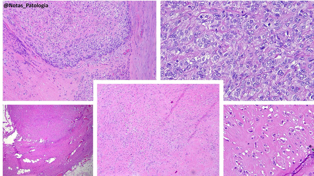
🔬YAP1::KMT2A‑rearranged sarcoma (with emphasis on diagnostic pitfalls)🔬
Rare fibroblastic sarcoma recently defined by YAP1::KMT2A fusion and a unique methylation profile, often mimicking sclerosing epithelioid fibrosarcoma (SEF), low‑grade fibromyxoid sarcoma (LGFMS), or even benign fibroblastic lesions.
Definition 📚
Soft tissue sarcoma characterized by YAP1–KMT2A/KMT2A–YAP1 fusions, SEF‑like/LGFMS‑like morphology, and an epigenetic cluster distinct from classic MUC4‑positive SEF and LGFMS.
Epidemiology 📊
Predominantly affects young to middle‑aged adults (median around 35–40 years, but a wide 9–90+ years range).
No clear sex predilection, with a slight male predominance in some series.
Sites 📍
Mainly arises in somatic soft tissues of trunk and extremities (deep or subcutaneous), including paraspinal regions, chest wall, axilla, and retroperitoneum.
Uncommon presentations include acral, suboccipital, and rare primary bone cases.
Pathogenesis 🧬
Driven by YAP1–KMT2A fusion, usually in a complex YAP1–KMT2A–YAP1 configuration, preserving the TEAD‑binding domain of YAP1 and the CXXC domain of KMT2A.
Unlike KMT2A‑rearranged leukemias, these tumors lack a strong HOXA signature, suggesting a distinct mechanism involving Hippo/YAP signaling and epigenetic remodeling.
Clinical Features 🩺
Presents as a slow‑growing mass, sometimes present for years, often becoming painful or clinically relevant only at later stages.
Initial pathology reports may label it as benign (fibromatosis, fibroma, “fibrous histiocytoma”) until recurrence or progression reveals malignant behavior.
Laboratory Diagnosis 🧪
No specific serum marker; diagnosis is histologic and molecular.
FISH for EWSR1/FUS is negative; FISH for KMT2A/YAP1 can be falsely negative, so broader DNA/RNA NGS panels or RNA‑seq are often required.
Histopathology 🔬
Architecture: grossly nodular tumors, but with infiltrative borders and “honeycomb” spread through subcutaneous fat and skeletal muscle.
Cytology: ovoid/epithelioid cells in cords, nests or solid sheets within dense collagenous stroma; mitotic activity usually low in hypocellular areas, higher in more solid/anaplastic foci.
Special patterns: perineurioma‑like areas (bland spindle cells in whorled/loose‑storiform pattern), fibroma‑like zones (very dense collagen with sparse cells), and foci of small round or rhabdoid cells in recurrences/metastases.
Additional findings: thin‑walled vessels (sometimes hemangiopericytoma‑like); typical absence of alternating fibromyxoid zones and classic collagen rosettes of LGFMS.
Immunohistochemistry 🧫
Positives:
EMA (diffuse or focal)
YAP1 (N‑ and C‑terminal)
Cyclin D1 in most cases
CD34 and SMA may be focally positive
Negatives:
MUC4 consistently negative (key point against classic SEF/LGFMS)
S100, SOX10, desmin, STAT6, most endothelial markers in typical cases
⚠️Notes:
Some tumors show strong ERG/CD31/D2‑40 and TFE3 expression, closely mimicking epithelioid hemangioendothelioma; in such cases, documentation of YAP1–KMT2A fusion and molecular clustering are crucial for correct classification.
Differential Diagnosis 🧠
Classic SEF – MUC4 strongly positive with EWSR1/FUS–CREB3L1 fusions, more frequent collagen rosettes and an overall more aggressive clinical course.
LGFMS – fibromyxoid pattern with arcades of vessels and collagen rosettes, MUC4 diffuse, FUS–CREB3L2/CREB3L1 fusions.
Perineurioma/DFSP – honeycomb infiltration and CD34 expression may overlap, but they lack SEF‑like morphology and YAP1–KMT2A fusion.
Epithelioid hemangioendothelioma – blister cells and endothelial markers; typical EHE harbors WWTR1/YAP1–TFE3 fusions and falls into a different molecular cluster.
Fibromatosis/scar – fibroma‑like areas may mislead; subtle atypia, transition to more cellular SEF‑like zones, and EMA/cyclin D1 expression favor sarcoma.
Prognosis 📈
Intermediate prognosis between LGFMS and classic SEF: local recurrence in roughly 20–30% and metastases in about 50% of patients with follow‑up, mainly to lungs/pleura.
Overall survival is worse than LGFMS but better than classic SEF; robust histologic or molecular risk stratifiers within this group are not yet well defined.
Treatment 💉
Mainstay is wide surgical excision with negative margins, often combined with adjuvant radiotherapy for deep‑seated or margin‑positive lesions.
Evidence for systemic chemotherapy is limited; extrapolation from SEF suggests modest responses to conventional sarcoma regimens, and no validated targeted therapies are currently established for this fusion.
Take-Home Messages @Notas_Patologia ✅
📌Consider YAP1::KMT2A‑rearranged sarcoma in SEF‑like/LGFMS‑like tumors that are MUC4‑negative.
MUC4 negativity + EMA/cyclin D1 positivity and a YAP1–KMT2A fusion define the diagnosis.
📌FISH for KMT2A/YAP1 may be falsely negative – broader NGS or RNA‑seq is often needed.
📌DNA methylation profiling shows a distinct cluster, separate from SEF and LGFMS.
📌Clinically, this is a malignant sarcoma with a real risk of lung metastasis, even when histology appears deceptively bland.
Selected References 📖
Warmke LM et al. YAP1::KMT2A‑rearranged sarcomas harbor a unique methylation profile and are distinct from sclerosing epithelioid fibrosarcoma and low‑grade fibromyxoid sarcoma. Virchows Arch, 2024.
Massoth LR et al. Pan‑sarcoma genomic analysis of KMT2A rearrangements reveals distinct subtypes defined by YAP1–KMT2A–YAP1 and VIM–KMT2A fusions. Modern Pathology, 2020.
Puls F et al. Recurrent fusions between YAP1 and KMT2A in morphologically distinct neoplasms within the spectrum of low‑grade fibromyxoid sarcoma and sclerosing epithelioid fibrosarcoma. American Journal of Surgical Pathology, 2020.
Agarwal A et al. YAP1–KMT2A fusion‑positive sarcoma: an emerging soft tissue tumor entity with morphological features resembling sclerosing epithelioid fibrosarcoma. Indian Journal of Pathology and Microbiology, 2024.
⚠️Disclaimer⚠️
“This text is an educational summary for healthcare professionals and students. It does not replace full pathology reports, local guidelines, or individualized clinical decision-making.”
Hashtags:
#MedicalEducation #NotasDePatologia #SoftTissuePathology #Sarcoma #PathTwitter
🖋️ Case and slide coloration courtesy of Dr. Alexandre Carneiro (@AmcarneiroMD), as part of an academic partnership project.
Last Seen Hashtags on Sotwe
Most Popular Users

Elon Musk 
@elonmusk
240.3M followers

Barack Obama
@barackobama
119.2M followers

Donald J. Trump
@realdonaldtrump
111.6M followers

Cristiano Ronaldo
@cristiano
109.7M followers

Narendra Modi
@narendramodi
106.9M followers

Rihanna
@rihanna
97.5M followers

NASA
@nasa
92.1M followers

Justin Bieber
@justinbieber
90.7M followers

KATY PERRY
@katyperry
87.2M followers

Taylor Swift
@taylorswift13
81.1M followers

Lady Gaga
@ladygaga
72.6M followers

Kim Kardashian
@kimkardashian
69.6M followers

Virat Kohli
@imvkohli
69.2M followers

YouTube
@youtube
68.6M followers

Bill Gates
@billgates
63.6M followers

The Ellen Show
@theellenshow
62.5M followers

Neymar Jr
@neymarjr
61.9M followers

CNN
@cnn
61.9M followers

X
@x
60.9M followers

Selena Gomez
@selenagomez
60.3M followers